Fisioter. Mov., Curitiba, v. 30, n. 3, p. 537-547, Jul./Sep. 2017 Licenciado sob uma Licença Creative Commons DOI: http://dx.doi.org/10.1590/1980-5918.030.003.AO12
Spastic diparetic does not directly affect the capacity to ascend
and descend access ramps: three-dimensional analysis
Diparesia espástica não afeta a capacidade de subir e
descer rampas de acesso: análise tridimensional
Tainá Ribas Mélo[a,b], Ana Tereza Bittencourt Guimarães[c], Vera Lúcia Israel[a]*
[a] Universidade Federal do Paraná (UFPR), Curitiba, PR, Brazi
[b] Scholarship holder REUNI/Ministry of Education and Culture, Brazil [c] Universidade Estadual do Oeste do Paraná (UNIOESTE), Cascavel, PR, Brazil
[R]
Abstract
Introduction: Diplegic children have difficulties in gait and therefore ramps are used as strategies of acces -sibility. Objective: The present study investigated the influence of an inclined surface (ascending and descend -ing) on the kinematic characteristics during gait of the diplegic group (DG) when compared to typically de -veloping children of the control group (CG). Methods: Study participants included 20 children (10 with DG and 10 CG) matched by age, which were evaluated in three experimental conditions (horizontal and inclined ascending and inclined descending surfaces of 7º) through an optoelectronic imaging system. Results: Among the linear kinematic variables, only step width differed among groups, however, without influence of the sur -face. The foot height differed among the groups only in the descending phase, where DG had greater difficulty in raising the foot. The 3-dimensional gait analyses could not provide more evidences of differences in kine -matics variables, especially in transverse plane, between DG and CG, but provide some evidence to support that hip range of motion (ROM) during the gait cycle, hip flexion-extension in initial contact, knee ROM and the 2nd anterior-posterior trunk peak amplitude of the DG were influenced on descent by their flexor pattern. Conclusion: The DG was most affected by the inclination plane than CG especially on descent. Although a hip
* TRM: PhD student, e-mail: [email protected]
and knee flexor pattern is evident for DG on inclination of 7º, this angle is accessible since it allows indepen -dent gait functional activity.
Keywords: Cerebral Palsy. Diplegia. Gait. Accessibility.
Resumo
Introdução:Crianças com diplegia apresentam dificuldades na marcha e rampas são utilizadas como estra -tégia de acessibilidade. Objetivo: O presente estudo investigou a influência da superfície inclinada (subida e
descida) sobre as variáveis cinemáticas durante a marcha no grupo com diplegia (GD) comparado ao grupo de crianças com desenvolvimento típico no grupo controle (GC). Métodos: Participaram do estudo 20 crianças
(10 GD e 10 GC) pareadas por idade, as quais foram avaliadas em 3 condições experimentais (horizontal, subi
-da e desci-da de 7º) por meio de um sistema de imagem optoeletrônico. Resultados: Nas variáveis cinemáticas
lineares apenas o comprimento do passo diferiu entre os grupos sem influência da inclinação do plano. A altura do pé diferiu entre os grupos na descida, com maior dificuldade do GD em elevar o pé. A análise tridimensional da marcha não permitiu identificar diferenças cinemáticas no plano transverso entre GD e GC, mas identificou que a ADM de quadril durante o ciclo de marcha, flexo-extensão de joelho no contato inicial, ADM de joelho e o 2º pico de amplitude de movimento (ADM) ântero-posterior de tronco do GD foram influenciadas na descida devido ao padrão flexor desse grupo. Conclusão: O GD foi mais afetado que o GC, especialmente na descida.
Embora sejam evidenciados padrões de flexão de quadril e joelhos do GD na inclinação de 7º, essa angulação é acessível ao permitir marcha funcional independente.
Palavras-chave: Paralisia Cerebral. Diplegia. Marcha. Acessibilidade.
Introduction
Gait is a complex motor task closely related to functional independence, which requires continu -ous adjustments of the lower limbs (LL) to accom -modate environmental challenges such as obstacles and uneven surfaces (1). Walking in challenging en-vironments poses a treat for healthy young and old controls and may represent an additional demand in subjects with motor and sensorial deficits or dis -abilities (2).
Cerebral palsy (CP) is a non-progressive neuro-logical condition that can cause sensory, cognitive, perceptual disorders with implications for functional performance, especially in diparesis or diparetic/ diplegic CP (3). These children generally adopted dif-ferent walking strategies, most of them with crouched gait, knee internal rotation, anterior pelvic tilt (4) with toe-walking (5). These individuals’ necessities of accessibility structures that not always were inves -tigated about yours biomechanics advantages, and the motion effectiveness.
Ramps have been recommended in order to pro -mote accessibility for individuals with physical im -pairments when level changes are required (2), and in an ecological approach (6) could be an additional challenge while walking in ramps.
539
Materials and methods
Sampling design
This project was approved by the Research Ethics Committee of the Health Sciences Sector/ UFPR, registration CEP/SD: 936.061.10.06, CAAE: 0037.0.091.000-10. The study was conducted after consent from the legal representative, stated in the Term of Free and Informed Consent.
The sample size was calculated in GPower 3.19®, assuming a 0.84 power analysis, effect size 0.80, and type I error of 0.05, defining a n=20 children. The authors Stott et al. (2) used a sample of similar size.
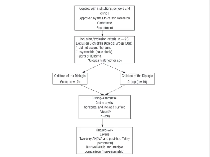
Figure 1 shows the experimental design of the study, described below.
Thus, the purpose of this study was to investigate the influence of the inclined surface (ascending and descending) on the 3D gait’s kinematic characteris -tics in children with spastic diplegic CP. In addition, diplegic subjects were compared to a control group with typically development (TD). It was hypothesized that: (1) 3-dimensional gait analyses could provide more evidences of differences in kinematics variables between DG and CG, especially in transverse plane; (2) the flexor pattern and the restrict foot strike ac -commodation of diplegic children could provide more difficulties and adaptations than the CG ascending and descending surfaces, mainly in angular variables on descending; (3) diplegic children could not have the same slope walking strategies that children with TD.
Children 7-13 years were selected from regular schools, institutions and/or clinical care units. Ten children were allocated for convenience in the diple -gic group (DG = 10.25 ± 1.76 years; 28.95 ± 5.39 kg;
1.34 ± 0.09 m) and 10 children in the control group (CG = 10.43 ± 1.86 years, 31.50 ± 5.37 kg; 1.40 ± 0.12 m) matched by age.
Contact with institutions, schools and clinics
Approved by the Ethics and Research Committee
Recruitment
Inclusion /exclusion criteria (n = 23) Exclusion 3 children Diplegic Group (DG): 1 did not ascend the ramp
1 asymmetric (case study) 1 signs of autismo
*Groups matched for age
Rating-Anamnese Gait analysis: horizontal and inclined surface
- Vicon® (n=20)
Shapiro-wilk Levene
Two-way ANOVA and post-hoc Tukey (parametric)
Kruskal-Wallis and multiple comparison (non-parametric) Children of the Diplegic
Group (n=10)
Children of the Diplegic Group (n=10)
motion compensations in this segment due to their motor difficulties and cerebral lesion (2). The coor -dinates were captured so that the movement could be reconstructed in 3D.
Data were filtered by Vicon® software through a low-pass Butterwortth filter with a frequency of 10 Hz (20). For comparison purposes between groups only the right lower limb data were used as a refer -ence when considering symmetry between partici -pants (15). The stride length and step width were normalized in relation to the height of each child (21).
The data were normalized temporally as a func -tion of the gait cycle dura-tion. Ten gait cycles were performed and the pooled average of 3 valid attempts was calculated for each experimental condition (20). Children were allowed to rest at any time to minimize the fatigue effects (15).
Data analysis
The data were analyzed with descriptive statistics (mean and standard deviation). Shapiro-Wilk and Levene tests tested the distribution normality and homogeneity of variances respectively. Data with normal distribution were treated with parametric statistics: Student t test for independent samples was used to compare the characteristics of the subjects (weight, height and age); Two-way ANOVA was used to compare the linear and angular kinematic data having as independent factors the groups (DG and CG) and experimental conditions (horizontal, ascend -ing and descend-ing surface). Tukey test for equal n’s was used to identify significant differences between means. The data with non-normal distribution were analyzed using the Kruskal-Wallis test, with Dunn Multiple Comparison test. Statistical tests were per -formed in Statistica® software adopting a significance level of p ≤ 0.05.
Results
Spatial and Temporal Linear Variables
Only the stride length differed between the groups being lower for the DG during ascent (p < 0.05). Negative values on foot elevation on ascent for the DG indicate that subjects drag the foot during the gait (Table 1).
Included in the DG were children with symmet -rical independent gait with Gross Motor Function Classification System (GMFCS) I and II and that could understand simple verbal instructions (9, 10). The chil -dren with GMFCS II of this study did not need aids to walk on ramp. Exclusion criteria for the DG included: visual changes and/or moderate to severe intellectual disability, other CP motor disorders such as ataxia, athetosis and dystonia, orthopedic disorders such as lower limb (LL) discrepancy, shortening and deformity or other situations that could prevent independent gait (11) and symmetry, application of botulinum toxin in the LL within the previous six months (12) and phenol within a time period of less than 36 months and/or surgery in the LL or trunk within a time period of less than one year (12 - 14).
The control group (CG) was composed of typically developing children, matched by age. For the CG, the exclusion criteria comprised of those displaying ortho -pedic, neuromuscular and/or cardiovascular (15) or visual impairment that could interfere with gait, and that could engage in systematic programs of physical/ sports activities besides those of regular school.
Data collection
The children were familiarized with the assess -ment’s climatized location and asked to walk barefoot on 3 experimental conditions: horizontal (10 m walk -way), ascending and descending surface (inclined surface with 7º). It was verified whether the children were able to perform the task unaided, to then af -terwards walk freely at a self-selected speed on the experimental conditions. The slope of the ramp was similar to that used in other studies on populations without diplegic CP (16 - 18) and with diplegic CP (2).
541
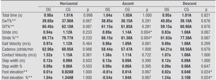
Table 1 - Gait spatial and temporal linear variables (mean) of children with spastic diplegic (DG) and control group (CG) during horizontal, ascent and descent surface
Horizontal Ascent Descent
DG CG p DG CG p DG CG p
Total time (s) 0.98a 1.01A 0.998 1.04a 1.05A 1.000 0.95a 1.01A 0.821 SwT%** 39.60a 37.90A 0.067 38.85a 38.15A 0.281 40.85a 39.10A 0.676 StT%** 60.40a 62.10A 0.067 61.15a 61.85A 0.281 59.15a 60.90A 0.676 Stride (m) 0.94a 1.12A 0.233 0.89a 1.14A 0.004* 0.83a 1.08A 0.067 Stride %** 69.77a 79.77A 0.233 66.15a 81.38A 0.004* 61.63a 77.38A 0.067 Gait Velocity (m/s) 0.97a 1.12A 0.464 0.86a 1.09A 0.061 0.89a 1.08A 0.269 Cadence (stride/min 62.06a 60.05A 0.988 58.44a 57.47A 1.000 64.21a 59.56A 0.679 Contact Velocity (m/s) 1.52a 1.15A 0.361 1.38a 1.11A 0.722 1.07a 1.08A 1.000 Step width (m) 0.12a 0.09A 0.503 0.13a 0.09A 0.395 0.12a 0.09A 1.000 Step width % 0.09a 0.06A 0.503 0.09a 0.06A 0.395 0.09a 0.06A 0.647 Foot elevation** 0.01a 0.02AB 1.000 -0.01a 0.01A 0.067 0.02a 0.04B 0.024* Foot elevation %** 1.04a 1.34AB 1.000 -0.54a 1.04A 0.067 1.24a 3.10B 0.024*
Note: DG: diplegic group; CG: control group; SwT: Swing Time; StT: Stance Time; Stride% and step width%= normalized with respect to height; SD = standard deviation.
a, b- comparison between surfaces for diplegic group (p< 0.05) A, B- comparison between surfaces for control group (p< 0.05) p-value of comparisons between groups in each surface. *p<0.05
** variables with non-parametric statistics (Kruskal-Wallis, p value).
The step width was similar between the groups for the 3 conditions. The observed difference was in rela -tion to the group, being significantly higher (p = 0.004) for the DG when compared to the CG.
Angular Variables
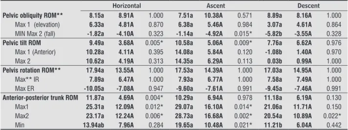
Table 2 - Gait angular variables of the lower limbs and trunk (mean) of children with spastic diplegic and control group dur-ing the horizontal, ascent and descent surface
Horizontal Ascent Descent
DG CG p DG CG p DG CG p
Hip Flex/Ext ROM 41.24a 42.66A 0.993 50.85b 52.08B 0.996 30.52c 37.60A 0.067 Max Flex 34.98a 26.38A 0.128 47.86b 37.72B 0.044* 29.82a 21.84A 0.186 Max Ext** -6.26a -16.28A 0.708 -2.99a -14.36A 0.666 -0.70a -15.77A 0.012* Initial contact 31.94a 23.64A 0.161 44.04b 35.08B 0.106 23.75c 16.43A 0.277
Hip Abd/Ad ROM** 13.70a 13.37A 1.000 13.72a 13.53A 1.000 14.33a 14.67A 1.000 Max (adduction) 9.62a 8.16A 0.969 8.47a 8.84A 1.000 9.08a 9.81A 0.999 Mín (abduction) -4.08a -5.21A 0.994 -5.26a -4.69A 1.000 -5.24a -4.86A 1.000
Hip RE/RI ROM 15.57a 20.41A 0.184 14.20a 17.83A 0.487 15.08a 21.99A 0.016* Max (IR) 8.42a 4.73A 0.901 8.59a 3.69A 0.737 8.01a 6.43A 0.998 Mín (ER) -7.14a -15.68A 0.225 -5.61a -14.14A 0.225 -7.07a -15.56A 0.230 Knee ROM 36.84a 53.77A 0.001* 37.85ab 51.14A 0.009* 41.48b 59.23A 0.000* Max 2 Flex 33.26a 15.26A 0.025* 41.62a 21.90A 0.010* 34.50a 20.35A 0.131 Max 1 Flex 53.15a 56.32A 0.978 55.74a 55.44A 1.000 60.60a 61.12A 1.000 Max Ext 16.32a 2.05A 0.223 17.89a 4.30A 0.271 19.12a 1.88A 0.082 Initial contact** 26.56a 3.04A 0.003* 37.68a 15.69A 0.297 23.1a 5.51A 0.073
Ankle ROM** 23.86a 31.93A 0.401 24.66a 33.35A 0.065 21.69a 28.44A 1.000
Max lex** (dorsilexion) 11.64a 14.24A 1.000 14.20a 14.60A 1.000 8.72a 17.96A 1.000
Max ext (plantarlexion) -12.23a -17.69A 0.674 -10.46a -18.75A 0.230 -12.97a -10.48A 0.984
Initial contact -1.73a 1.56A 0.896 0.67a 3.64A 0.930 -7.27a -2.56A 0.656
Horizontal Ascent Descent
Pelvic obliquity ROM** 8.15a 8.91A 1.000 7.51a 10.38A 0.571 8.89a 8.16A 1.000 Max 1 (elevation) 6.33a 4.81A 0.870 6.38a 5.46A 0.984 3.07a 4.61A 0.864 MIN Max 2 (fall) -1.82a -4.10A 0.323 -1.14a -4.92A 0.015* -5.82b -3.55A 0.328
Pelvic tilt ROM 9.49a 3.68A 0.005* 10.58a 5.06A 0.009* 7.76a 6.62A 0.976 Max 1 (Anterior) 10.28a 4.11A 0.395 14.08a 5.84A 0.120 -1.08b 1.40A 0.970 Max 2 10.62a 4.19A 0.313 14.35a 6.29A 0.113 0.03b 0.99A 1.000
Pelvis rotation ROM** 17.94a 13.55A 1.000 17.53a 14.39A 1.000 17.03a 14.95A 1.000 Max** IR 7.89a 6.47A 1.000 7.93a 6.77A 1.000 7.58a 7.49A 1.000 Max ER -10.05a -7.08A 0.947 -9.60a -7.61A 0.991 -9.45a -7.46A 0.991
Anterior-posterior trunk ROM 11.87a 4.69A 0.004* 10.29a 6.94A 0.978 11.18a 6.19A 0.130 Max1 25.31a 12.09A 0.012* 29.07a 16.10A 0.014* 21.06a 11.71A 0.150 Max2 23.17a 12.24A 0.006* 28.73a 16.68A 0.002* 20.54a 10.89A 0.022* Min 13.94ab 7.96A 0.284 19.65a 10.48A 0.021* 11.21b 6.04A 0.442
Note: DG: diplegic group; CG: control group; ROM: range of motion (in degrees °); Flex: lexion; Max: maximum; Ext: extension; Abd: abduction;
Ad: adduction; IR: internal rotation; ER: external rotation. a, b- comparison between surfaces for diplegic group (p <0.05). A, B- comparison between surfaces for the control group (p <0.05). p-value of comparisons between groups in each surface.
* p <0.05. ** Variables with non-parametric statistics (Kruskal-Wallis, p value).
The angular variables for lower limbs (LL) (Table 2) showed differences during the range of motion (ROM) of hip flexion and extension was influenced by the experimental conditions, but not of the group, that is, there was a change of hip flexion and exten -sion values in the horizontal surface conditions (41.24 ± 5.99 vs 42.66 ± 4.17) and on ascent (50.85 ± 6.83 vs 52.08 ± 2.64), both in the DG as well in the CG respectively. However, the values of maximum hip flexion during ascent were higher in the DG. On ini -tial contact, both groups increased hip flexion during ascent and decreased during descent, with higher values for the DG than for the CG.
For hip extension the DG showed lower values in all the experimental conditions, with significant dif -ferences between the groups only on descent (-0.70 ± 9.63 vs -15.77 ± 5.02).
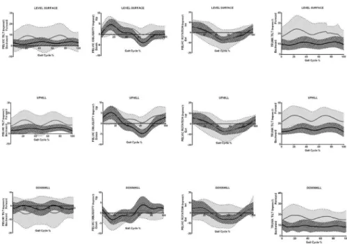
In the transverse plane, the hip rotation amplitude in the control group (CG) was higher than in the DG during descent. In the frontal plane, no differences between groups were found for maximum hip ad -duction and ab-duction or between the experimental conditions. Figures 2 and 3 show the angular dis -placements (motor behavior) of both groups in the 3 experimental conditions.
543
Figure 3 - Evolution of the angular variables of the pelvis and trunk Control Group and Diplegic Group over time, the horizontal, ascending and descending surfaces.
The knee ROM was lower in the DG than in the CG in all experimental conditions. On initial contact, the DG presented greater knee flexion and in the stance phase, no differences were found between groups. During the swing phase, this variable was greater for the DG compared to the CG in the horizontal and ascent surface.
For the pelvic tilt range differences were found between the DG and the CG in the horizontal and as -cent surface. The differences were related to groups and not to the experimental conditions.
On the horizontal surface the DG presented great -er trunk ant-erior tilt and high-er variability in this pattern. On the ascent and descent the groups did not differ between themselves for trunk ROM. While for the 1st and 2nd anterior trunk tilt peaks, the DG presented greater anterior tilt, with no differences between the experimental conditions.
Pelvic obliquity and rotation did not differ be -tween groups or conditions evaluated.
Discussion
Spatial and Temporal Linear Variables
In this study there was no difference in the stride length in the horizontal surface between the DG and CG, contrasting Carriero et al. (4) in studying children
with diplegic CP (0.80 ± 0.26 vs 1.16 ± 0.16), with -out previous surgery, probably with greater muscle shortening than in the present study, that accepted children at least one year of previous surgery.
Although no significant differences were found as mentioned by Stott et al. (2) during descent, the DG had a smaller stride length tendency in the 3 experi -mental conditions, probably due to the subjects’ mo -tor control deficit, considering that CP causes changes in posture and voluntary movement execution (22) and step reduction can be a strategy (23) conse-quently of the brain lesions and difficulties of the movement control (2). The lack of difference among experimental conditions for both groups could be due to the low inclination (7º) which is considered within the standards for accessibility and would fa -cilitate movement execution. This is also defended by Kawamura et al. (23) that identified changes on this variable from an inclination of 12º.
knee and peak knee flexion ROM in swing phase. In this study, this difference between the groups, when the experimental conditions of the horizontal surface to the inclined surface were not considered, differs from what was reported by Stott et al. (2).
In evaluating the step width the DG showed higher values than the CG in the 3 conditions, as a strategy to improve the base of support and their center of mass displacement difficulties (24, 25). The experimental conditions did not influence this variable, which cor -roborates with other studies (23).
The differences between this study and what was reported by Stott et al. (2) may be due to the inclusion criteria. In our study, we evaluated children of level I and II of the GMFCS and the other authors assessed children only from level II; also the difference may be due to the type of motion analysis, this research used three-dimensional analysis whereas the above authors used two-dimensional analysis.
Angular Variables
On ascent, similar to what some studies (17, 23, 26) have observed both the DG as well as the CG in -creased hip flexion during gait to adjust the foot to surface inclination, from the initial contact, and this would be directly related to the degree of inclination. This strategy was observed as being influenced by the inclination surface but not by the group type, which for some researchers (4, 5), is due to the DG flexor pattern. This would lead to less dynamic hip exten -sion, evidenced for the DG in the 3 conditions, with notable difference during descent.
There was no difference between the maximum hip adduction and abduction for the experimen -tal conditions between groups, which agrees with Steinwender et al. (27) for which only in the crouched gait pattern would the diplegic child present instabil -ity to activate the hip abductors on the swing due to selective control difficulty (27). While for Carriero et al. (4), and Kirkwood et al. (28) in the horizontal surface there would be a difference in these variables for diplegic children.
For hip rotation ROM only on descent were the groups different and the CG presented higher ROM than the DG. Despite the statistical similarity of maxi -mum external rotation between the groups, on the three inclined surfaces, it was observed that the DG always presented lower values during gait. Children
with diplegic CPmay present higher maximum inter -nal rotation values in the horizontal surface when compared to the CG (4), as a musculoskeletal adapta -tion to the gait’s func-tional movement.
For knee flexion/extension ROM the DG showed lower values than the CG, differing from each other in the 3 conditions. Hamstring tightness of children with diplegic CP can justify knee extension difficulty (4) and is therefore an effect of the group, not from surface inclination.
When evaluating initial contact in the horizontal surface the DG showed higher knee flexion values, possibly due to the flexor pattern evidenced in chil -dren with diplegic CP (4, 29). Even higher values were observed during ramp ascent as mentioned by Stott et al. (2).The increase on this variable is reported for typical gait (22) as well as for gait with diplegic CP in surface change as a neuromotor adjustment strategy to accommodate the lower member to the inclination. Maximum knee flexion occurring in the swing phase was not different between the groups on the horizontal surface, like the results of Carriero et al. (4), nor among the inclined surfaces.
The value of the maximum knee flexion in the stance phase was higher for the DG on the hori -zontal surface and on ascent (2), and consequently showed lower extension. Although significant differ -ences were not observed, there was a trend of the CG to perform the extension movement, while diplegic children remain in flexion, this event is also demon -strated on the horizontal surface (4, 29) and inclined surface (2) due to the flexor pattern of these children.
Hyperactivity and/or shortening of the gastrocne -mius muscle, which is biarticular, could have greater action on the knee joint, with increase in its flexion, than on the ankle joint (30) and this was also dem -onstrated in this study.
545
In the analysis of foot angle relative to the surface it was found that there was inter and intra-group differences. The DG does not accommodate the foot to the surface as does the CG making contact in plan -tarflexion due to the difficulty of making it in dorsi -flexion due to the shortened gastrocnemius muscle (30). This difficulty associated with knee flexor pat -tern also influenced the foot elevation variable. The negative value for ascent represents that the DG drags the foot. Even though the methodological analysis of van der Krogt et al. (31) was different, the authors also demonstrated this difficulty of raising the foot off the ground (pre-swing).
There was a pelvic tilt ROM difference between the groups on the horizontal surface and on ascent, agreeing with Carriero et al. (4) who found greater anterior pelvic tilt for children with diplegic CP. This would happen as a compensation due to the decrease in extension capability of these children (5, 32) and in the flexed knee gait by the gait pattern and the weakness of the extensor muscles of knee and hip joint (32). The proportional increase to the ramp’s in -clination would occur as a means to follow the move -ment and allow increased hip flexion, coinciding with simple support on each side (17, 26).
Regarding pelvic obliquity a difference was ob -served in minimum obliquity (fall) when comparing the inclination of surfaces, with a predominance of negative values between DG during descent. These findings are in agreement with other authors (4, 27) who explained this by the fact that children with diplegic CP present functional symmetry between the LL just as typical children. The increased pelvic obliquity would be used only as compensation for people with asymmetry (discrepancy) in the length of the LL or functional discrepancy, as in the case of hemiparetic.
There was no difference between the DG and CG in the horizontal surface for pelvic rotation. With respect to the trunk the DG has more trunk anterior displacement than the CG, like the children evaluated by Heyrman et al. (10) in the horizontal surface and by Stott et al. (2) during ascent. For these authors this may be due to postural instability of these children and as compensation of changes in the LL and pel -vis. In addition, it could be compensation due to hip extensor weakness of the DG and a lack of eccentric control of the quadriceps (2). The great variability of movement shown in the DG makes the comparison between groups difficult.
It’s worth mentioning that the high variability of values for all joints among children with diplegic CP were also observed in the study by Stott et al. (2) and lead to consider an immaturity of movements, corroborating the findings of Hodapp et al. (33) and Prosser et al. (15). Immaturity in the gait for the DG would be a consequence of encephalic lesion and inhibition of the tonic muscular development that occurs over the children’s growth (33).
This variability, however, was observed in all three inclined surfaces in a similar manner.
Study limitations
Some limitations in the study were considered: 1 - The small sample size. Randomized studies with larger populations in child care centers with CP are suggested.
2 - The gait analysis in this study was not catego -rized by groups according to gait patterns. Studies which categorize these variables are suggested.
Conclusion
Despite the initial hypothesis that 3-dimensional gait analyses could provide more evidences of differ -ences in kinematics variables between DG and CG, especially in transverse plane it was not observed due the great variability of movement shown in the DG.
The findings provide some evidence to support the second hypothesis and some variables were more influenced on descent by their flexor pattern. This fact occurs on foot elevation in the GC, and for the variables: hip ROM during the gait cycle; hip flexion-extension in initial contact; knee ROM and the 2nd anterior-posterior trunk peak amplitude of the DG, and therefore, being more influenced by the descent condition than by the CG. Probably on descent places high demands on the knee extensors and the exces -sive knee flexion seen in DG may reflect underlying poor eccentric quadriceps control, necessitating greater compensations at the trunk, hip and knee.
The implications of the findings of this study pro -vide e-vidence about acessibility and understanding for new investigations about the environment influ -ences on the motor strategies of diplegic children.
Acknowledgements
The authors thank the Support Program for the Restructuring and Expansion of Federal Universities (Reuni), established by Decree N.o 6.096 of April 24, 2007 by the Ministry of Education and Culture (MEC) of Brazil, for granting the master’s scholarship.
References
1. Figueiredo PR, Silva PL, Avelar BS, Chagas PS, Oliveira LC, Mancini MC. Assessment of gait in toddlers with normal motor development and in hemiplegic children with mild motor impairment: a validity study. Braz J Phys Ther. 2013;17(4):359-66.
2. Stott NS, Reynolds N, McNair P. Level versus inclined walking: ambulatory compensations in children with cerebral palsy under outdoor conditions. Pediatr Phys Ther. 2014;26(4):428-35.
3. Tosun A, Gökben S, Serdaroglu G, Polat M, Tekgöl H. Changing views of cerebral palsy over 35 years: the experience of a center. Turk J Pediatr. 2013;55(1):8-15.
4. Carriero A, Zavatsky A, Stebbins J, Theologis T, Shefelbine SJ. Correlation between lower limb bone morphology and gait characteristics in children with spastic diplegic cerebral palsy. J Pediatr Orthop. 2009;29(1):73-9.
5. Hicks JL, Schwartz MH, Arnold AS, Delp SL. Crouched postures reduce the capacity of muscles to extend the hip and knee during the single-limb stance phase of gait. J Biomech. 2008;41(5):960-7.
6. Ahl LE, Johansson E, Granat T, Carlberg EB. Functional therapy for children with cerebral palsy: an ecological approach. Dev Med Child Neurol. 2005;47(9):613-9.
7. Noble JW, Prentice SD. Intersegmental coordination while walking up inclined surfaces: age and ramp angle effects. Exp Brain Res. 2008;189(2):249-55.
8. Carriero A, Zavatsky A, Stebbins J, Theologis T, Shefelbine SJ. Determination of gait patterns in children with spastic diplegic cerebral palsy using principal components. Gait Posture. 2009;29(1):71-5.
9. Fowler EG, Goldberg EJ. The effect of lower extremity selective voluntary motor control on interjoint coordination during gait in children with spastic diplegic cerebral palsy. Gait Posture. 2009;29(1):102-7.
10. Heyrman L, Feys H, Molenaers G, Jaspers E, Monari D, Meyns P, et al. Three-dimensional head and trunk movement characteristics during gait in children with spastic diplegia. Gait Posture. 2013;38(4):770-6.
11. Rodda JM, Graham HK, Carson L, Galea MP, Wolfe R. Sagittal gait patterns in spastic diplegia. J Bone Joint Surg Br. 2004;86(2):251-8.
12. Wingert JR, Sinclair RJ, Dixit S, Damiano DL, Burton H. Somatosensory-evoked cortical activity in spastic diplegic cerebral palsy. Hum Brain Mapp. 2010;31(11):1772-85.
13. Eek MN, Beckung E. Walking ability is related to muscle strength in children with cerebral palsy. Gait Posture. 2008;28(3):366-71.
14. Mélo TR, Rodacki ALF, Guimarães ATB, Israel VL. Repeatability and comparison of clinical tests in children with spastic diplegia and with typical development. Fisioter Mov. 2015;28(1):13-22. 15. Prosser LA, Lauer RT, VanSant AF, Barbe MF, Lee SC.
Variability and symmetry of gait in early walkers with and without bilateral cerebral palsy. Gait Posture. 2010;31(4):522-6.
16. Carriero A, Zavatsky A, Stebbins J, Theologis T, Shefelbine SJ. Determination of gait patterns in children with spastic diplegic cerebral palsy using principal components. Gait Posture. 2009;29(1):71-5.
17. McIntosh AS, Beatty KT, Dwan LN, Vickers DR. Gait dynamics on an inclined walkway. J Biomech. 2006;39(13):2491-502.
547
19. Melanda AG, Kawamura CM, Freitas CD, Lucareli PRG, Pinheiro PO. Laboratório de Marcha. In: Borges D, Moura EW, Lima E, Silva PAC. Fisioterapia: aspectos clínicos e práticos da reabilitação. São Paulo: Artes Médicas; 2007. p. 615-40.
20. Hsue B-J, Miller F, Su F-C. The dynamic balance of the children with cerebral palsy and typical developing during gait. Part I: Spatial relationship between COM and COP trajectories. Gait Posture. 2009;29(3):465-70.
21. Saraph V, Zwick E-B, Auner C, Schneider F, Steinwender G, Linhart W. Gait improvement surgery in diplegic children: how long do the improvements last? J. Pediatr. Orthop. 2005;25(3):263-7.
22. Bax M, Goldstein M, Rosenbaum P, Leviton A, Paneth N, Dan B, et al. Proposed definition and classification of cerebral palsy, April 2005. Dev Med Child Neurol. 2005;47(8):571-6.
23. Kawamura K, Tokuhiro A, Takechi H. Gait analysis of slope walking: a study on step length, stride width, time factors and deviation in the center of pressure. Acta Med Okayama. 1991;45(3):179-84.
24. Hsue BJ, Miller F, Su FC. The dynamic balance of the children with cerebral palsy and typical developing during gait: Part II: Instantaneous velocity and acceleration of COM and COP and their relationship. Gait Posture. 2009;29(3):471-6.
25. Stackhouse C, Shewokis PA, Pierce SR, Smith B, McCarthy J, Tucker C. Gait initiation in children with cerebral palsy. Gait Posture. 2007;26(2):301-8.
26. Leroux A, Fung J, Barbeau H. Postural adaptation to walking on inclined surfaces: I. Normal strategies. Gait Posture. 2002;15(1):64-74.
27. Steinwender G, Saraph V, Zwick EB, Steinwender C, Linhart W. Hip locomotion mechanisms in cerebral palsy crouch gait. Gait Posture. 2001;13(2):78-85.
28. Kirkwood RN, Franco RdLLD, Furtado SC, Barela AMF, Deluzio KJ, Mancini MC. Frontal Plane motion of the pelvis and hip during gait stance discriminates children with diplegia levels I and II of the GMFCS. ISRN Pediatrics. 2012;2012: 163039.
29. Brunner R, Dreher T, Romkes J, Frigo C. Effects of plantarflexion on pelvis and lower limb kinematics. Gait Posture. 2008;28(1):150-6.
30. Maas JC, Huijing PA, Dallmeijer AJ, Harlaar J, Jaspers RT, Becher JG. Decrease in ankle–foot dorsiflexion range of motion is related to increased knee flexion during gait in children with spastic cerebral palsy. J Electromyogr Kinesiol. 2015;25(2):339-46.
31. van der Krogt MM, Bregman DJ, Wisse M, Doorenbosch CA, Harlaar J, Collins SH. How crouch gait can dynamically induce stiff-knee gait. Ann Biomed Eng. 2010;38(4):1593-606.
32. Shin HI, Sung KH, Chung CY, Lee KM, Lee SY, Lee IH, et al. Relationships between Isometric Muscle Strength, Gait Parameters, and Gross Motor Function Measure in Patients with Cerebral Palsy. Yonsei Med J. 2016;57(1):217-24.
33. Hodapp M, Klisch C, Mall V, Vry J, Berger W, Faist M. Modulation of soleus H-reflexes during gait in children with cerebral palsy. J. Neuroph. 2007;98(6):3263-8.
34. Associação Brasileira de Normas Técnicas. Acessibilidade a edificações, mobiliário, espaços e equipamentos urbanos. ABNT NBR 950:2004. 97 p.
Received in 05/01/2016 Recebido em 01/05/2016
Approved in 02/07/2017