Fisioter. Mov., Curitiba, v. 30, n. 3, p. 549-558, Jul./Sep. 2017 Licenciado sob uma Licença Creative Commons DOI: http://dx.doi.org/10.1590/1980-5918.030.003.AO13
Postural alignment of patients with Chronic
Obstructive Pulmonary Disease
Alinhamento postural de pacientes com Doença
Pulmonar Obstrutiva Crônica
Márcia Aparecida Gonçalves, Davi de Souza Francisco, Caroline Semprebom de Medeiros, Ana Karla Vieira Brüggemann, Giovana Zarpellon Mazo, Elaine Paulin*
Universidade do Estado de Santa Catarina (UDESC), Florianópolis, SC, Brazil
[R]
Abstract
Introduction: In chronic obstructive pulmonary disease (COPD), airflow resistance impairs respiratory me -chanics that may compromise postural alignment. There is a lack of studies that have investigated
compro-mised postures and their possible associations with pulmonary function. Objectives: To compare the postural
alignment of COPD patients with apparently healthy individuals; To correlate pulmonary function with pos -tural alignment in the COPD group. Methods: 20 COPD patients and 20 apparently healthy individuals
per-formed: anthropometry, spirometry and postural evaluation. The following postural changes were assessed:
lateral head tilt (LHT), shoulder asymmetry (SA1), anterior pelvic asymmetry (APA), lateral trunk tilt (LTT), scapular asymmetry (SA2), posterior pelvic asymmetry (PPA), head protrusion (HP), shoulder protrusion (SP), anterior pelvic tilt (APT) and thoracic kyphosis (TK). Results: There was a statistically significant dif
-ference between COPD patients and apparently healthy individuals in the following variables: PPT (p= 0.021), APT (p=0.014) and TK (p=0.011). There was a correlation between pulmonary variables and postural align
-ment in the COPD group: Forced Volume in one second (FEV1% pred) and HP (°) (r=0.488, p=0.029), FEV1 (% pred) and APT (°) (r= -0.472, p= 0.036); Forced Vital Capacity (FVC % pred) and HP (°) (r=0.568, p=0.009); FVC (% pred) and APT (°) (r=-0.461, p=0.041). Conclusion: Postural alignment of the anterior tilt of the right
* MAG: MS, e-mail: [email protected]
DSF: undergrad, e-mail: [email protected] CSM: undergrad, e-mail: [email protected] AKVB: MS, e-mail: [email protected]
550
and left pelvis and thoracic kyphosis is different when compared with COPD patients and healthy individuals. There is a relationship between pulmonary function and postural alignment in COPD patients.
Keywords: Chronic Obstructive Pulmonary Disease. Posture. Photogrammetry.
Resumo
Introdução: Na doença pulmonar obstrutiva crônica (DPOC), a resistência ao fluxo aéreo prejudica a mecânica
respiratória que pode comprometer o alinhamento postural. Existe uma escassez de estudos que tenham inves -tigado os comprometimentos posturais e suas possíveis relações com a função pulmonar. Objetivos: Comparar o alinhamento postural entre pacientes com DPOC e indivíduos aparentemente saudáveis; correlacionar a fun-ção pulmonar com o alinhamento postural no grupo DPOC. Métodos: 20 pacientes com DPOC e 20 indivíduos aparentemente saudáveis realizaram: antropometria, espirometria e avaliação postural. Foram analisadas as alterações posturais: inclinação lateral da cabeça (ILC), desnivelamento dos ombros (DO), desnivelamento pélvico anterior (DPA), inclinação lateral do tronco (ILT), desnivelamento das escápulas (DE), desnivelamento pélvico posterior (DPP), protrusão da cabeça (PC), protrusão de ombro (PO), báscula anterior da pelve (BAP) e cifose torácica (CT). Resultados: Houve diferença estatisticamente significante entre os pacientes com DPOC e
os indivíduos aparentemente saudáveis nas variáveis: DPP (0,021), BAP (p=0,014) e CT (p=0,011). Houve cor -relação entre as variáveis pulmonares e o alinhamento postural no grupo DPOC: volume forçado no primeiro segundo (VEF1 %prev) e PC (°) (r= 0,488, p=0,029), VEF1 (%prev) e BAP (°) (r= -0,472; p= 0,036); Capacidade Vital Forçada (CVF %prev) e PC (°) (r= 0,568; p= 0,009); CVF %prev) e BAP (°) (r= -0,461; p=0,041). Conclusão:
O alinhamento postural da báscula anterior da pelve direita e esquerda e da cifose torácica é diferente quando comparados pacientes com DPOC e saudáveis. Existe relação entre a função pulmonar e o alinhamento pos -tural no paciente com DPOC.
Palavras-chave: Doença Pulmonar Obstrutiva Crônica. Postura. Fotogrametria.
Introduction
Chronic obstructive pulmonary disease (COPD)
is characterized by airflow limitation. It is usually progressive and associated with a chronic inflamma
-tory response in the airways due to noxious particles
or gases (1). COPD is increasingly recognized as sys-temic and heterogeneous, and may involve skeletal
muscle dysfunction (2, 3), heart diseases (4), nutri
-tional dysfunctions (5), biochemical changes (6), os
-teoporosis (7), psychological disorders (8), as well as the impairment of pulmonary mechanics (9).
Some changes in pulmonary mechanics include
lung hyperinflation and air trapping, which are gradu
-ally identified in COPD patients. These factors may
contribute to increase the anteroposterior diameter
of the thorax (10, 11, 12), horizontalization of the ribs (11, 13), as well as to compromise the postural align -ment due to compensations in the scapular pelvic girdles and especially in the thoracic spine.
Additionally, COPD patients usually show rec
-tification and shortening of the diaphragm, which
may trigger changes in the endothoracic fascia. This shortening may lead to pelvic anteversion and psoit-ic diaphragmatpsoit-ic hyperlordosis due to the connec-tions of the muscle-aponeurotic connecconnec-tions of the
diaphragm muscle with the iliopsoas muscles, the
transverse abdominis muscle and the lumbar square
muscle (14).
Few studies have evaluated postural alignment of COPD patients (14, 15). Dias et al. (15) evaluated the
kinematics of the scapular girdle, cervical and
tho-racic spines of 19 COPD patients and 19 healthy in
-dividuals. The study showed a higher elevation of the
scapula among COPD patients, compared to healthy individuals. The authors report that this change is
likely to occur due to pulmonary hyperinflation that
changes the position of the sternum and the scapula.
However, they did not find significant differences in the regions of the cervical and thoracic spine between
551
COPD according to the Global Initiative for Chronic
Obstructive Lung Disease (GOLD) classification
(1). A diagnostic record, developed by researchers linked to the Laboratory of Respiratory Physiotherapy (LAFIR) at the State University of Santa Catarina
(UDESC), was used in order to identify the charac -teristics of the individuals participating in the study.
For COPD patients, the following inclusion criteria were considered: 1) clinical stability in the last month and at the beginning of the protocol of evaluation; 2) patients who did not use supplemental oxygen; 3) inexistence of other associated respiratory or cardio
-vascular diseases; 4) patients without involvement in pulmonary rehabilitation programs in the 6 months prior to the start of this study; 5) patients who did not undergo recent spinal or lower limb surgeries and/or who hadn´t had fractures in the past 6 months. The exclusion criteria were as follows: 1) presence of ex
-acerbations of the disease during the study; 2) clinical
intercurrences related cardiorespiratory
abnormali-ties during the evaluations; 3) inability to perform
any of the study evaluations (lack of understanding
or cooperation); and 4) patients´ with drawal during
the evaluation period.
The group of apparently healthy individuals
in-cluded: 1) individuals with normal spirometry (FEV1/ FVC ≥ 0.7; FEV1 ≥ 80% of predicted, FVC ≥ 80% of predicted); 2) age, body mass and BMI compatible with COPD group; and 3) inexistence of associated comorbidities. Exclusion criteria were as follows:
1) inability to perform any of the study evaluations
(lack of understanding or cooperation); 2) individu
-als´ withdrawal during the evaluation period.
Evaluated parameters
Anthropometry
For the anthropometric measurements, a
previ-ously calibrated scale and a stadiometer were used
for measuring body mass and height, respectively (Welmy® model W200/5, São Paulo, SP, Brazil). The Body Mass Index (BMI) was determined through the equation: body mass/height2. The individuals were classified as underweight (<18.5 kg/m2), adequate or eutrophic (≥18.5 < 25 kg/m2), overweight (≥ 25 < 30 kg/m2) and obesity (≥ 30 kg/m2) (16). The individu
-als were instructed to wear light clothes, to remove
their shoes and to remain in an upright position.
Pachioni et al. (14) compared the posture of 15 COPD patients with 15 healthy individuals. The
authors observed the presence of three important postural changes in COPD patients: posterior pel-vic asymmetry, anterior pelpel-vic asymmetry and in-creased thoracic kyphosis. The study suggests that
such changes could be related to the disease itself; however, the authors did not conduct a correlational analysis between pulmonary function variables and
postural alignment.
With the progression of COPD, postural changes
may become increasingly evident and it will be more difficult for patients to perform their daily life activi
-ties, physical exercises. Also, these changes will main -ly interfere in the pulmonary function of these
indi-viduals, which is already compromised. Therefore,
postural evaluation is of utmost importance in order
to identify the existence of changes in the postural alignment and its relation with the pulmonary func -tion in COPD patient. The results obtained in this
current study may offer significant contributions to the relationship between posture and respiration, since there are only a few studies in the literature
about the presence of postural imbalances in COPD
patients and how much this may further compromise
their pulmonary function. Moreover, these results may provide further information to develop more appropriate therapy programs combining
respira-tory strategies, which are already part of pulmonary
rehabilitation programs.
Thus, this study aimed: 1) to compare postural
alignment of COPD patients with apparently healthy individuals; 2) to correlate pulmonary function vari
-ables with postural alignment in the COPD group.
Methods
Study Population and Sample
This is an analytical cross-sectional study with a quantitative approach. It was approved by the Ethics
and Research Committee involving Human Subjects
under the number CAEE: 08857612.2.0000.0118 and all participants were previously informed about
the study and signed an Informed Consent Form, as
determined by Resolution 466/12 of the National
Health Council.
The sample consisted of 20 “apparently” healthy
552
In the posterior view, two postural changes were
evaluated: the scapular asymmetry (SA2), evaluated
by the angle formed between the inferior angles of the scapulae and the horizontal axis; and posterior pelvic asymmetry (PPA) was measured by the angle between the two posterior superior iliac spines and the horizontal axis.
In the right lateral view, two postural changes were evaluated: head protrusion (HP), by the angle between the spinous process on the C7/tragus and horizontal axis; and shoulder protrusion (SP), by measuring the angle between the spinous process of C7/acromion and the horizontal axis.
In the left lateral view, two postural changes were
evaluated: the anterior pelvic tilt (APT), obtained by
the angle between the anterior-superior/posterior-superior iliac spine and the horizontal axis; and tho
-racic kyphosis (TK), which was measured by the angle between the vertebra T3 and T12 with vertex in the
most prominent vertebra.
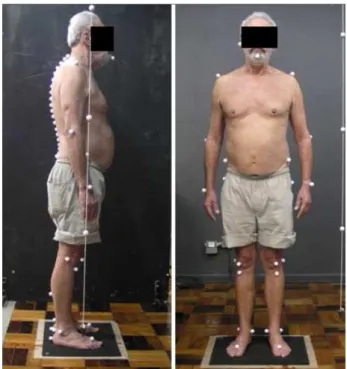
Next, the individual was placed in a static ortho
-static position at a distance of 50 cm in front of a black wall and next to a plumb line marked with three Styrofoam balls 50 cm apart from each other them, to allow calibration for the photograph. His/her feet were positioned loosely on top of a black EVA foam mats to shoot the first photograph. The individual was informed, through a verbal command, to remain in a comfortable position, with the eyes fixed at a line, in a relaxed posture. Subsequently, with a white chalk, the foot outlines were drawn so that the in
-dividual could stand at the same point for the next photo shoot. After the front and left side views were photographed, the mat was rotated 180º to obtain
the photographs in the posterior and right lateral
views (Figure 1).
Two photo cameras (Sanyo BD 200 14.1 mega pix
-els, DSC - W610) were used, which were placed on a tripod (97 cm high). One camera was placed in the
front of the individual and the other camera at the
left side. However, both cameras were positioned at a distance of 2.30m from the participant. The distance and height of the camera was adapted, as previously
mentioned, to facilitate visualization of the individual. According to Mota et al. (20), the distance should be
sufficient to position the entire body in the center of the image and the resolution should be sufficient to show each of the markers clearly.
The photos were digitalized and analyzed with the PAS software. The analysis of the photos and Spirometry
Spirometry was performed to verify the sub
-jects´ pulmonary capacity, using a portable digital
spirometer (EasyOne®, ndd Medical Technologies, Zurich, Switzerland), previously calibrated accord -ing to the methods and criteria recommended by the American Thoracic Society and the European
Respiratory Society (17). The following parameters were measured: forced vital capacity (FVC), forced expiratory volume in one second (FEV1) and FEV1/ FVC ratio before and 15 minutes after inhalation of bronchodilator (BD) salbutamol (400μg) in COPD patients. At least three acceptable maneuvers and two reproducible maneuvers were performed. Spirometry variables are expressed in absolute values and in per -cent of predicted values of normality, as determined
by Pereira et al. (18). The normal lung function test criteria consist of FVC and FEV1 ≥ 80% of predicted
and FEV1/FVC ≥ 0.7.
Postural Alignment
For the evaluation of the postural alignment, the
Postural Assessment Software (PAS/SAPO) validated by Ferreira et al. (19) was used. Before the photo
-graphs were taken, male subjects were instructed to wear bermuda shorts, and female subjects to wear
bermuda shorts and a top. Initially, anatomical points
were identified through the palpatory anatomy in the following regions of the body: head, shoulders, pelvis, spine, upper limbs and lower limbs. After the points had been identified, they were marked with Styrofoam balls, with a diameter of 20 mm, fixed to the body parts with double-sided adhesive tape.
In order to mark the points and define the pos
-tural changes that would be evaluated, the protocol developed by Pachioni et al. (14) was used.
In the anterior view, four postural changes were
evaluated: lateral head tilt (LHT) by measuring the
angle formed between the glabella/mento and the horizontal axis; the shoulder asymmetry (SA1) by the angle formed between the two acromions and the horizontal axis; anterior pelvic asymmetry (APA) measured by the angle between the two anterior su
-perior iliac spines and the horizontal axis; and finally,
the lateral trunk tilt (LTT) measured by the angle
553 pelvic tilt, the Mann-Whitney U test was used. The Pearson Correlation Coefficient was used to com
-pare the pulmonary variables with lateral head tilt,
shoulder asymmetry, scapular asymmetry, head pro-trusion, shoulder propro-trusion, anterior pelvic tilt and thoracic kyphosis. The Spearman's correlation
coef-ficient correlated pulmonary function with anterior
pelvic asymmetry, lateral trunk tilting and posterior
pelvic asymmetry. The significance level for all the tests was set at an alpha of 5%.
Results
Participants in the study were 40 subjects of both genders (20 men and 20 women), who were divided into two groups: group 1 with 20 COPD patients, aged 65.35 (± 6.76) years and group 2 with 20 apparently healthy individuals, aged 65.55 (± 4.57) years.
Considering age and anthropometric and pulmonary
characteristics between the studied groups (i.e., COPD
patients and apparently healthy individuals), Table 1
shows no statistically significant difference between
age, body mass, height and BMI, demonstrating that both are homogenous groups for these variables.
Regarding pulmonary capacity, a significant differ
-ence (p < 0.001) was observed between the groups
in the predicted percentage of FEV1 and in the FVC
percent of predicted. The best results, on average,
were for apparently healthy individuals.
According to GOLD staging system, the patients showed severe COPD and are classified as Stage III (FEV1 = 46.25 ± 14.52% of predicted).
Regarding the postural alignment of the groups with
COPD patients and apparently healthy individuals, table
2 shows a statistically significant difference between the groups for posterior pelvic asymmetry (p = 0.021), anterior pelvic tilt (p = 0.014) and in the thoracic kyphosis angle (p = 0.011). Therefore, it can be observed that COPD pa -tients have a greater posterior pelvic asymmetry, anterior pelvic tilt of the left and right pelvis, and greater thoracic kyphosis angle.
When comparing men and women in the COPD group, the results showed that there was no significant
difference in any of the measurements performed by
the postural evaluation. Both groups of genders showed
similar degrees for the variables of postural alignment
(Table 3).
measurements of the angles of the variables of the
postural alignment were based on the coordinates of
the anatomical points.
Figure 1 - Evaluation of the postural alignment using PAS. Note: Produced by the author.
Statistical Analysis
The data was analyzed with the SPSS (Statistical Package for the Social Sciences) for Windows, ver
-sion 20.0 (IBM Corporation, Armonk, NY, USA). The
descriptive analysis as mean and standard deviation
was applied to all variables. For the sample calcula
-tion, the statistical power for 20 subjects per group was calculated, a post hoc sample calculation was performed, using Student´s t test with the GPower 3.1 program. The mean values of thoracic kyphosis were determined in both groups: COPD - mean (207.13 ± 4.83); healthy - mean (202.81 ± 5.41). The calculated effect was 1.104315 and, using a margin of error of 5%, the power of the calculated sample was 80 for
20 subjects in each group.
To verify the normality of the data, the
Shapiro-Wilk test was applied; to compare shoulder asym -metry, head protrusion, shoulder protrusion and
thoracic kyphosis between the groups, the Student´s t test was used; and to compare the lateral head tilt,
554
Table 1 - Characteristics of the studied groups (n = 40)
Variables COPD
(n = 20)
Apparently healthy
(n = 20) p-value
Gender (M/F) 10/10 10/10
-Age (years) 65.35 ± 6.76 65.55 ± 4.57 0.913
Body Mass (kg) 74.95 ± 19.33 76.60 ± 12.55 0.751
Height (cm) 164.25 ± 0.09 165.95 ± 0.11 0.613
BMI (kg/m2) 27.65 ± 6.10 27.75 ± 3.09 0.946
FEV1/FVC (L) 0.53 ± 0.16 0.79 ± 0.04 < 0.001*
FEV1 (L) 1.35 ± 0.58 2.68 ± 0.66 < 0.001*
FEV1 ( pred) 46.25 ± 14.52 92.70 ± 8.57 < 0.001*
FVC (L) 2.34 ± 0.82 3.41 ± 0.82 < 0.001*
FVC ( pred) 64.30 ± 12.26 92.55 ± 8.40 < 0.001*
Note: The values are expressed as mean ± standard deviation; M: male; F: female; BMI (kg/m2): body mass index in kilograms per square meter; FEV1 (% pred): percent of predicted of forced expiratory volume in one second; FVC (% pred): percent of predicted of forced vital capacity; *p < 0.05.
Table 2 – Comparison between the postural alignment of COPD patients and apparently healthy individuals (n = 40)
Variables COPD
(n = 20)
Apparently healthy
(n = 20) p-value
LHT (°) 2.87 ± 2.47 3.28 ± 2.20 0.616
SA1 (°) 2.72 ± 1.63 2.77 ± 1.62 0.923
APA (°) 2.19 ± 1.48 1.47 ± 1.25 0.065
LTT (°) 2.90 ± 2.52 2.84 ± 1.47 0.598
SA2 (°) 2.49 ± 1.70 2.39 ± 1.85 0.655
PPA (°) 3.03 ± 1.99 1.80 ± 1.25 0.021*
HP (°) 42.41 ± 6.91 43.44 ± 6.66 0.637
SP (°) 157.28 ± 12.52 150.04 ± 13.57 0.088
APT (°) 7.55 ± 4.29 4.55 ± 2.92 0.014*
TK (°) 207.13 ± 4.83 202.81 ± 5.41 0.011*
Note: The values are expressed as mean ± standard deviation. LHT: lateral head tilt; SA1: shoulder asymmetry; APA: anterior pelvic asym-metry; LTT: lateral trunk tilt; SA2: scapular asymasym-metry; PPA: posterior pelvic asymasym-metry; HP: head protrusion; SP: shoulder protrusion; APT: anterior pelvic tilt; TK: thoracic kyphosis; (°): measurements in degrees; * p < 0.05.
Table 3 – Comparison of postural alignment between men and women in the COPD group (n = 20)
Variables COPD
(n = 20)
Apparently healthy
(n = 20) p-value
LHT (°) 2.27 ± 2.38 3.47 ± 2.54 0.290
SA1 (°) 2.72 ± 1.75 2.73 ± 1.59 0.989
APA (°) 1.82 ± 1.21 2.57 ± 1.69 0.268
LTT (°) 2.51 ± 2.55 3.29 ± 2.56 0.504
SA2 (°) 2.86 ± 2.17 2.14 ± 1.04 0.360
PPA (°) 2.43 ± 1.07 3.64 ± 2.53 0.189
HP (°) 40.71 ± 6.75 44.12 ± 6.98 0.283
SP (°) 160.88 ± 11.86 153.68 ± 12.70 0.207
APT (°) 7.13 ± 4.12 7.98 ± 4.64 0.670
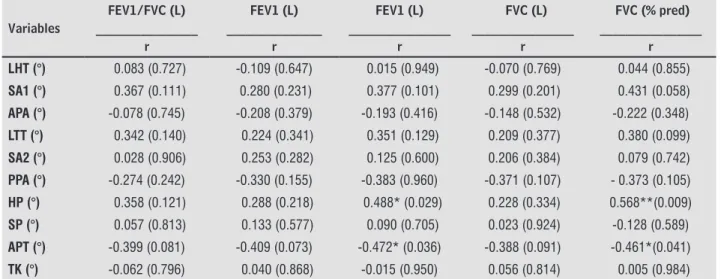
555 This study showed a relationship between pulmo
-nary variables and measures of postural alignment in
the COPD patients group. A positive correlation was found between FEV1 (% pred) and head protrusion (r = 0.488, p = 0.029); FVC (% pred) and head protru
-sion (r = 0.568; p = 0.009). However, there was a nega
-tive correlation between FEV1 (% pred) and anterior pelvic tilt (r = -0.472; p=0.036); FVC (% pred) and anterior pelvic tilt (r = -0.461; p = 0.041). There was no correlation between the other variables related to pulmonary function with measures of postural alignment (Table 4).
Discussion
The current study identified two major postural
changes in COPD patients compared to apparently healthy individuals: greater angle in thoracic ky-phosis, in the anterior pelvic tilt and posterior
pel-vic asymmetry. Our results are in agreement with the study conducted by Pachioni et al. (14), who also compared the postural alignment of 15 COPD patients and 15 healthy individuals and found a statistically significant difference in these same variables.
Table 4 - Correlation between pulmonary function and measures of postural alignment of COPD group (n = 20)
Variables
FEV1/FVC (L) ________________
r
FEV1 (L) _______________
r
FEV1 (L) ________________
r
FVC (L) ________________
r
FVC ( pred) ________________
r
LHT (°) 0.083 (0.727) -0.109 (0.647) 0.015 (0.949) -0.070 (0.769) 0.044 (0.855)
SA1 (°) 0.367 (0.111) 0.280 (0.231) 0.377 (0.101) 0.299 (0.201) 0.431 (0.058)
APA (°) -0.078 (0.745) -0.208 (0.379) -0.193 (0.416) -0.148 (0.532) -0.222 (0.348)
LTT (°) 0.342 (0.140) 0.224 (0.341) 0.351 (0.129) 0.209 (0.377) 0.380 (0.099)
SA2 (°) 0.028 (0.906) 0.253 (0.282) 0.125 (0.600) 0.206 (0.384) 0.079 (0.742)
PPA (°) -0.274 (0.242) -0.330 (0.155) -0.383 (0.960) -0.371 (0.107) - 0.373 (0.105)
HP (°) 0.358 (0.121) 0.288 (0.218) 0.488* (0.029) 0.228 (0.334) 0.568**(0.009)
SP (°) 0.057 (0.813) 0.133 (0.577) 0.090 (0.705) 0.023 (0.924) -0.128 (0.589)
APT (°) -0.399 (0.081) -0.409 (0.073) -0.472* (0.036) -0.388 (0.091) -0.461*(0.041)
TK (°) -0.062 (0.796) 0.040 (0.868) -0.015 (0.950) 0.056 (0.814) 0.005 (0.984) Note: The values are expressed as correlation (r); * p < 0,05; ** p < 0,001; FEV1 (% pred): percent of predicted of forced expiratory volume in one second; FVC (% pred): percent of predicted of forced vital capacity; LHT: lateral head tilt; SA1: shoulder asymmetry; APA: anterior pelvic asymmetry; LTT: lateral trunk tilt; SA2: scapular asymmetry; PPA: posterior pelvic asymmetry; HP: head protrusion; SP: shoulder protrusion; APT: anterior pelvic tilt; TK: thoracic kyphosis; (°): measurements in degrees.
The greater angle in the thoracic kyphosis of the COPD group may be caused by increased
anterior-posterior diameter of the thorax (10, 11, 12) and horizontalization of the ribs (11, 13), possibly due to pulmonary hyperinflation and air trapping, which
is characteristic of COPD (21).
In COPD patients, the disease itself and other fac-tors, such as, aging, may be related to the increase in thoracic kyphosis. Usually, thoracic kyphosis
un-dergoes changes with aging, as it is often related to
aspects such as, osteoporosis, obesity, sedentary
life-style and muscular weakness (22, 23, 24, 25), which
are commonly observed in elderly individuals.
However, in this study, there was no statistically significant difference in mean age between the evalu
-ated groups, which reinforces the hypothesis that
this postural change may be related to the progress
and the systematic changes caused by COPD and not only due to the aging process.
Another change found in COPD patients was a
greater angle in the anteriorization of the tilt and greater posterior pelvic asymmetry, on the right and
left sides, when compared with apparently healthy individuals, which was also reported by Pachioni et al. (14). Thus, this change shows a possible relationship between this postural imbalance and the progress
of the disease. Considering that participants of this study are elderly subjects and that body posture may change over the years (22), an increase in anterior pelvic tilt is likely to be caused by the aging process.
However, the mean age between both groups is very similar and this postural change was more rel
-evantly identified in COPD patients. Consequently, the
556
aging, but also to physiopahtological factors related to
the disease as these patients often show respiratory
distress that potentiate the recruitment of accessory
muscles and thoracic cavity muscles (26), trigger -ing the apical respiratory pattern. This respiratory pattern elevates the muscle action potentials, such as the sternocleidomastoid, resulting in shortening,
loss of flexibility and changes in the position of the
head and compensations in the scapular girdle, pelvic
spine and thoracic spine (27, 28).
In order to verify if these changes in postural
alignment were related to pulmonary function, a cor
-relation between these variables was performed. The results showed that there is a positive correlation of FEV1 (% pred) and FVC (% pred) with the head
protrusion angle and a moderate negative correlation
with the anterior pelvic tilt. However, no correlation was found between the other pulmonary variables.
According to the results, postural imbalances found in the head position of COPD patients may be related to the impairment of respiratory function,
as well as to the compensatory postures adopted by
the patient during respiratory attacks. According to
Brech et al. (29), obstruction or narrowing of phar
-ynx, airspace has been associated with anterioriza
-tion and extension of the head posi-tion in order to rectify the path for airflow passage and facilitate the entry of air to the lower airways.
Due to these factors, COPD patients adopt pos-tures that may facilitate the action of respiratory
mus-cles. This is extremely worrying. According to Okuro et al. (30), the anteriorization of the head increases
the activity of the sternocleidomastoid muscle and causes elevation of the thoracic cavity. Consequently,
the thoracoabdominal mobility will be decreased and the ventilatory efficiency promoted by the diaphragm will be compromised. This mechanical disadvantage intensifies the inspiratory effort and generates a vi -cious circle of muscular tension, postural alteration
and increased respiratory work.
Thus, it is observed that the changes resulting from the disease process can trigger the postural
im-balances that were found in these patients. However, it is difficult to quantify these imbalances because the
literature does not offer reference values for postural alignment for elderly people. For this reason, this
study was careful to compare the postural alignment
of COPD patients and apparently healthy individuals
within the same age group.
To further analyze the postural imbalances in
COPD patients, a comparative analysis was carried out between men and women. The results showed
similarities in the degrees of the angles of the postural
alignment between the groups. These results may
be due to the similarity of age of the sample studied
as both groups showed typical natural aging, which
causes changes and gradual reductions in the capa-bilities of the various systems of the human body.
There is a lack of studies comparing postural
align-ment between elderly women and men. However, some studies have shown that postural changes in the
region of the thoracic curvature are generally more
pronounced in women due to hormonal factors and muscle weakness of the spinal extenders (31, 32).
A limitation of this study is the evaluation of tho-racic kyphosis using the PAS method because the
visualization of the markers may be difficult in the lateral view, which compromises accurate measure
-ment of the individual´s thoracic kyphosis. The lit
-erature has already reported that the PAS software
is not the best tool to evaluate thoracic kyphosis. It
proposes other instruments such as the Cobb index (gold standard) and measurement using the flexi
-curve ruler (33). However, the main objective of this study was to assess not only thoracic kyphosis, but also the postural alignment as a whole.
To date, the literature has not presented values for postural alignment for this population. Therefore, it
was not possible to establish a comparison between the values found in this study with values consid
-ered as “normal”. For this reason, we compared the values found in COPD group with those in healthy individuals. However, it is believed that the results of
the present study may positively contribute as a basis for further studies aiming to establish these reference values and to investigate the major changes that may
occur in postural alignment with advancing age.
Conclusion
COPD patients assessed by the Postural
Assessment Software (PAS) showed changes in the
postural alignment, namely, greater angle in the tho-racic kyphosis, in the anteriorization of the pelvic tilt
and in the posterior pelvic asymmetry when com
-pared with apparently healthy individuals.
In the COPD group, there was a relationship be
557 postural alignment. However, it cannot be stated that
the physiopathological factors of the disease are able
to influence postural alignment or that posture can
further compromise pulmonary function. Thus, the lack of published studies on postural alignment
asso-ciated with the compromised respiratory function of
COPD patients reinforces the need for further studies.
References
1. Global initiative for chronic obstructive lung disease (GOLD). Global Strategy for the diagnosis, manage-ment and prevention of chronic pulmonary disease.
2016 [cited 2016 Nov 6]. Available from: https://ti
-nyurl.com/jeuvymu.
2. Maltais F, Decramer M, Casaburi R, Barreiro E, Burelle
Y, Debigaré R, et al. An official American Thoracic Society/European Respiratory Society statement: up -date on limb muscle dysfunction in chronic obstruc-tive pulmonary disease. Am J Respir Crit Care Med.
2014;189(9):e15-62.
3. Cebollero P, Zambom-Ferraresi F, Hernández M, Hue-to J, Cascante J, AnHue-ton MM. InspiraHue-tory fraction as a
marker of skeletal muscle dysfunction in patients with COPD. Rev Port Pneumol. 2017;23(1):3-9.
4. Chen W, Thomas J, Sadatsafavi M, FitzGerald JM.
Risk of cardiovascular comorbidity in patients with
chronic obstructive pulmonary disease: a
system-atic review and meta-analysis. Lancet Respir Med. 2015;3(8):631-9.
5. Schols AM, Ferreira IM, Franssen FM, Gosker HR,
Jans-sens W, Muscaritoli M, et al. A Nutritional assessment
and therapy in COPD: a European Respiratory Society
Statement. Eur Respir J. 2014;44(6):1504-20. 6. Agustí A, Edwards LD, Rennard SI, MacNee W, Tal-Sing
-er R, Mill-er BE, et al. P-ersistent systemic inflammation is associated with poor clinical outcomes in COPD: a novel phenotype. PLoS One. 2012;7(5):e37483. 7. Romme EA, Smeenk FW, Rutten EP, Wouters EF.
Os-teoporosis in chronic obstructive pulmonary disease.
Expert Rev Respir Med. 2013;7(4):397-410.
8. Rzadkiewicz M, Bråtas O, Espnes GA. What else should we know about experiencing COPD? A nar
-rative review in search of patients’ psychological
burden alleviation. Int J Chron Obstruct Pulmon Dis.
20166;11:2295-2304.
9. O’Donnell DE, Webb KA, Neder JA. Lung hyperinfla -tion in COPD: applying physiology to clinical practice.
COPD Res Pract. 2015;1(1):4.
10. Oliva IB, Cortopassi F, Rochester CL, Rubinowitz AN.
Combined pulmonary fibrosis and emphysema syn -drome: a radiologic perspective. Monaldi Arch Chest
Dis. 2011;75(4):220-34.
11. Aliverti A, Quaranta M, Chakrabarti B, Albuquerque
AL, Calverley PM. Paradoxical movement of the lower ribcage at rest and during exercise in COPD patients. Eur Respir J. 2009;33(1):49-60.
12. Oliveira PC. Apresentações clínicas da DPOC. Pulmão
RJ. 2013;22(2):15-8.
13. Soares SMTP, Carvalho CRR. Intolerância ao exercí -cio em pacientes com doença pulmonar obstrutiva
crônica. Rev Cienc Med. 2009;18(3):143-51.
14. Pachioni CAS, Ferrante JA, Panissa TSD, Ferreira DMA,
Ramos D, Moreira GL, et al. Avaliação postural em
pacientes com doença pulmonar obstrutiva crônica.
Fisioter Pesqui. 2011;18(4):341-5.
15. Dias CS, Kirkwood RN, Parreira VF. Orientation and
position of the scapula, head and kyphosis
thorac-ic in male patients with COPD. Can J Respir Ther. 2009;45(2):30-4.
16. Brasil. Ministério da Saúde. Orientações para coleta e análise de dados antropométricos em serviços de saúde: norma técnica do sistema de Vigilância Ali
-mentar e Nutricional - SISVAN. Brasília: Ministério da Saúde; 2011.
17. Miller MR, Hankinson J, Brusasco V, Burgos F, Casaburi R, Coates A, et al. Standardisations of spirometry. Eur
Respir J. 2005;26:319-38.
18. Pereira CA, Sato T, Rodrigues SC. New reference values for forced spirometry in white adults in Brazil. J Bras Pneumol. 2007;33(4):397-406.
19. Ferreira EA, Duarte M, Maldonado EP, Burke TN, Marques AP. Postural assessment software (PAS/
SAPO): validation and reliability. Clinics (Sao Paulo).
2010;65(7):675-81.
20. Mota YL, Mochizuki L, Carvalho GA. Influência da res
558
28. Corrêa ECR, Bérzin F. Efficacy of physical therapy
on cervical muscle activity and on body posture in school-age mouth breathing children. Int J Pediatr
Otorhinolaryngol. 2007;71(10):1527-35.
29. Brech GC, Augusto CS, Ferrero P, Alonso AC.
Alter-ações posturais e tratamento fisioterapêutico em respiradores bucais: revisão de literatura. Acta ORL. 2009;27(2):80-4.
30. Okuro RT, Morcillo AM, Ribeiro MAGO, Sakano E, Conti
PBM, Ribeiro JD. Respiração bucal e anteriorização
da cabeça: efeitos na biomecânica respiratória e na
capacidade de exercício em crianças. J Bras Pneumol. 2011;37(4):471-9.
31. Katzman B, Wanek L, Shepherd JA, Sellmeyer DE. Age-Related Hyperkyphosis: Its Causes, Conse-quences, and Management. J Orthop Sports Phys Ther.
2010;40(6):352-60.
32. Bandeira FM, Delfino FC, Carvalho GA, Valduga R. Comparação entre a cifose torácica de idosos seden
-tários e praticantes de atividade física pelo método flexicurva. Rev Bras Cineantropom Desempenho Hum. 2010;12(5):381-6.
33. Teixeira FA, Carvalho GA. Confiabilidade e validade das medidas da cifose torácica através do método flexicurva. Rev Bras Fisioter. 2007;11(3):199-204 .
Received in 11/06/2015 Recebido em 06/11/2015
Approved in 02/07/2017
Aprovado em 07/02/2017
21. Yamaguti WPS, Paulin E, Shibao S, Chammas MC, Salge JM, Ribeiro M, et al. Air trapping: The major factor limiting diaphragm mobility in chronic ob-structive pulmonary disease patients. Respirology.
2008;13(1):138-44.
22. Gasparotto LPR, Reis CCI, Ramos LR, Santos JFQ.
Autoavaliação da postura por idosos com e sem hipercifose torácica. Ciênc & Saúde Coletiva. 2012;17(3):717-22.
23. Roghani T, Zavieh MK, Manshadi FD, King N, Katzman
W. Age-related hyperkyphosis: update of its potential
causes and clinical impacts - narrative review. Aging Clin Exp Res. 2016;1-11.
24. Kado DM, Huang MH, Karlamangla AS, Cawthon P, Katzman W, Hillier TA, et al. Factors associated with kyphosis progression in older women: 15 years’ ex -perience in the study of osteoporotic fractures. J Bone
Miner Res. 2013;28(1):179-87.
25. Katzman WB, Wanek L, Shepherd JA, Sellmeyer DE. Age-Related Hyperkyphosis:Its Causes, Consequenc-es, and Management. J Orthop Sports Phys Ther.
2010;40(6):352-60.
26. Miranda EF, Malaguti C, Corso SD. Peripheral
mus-cle dysfunction in COPD: lower limbs versus upper limbs. J Bras Pneumol. 2011;37(3):380-8.
27. Pasinato F, Corrêa ECR, Peroni ABF. Avaliação da mecânica ventilatória em indivíduos com disfunção têmporo-mandibular e assintomáticos. Rev Bras Fi