BASILAR IMPRESSION, CHIARI MALFORMATION
AND SYRINGOMYELIA
A retrospective study of 53 surgically treated patients
José Alberto Gonçalves da Silva
1, Maurus Marques de Almeida Holanda
2ABSTRACT - The present study shows the results of 53 patients who have been treated surgically for basilar impression (BI), Chiari malformation (CM), and syringomyelia (SM). The patients were divided into two groups. Group I (24 patients) underwent osteodural decompression with large inferior occipital craniectomy, laminectomy from C 1 to C 3, dural opening in Y format, dissection of arachnoid adhesion between the cerebellar tonsils, medulla oblongata and spinal cord, large opening of the fourth ventricle and dural grafting with the use of bovine pericardium. Group II patients (29 patients) underwent osteodural-neural decompression with the same procedures described above plus dissection of the arachnoid adherences of the vessels of the region of the cerebellar tonsils, and tonsillectomy (amputation) in 10 cases, and as for the remainning 19 cases, intrapial aspiration of the cerebellar tonsils was performed. The residual pial sac was sutured to the dura in craniolateral position. After completion of the suture of the dural grafting, a thread was run through the graft at the level of the created cisterna magna and fixed to the cervical aponeurosis so as to move the dural graft on a posterior- caudal direction, avoiding, in this way, its adherence to the cerebellum.
KEY WORDS: basilar impression, Chiari malformation, syringomyelia, tonsillectomy, dura graft.
Impressão basilar Impressão basilarImpressão basilar
Impressão basilarImpressão basilar, malformação de Chiari e siringomielia: estudo retrospecivo de 53 casos operados, malformação de Chiari e siringomielia: estudo retrospecivo de 53 casos operados, malformação de Chiari e siringomielia: estudo retrospecivo de 53 casos operados, malformação de Chiari e siringomielia: estudo retrospecivo de 53 casos operados, malformação de Chiari e siringomielia: estudo retrospecivo de 53 casos operados
RESUMO - São analisados os resultados obtidos com o tratamento cirúrgico de 53 casos de impressão basilar (IB), malformação de Chiari (MC) e siringomielia (SM). Os pacientes foram divididos em dois grupos: no grupo I (24 casos) foi realizada a descompressão osteodural, caracterizada por craniectomia ampla occipital inferior, laminectomia variável de C 1 a C 3, abertura da dura-máter em forma de Y, dissecção das aderências aracnóideas das tonsilas cerebelares com o bulbo e medula cervical, abertura ampla do quarto ventrículo e enxerto dural; no grupo II (29 casos) foi utilizada a descompressão osteodural-neural, caracterizada pelos mesmos detalhes técnicos empregados no grupo I, acrescidos da tonsilectomia por amputação (10 casos) e por aspiração intrapial (19 casos). Nestes casos, o saco pial residual, resultante da aspiração intrapial das tonsilas cerebelares, foi suturado à dura-máter lateral e em posição cranial. Após o término do enxerto dural, foi passado um fio através do enxerto dural, à altura da cisterna criada, e fixado sobre a aponeurose cervical, com a finalidade de deslocar a plástica em direção caudal, evitando, desta forma, sua aderência ao cerebelo.
PALAVRAS-CHAVE: impressão basilar, malformação de Chiari, siringomielia, tonsilectomia, enxerto da dura-máter.
1Serviço de Neurologia e Neurocirurgia do Hospital Santa Isabel; 2 Serviço de Neurocirurgia do Hospital Santa Paula - João Pessoa PB, Brazil.
Received 16 August 2002, received in final form 4 December 2002. Accepted 10 December 2002. Dr. José Alberto Gonçalves da Silva - Avenida Minas Gerais 1150 - 58030-092 João Pessoa PB – Brasil.
Basilar Impression (BI), Chiari Malformation (CM) and Syringomyelia (SM) are malformations of the occipito cervical transition of clinical importance. BI was originally described by Ackermann1 (1790) in
cretins from the Alps. BI is characterized by a projection of the border of the foramen magnum into the posterior fossa. The CM, on the other hand, is a rhombencephalom abnormality resulting from the caudal migration of the inferior cerebellar portions, mainly the tonsils (CM I) or herniation of the pons, medulla and the fourth ventricle (CM II).
The association of BI, CM and SM is frequently described in the literature2-4-10. Surgical treatment of
BI was first mentioned by Ebenius11 (1934) when he
described the surgical procedure performed by Olivecrona in September, 1932.
Penfield and Coburn12 (1938) introduced a
(1941) described 7 cases of occipitocervical malfor-mations, among them that of a patient with BI and CM associated. The patient was treated surgically by Kahn on October 11, 1937. A complete resection of the herniated tonsils was made, and later an anatomopathologic examination revealed the presence of hypoplastic tissue and thickening of leptomeninges. According to Hankinson8, myelotomy as a form
of treatment for SM was described for the first time in the literature by Abbe and Cole (1892). Gustafson and Oldberg14 (1940) described the surgical
out-comes of 5 patients with occipitocervical malforma-tion – two of whom presented BI, CM and SM. One patient was surgically treated on May 6, 1939. To our knowledge, this was the first surgical intervention performed as a treatment for all these dimorphisms. The sole objective of this paper is to analyze the surgical results brought about by the use of two types of craniocervical decompression: osteodural, performed from 1972 to 1988; and osteodural-neural, in use from 1988 to 1997.
METHOD
For this study, 53 out of 305 recorded patients with occipitocervical malformation have been analyzed, 45 (84,9 %) of whom with BI, CM and SM – the remaining 8 (15 %) presented CM and SM. The average age was 34,4 years with a range of 19 to 60 years. The follow-up varied from 2 to 19 years.
The clinical symptoms observed during the preopera-tive period are shown on Tables 1 and 2, and the clinical signs on Tables 3 and 4.
The findings of muscular strength, atrophy and syringo-myelic dissociation are shown on Table 5, whereas Table 6 exhibits the surgical findings. Six patients were excluded from the present study – 4 from group I (2 died, and the other two had no follow-up) and 2 on group II who had no follow-up.
The diagnosis of BI was based on a plain skull X-rays, using the lines of Chamberlain and McGregor7. SM was diagnosed on 21 patients by means of cervical myelogra-phy, 17 by myelotomography and 5 by magnetic resonance imaging (MRI). Most of the time, the diagnosis of CM was made during the surgical procedure.
As far as to the surgical technique is concerned, the patients were distributed in two groups. Group I (24 pa-tients) underwent osteodural decompression with large inferior occipital craniectomy, laminectomy from C 1 to C 3, dural opening in Y format, dissection of arachnoid adhesion in between the cerebellar tonsils, medulla oblon-gata and spinal cord, large opening of the fourth ventricle and dural grafting with the use of bovine pericardium. On Group II (29 patients), osteodural-neural decompres-sion was performed, following the same procedures des-cribed above plus incision of arachnoid adherences at
re-gional vasculature and tonsillectomy (amputation) in 10 patients, and for the remaining 19 cases, intrapial aspira-tion of the cerebellar tonsils was performed. The residual pial sac was sutured to the dura in a craniolateral position. After completion of the dural grafting, the dura was hooked up by a stich through the graft at the level of the foramen magnum and fixed to the cervical aponeurosis, so as to move the dura to a posterior-caudal position, avoi-ding, in this way, its adherence to the cerebellum.
Other surgical procedures were conducted on 22 patients as say: median myelotomy (1 case), median myelotomy and Gardner technique (1 case), lateral myelotomy and Gardner technique (2 cases) and Gardner technique (18 cases).
The cerebellar tonsils herniation extended as far as C 1 on 6 patients, and as far as C 2 – C 3 on 47 patients.
RESULTS
As shown on Tables 1 and 2, clinical symptoms have improved after surgery in both groups, though slightly better in group II. On analyzing results on Table 3 and 4, one can see that the pyramidal signs and the cerebellar signs have improved better in group II than in group I. Table 5 demonstrates that the muscular strengh and amiothrophy as well as the syringomyelic dissociation improved more in group II that in group I. Table 6 presents the surgical findings related to the occipital bone, atlantoccipital ligament, dura-mater, arachnoid membrane, cere-bellum, vessels, Magendie foramen and the epen-dymal canal.
Concerning postoperative complications we have observed respiratory distress, CSF fistulae and hallu-cination on 3 (5.6 %) patients (in each complication), hiccups on 2 (3.7 %), superficial skin infection on 2 (3.7 %) others Gastrointestinal (G I) tract hemorrhage in another (1.8 %) and finally hypertensive pneu-mocephalus on one patient. There occurred two deaths among group I patients: one of respiratory complications, and another due to uncontrollable G I tract hemorrhage.
DISCUSSION
BI is classified as primary and secondary. Primary BI is a neurodysplasia caused by abnormal develop-ment of the neuroskeleton axis. It frequently comes in association with other congenital anomalies such as atlas assimilation, Klippel-Feil syndrome, Sprengel’s deformity, CM, platybasia, SM, syringo-bulbia1,4,13 – among others. Its association with others
Many theories have been presented in an attempt to explain the genesis of CM. The most important ones are based on the presence of hydrocephalus, mechanic factors and on the disembryogenesis of rhombencephalom. Experimental studies conducted by Marin Padilla and Marin Padilla 15 (1985) revealed
that in CM, the basiochondrocranium is smaller due to an underdevelopment of the basiooccipital. With a small posterior fossa, the ulterior development of the cerebellum will cause a herniation of the cere-bellar tonsils. According to those authors, the deve-lopment of CM happens as a result of para-axial mesodermal insufficiency after the closure of the neural tube.
With respect to SM, Simon16 (1895) introduced
the term hydromyelia to designate the dilation of the ependymal canal by CSF and keeping the term SM to cavities that develop independent of the cen-tral canal of the spinal cord. It has been unanimously
agreed – as one can see in present day literature – that both are but different stages of the same pathologic process.However, hydromyelia is considered to be a disturbance of congenital origin due to an incomplete regression of the fetus ependymal canal, whereas SM can be congenital or acquired 17.
Gardner and Angel 18 (1958) and Gardner 19
(1965) based their explanation of SM on the hydro-dynamic theory. They say that the water hammer effect brought about by pressure waves coming from the choroid plexuses pulsation is transmitted to the CSF caused an enlargement of the central canal of the spinal cord.
Williams20-23 takes the gradient that exists
bet-ween the intracranial and the intraspinal pressure as the cause of syringomyelic cavitation. Cough and sneeze reflexes would increase the intraabdominal and intrathoracic pressures increasing the venous
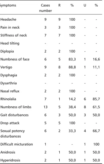
Table 1. Clinical symptoms observed in 20 cases of osteodural decompression.
Symptoms Cases R % U %
number
Headache 9 9 100 -
-Pain in neck 3 3 100 -
-Stiffness of neck 7 7 100 -
-Head tilting - - - -
-Diplopia 2 2 100 -
-Numbness of face 6 5 83,3 1 16,6
Vertigo 9 8 88,8 1 11,1
Dysphagia 2 2 100 -
-Dysarthria - - - -
-Nasal reflux 2 2 100 -
-Rhinolalia 7 1 14,2 6 85,7
Numbness of limbs 13 5 38,4 8 61,5 Gait disturbances 6 3 50,0 3 50,0
Drop attack 5 5 100 -
-Sexual potency 6 2 33,3 4 66,7
disturbances
Difficult micturation 1 - - 1 100
Anidrosis 2 1 50,0 1 50,0
Hyperidrosis 2 1 50,0 1 50,0
R, regressed; U, unchanged.
Table 2. Clinical symptoms observed in 27 cases of osteodural-neural decompression.
Symptoms Cases R % U %
number
Headache 7 7 100 -
-Pain in neck 12 11 91,6 1 8,3
Stiffness of neck 13 12 92,3 1 7,6
Head tilting 8 6 75,0 2 25,0
Diplopia 1 1 100 -
-Numbness of face 5 3 60,0 2 40,0
Vertigo 11 10 90,9 1 9,0
Dysphagia 9 9 100 -
-Dysarthria 1 1 100 -
-Nasal reflux 5 5 100 -
-Rhinolalia 16 - - 16 100
Numbness of limbs 22 10 45,4 12 54,5 Gait disturbances 9 5 55,5 4 44,4
Drop attack - - - -
-Sexual potency 12 4 33,3 8 66,7
disturbances
Difficult micturation 3 2 66,6 1 33,3
Anidrosis 6 3 50,0 3 50,0
Hyperidrosis 7 5 71,4 2 36,3
pressure and further dilating the epidural venous plexus. This would compress the dural sac, displa-cing, as a result, the CSF, and pushing it into the cranial cavity which returns rapidily to the spinal subaracnoid space, as soon as the pressure brought down to normal levels. In the case of tonsil hernia-tion, the CSF return, would be blocked by the cere-bellar tonsils which would then act as a valve, occlu-ding the foramen magnum. At this point, the spinal cord central canal – under a lower pressure than that of the intracranial cavity – would become the ideal place to accommodate CSF. The perpetuation of phases of craniospinal pressure dissociation cause the formation and maintenance of syringomyelia.
According to Taricco10 (1994), Ball and Dayan (1972)
and Aboulker (1979), respectively, admitted that CSF penetrates in the central canal of the spinal cord though Virchow-Robin spaces or through the dorsal roots, creating, in this way, the syringomyelic cavity. Gardner, Williams, Ball and Dayan, and Aboulker have with their theories, contributed enormouly for the explanation of the different stages surrounding the formation of the syringomyelic cavity.
Concerning the clinical aspects, Schultze24 (1882)
was probably the first author to make a clinical ana-tomopathologic correlation in SM. Later, many au-thors described clinical aspects of this condi-tion2,6,10,17,23,25,26. SM symptomology comes on as the
Table 3. Clinical signs observed in 20 cases of osteodural decompression.
Signs Cases R % U %
number
Lesion of V th nerve 11 8 72,7 3 27,2
Lesion of VI th nerve 1 1 100 -
-Facial spasm 1 1 100 -
-Nystagmus 8 4 50,0 4 50,0
Paresis of soft palate 2 2 100 7 70,0
Abolition of gag and palatal reflexes 10 3 30,0 20 100
Lesion of XI th nerve 18 6 33,3 12 66,7
Lesion of XII th nerve 2 1 50,0 1 50,0
Hypotonia 10 2 20,0 8 80,0
Spasticity 11 5 45,4 6 54,5
Cerebellar disturbances 2 1 50,0 1 50,0
Hyperreflexia 15 1 6,7 14 93,3
Clonus 2 - - 2 100
Hyporeflexia 16 - - 16 100
Hoffmann sign 2 - - 2 100
Babinski sign 7 2 28,5 5 71,4
Rossolimo sign - - - -
-Abolition of abdominal reflexes 14 - - 14 100
Unsteady gait 1 - - 1 100
Paresis of gait 5 3 60,0 2 40,0
Hypopallesthesia 19 - - 19 100
Fasciculations 1 - - 1 100
Claude Bernard - Horner 1 - - 1 100
result of the expansion of the syringomyelic cavity and the gliosis affecting the intramedullary and/or in the brain stem structures.
Milhorat et al.26 (1997) measured the pressure
in the interior of the syringomyelic cavity, and sug-gested that the distention would depended on dif-ferent degrees of intramedullary pressure, causing lesion of long tracts, gray matter and microcircula-tion. The involvement of the anterior horns gives rise to fibrillation, fasciculation, muscular weakness and atrophy. On the one hand, the compression of the posterior horn and ventral decussation will give origin to syringomyelic dissociation, and, on the other hand, a commitment of the sympathetic connections will result in Claude Bernard-Horner syndrome. With the expansion of the cavity, the spinal cord white matter
will be compressed causing lesion of the pyramidal and extrapyramidal tracts and dorsal columns.
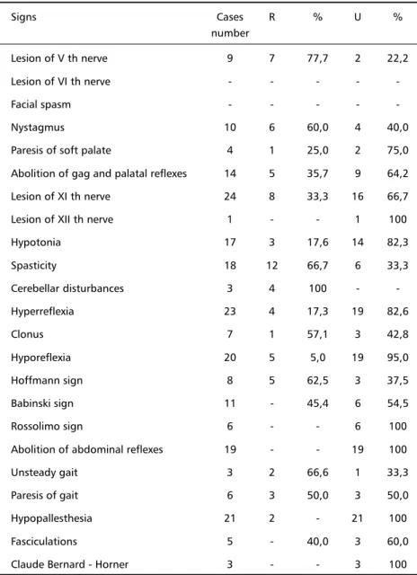
Tables 1 and 2 show the clinical symptoms pre-sented by our patients. In general, after surgery the majority of the symptoms improved in both groups. After analyzing the clinical signs (Tables 3 and 4), improvement of patients condition was far more noticeable in group II patients, especially where the lesion of trigeminal, vestibular, glossopharyngeal and vagus nerve are concerned. Improvement was also no-ticed to have occurred on the pyramidal signs of li-beration as hyperreflexia, spasticity, Babinski and Rossolimo’s sign.
As to the neurological deficits caused by the syringomyelic cavity itself (Table 5) there occurred
Table 4. Clinical signs observed In 27 cases of osteodural-neural decompression.
Signs Cases R % U %
number
Lesion of V th nerve 9 7 77,7 2 22,2
Lesion of VI th nerve - - - -
-Facial spasm - - - -
-Nystagmus 10 6 60,0 4 40,0
Paresis of soft palate 4 1 25,0 2 75,0
Abolition of gag and palatal reflexes 14 5 35,7 9 64,2
Lesion of XI th nerve 24 8 33,3 16 66,7
Lesion of XII th nerve 1 - - 1 100
Hypotonia 17 3 17,6 14 82,3
Spasticity 18 12 66,7 6 33,3
Cerebellar disturbances 3 4 100 -
-Hyperreflexia 23 4 17,3 19 82,6
Clonus 7 1 57,1 3 42,8
Hyporeflexia 20 5 5,0 19 95,0
Hoffmann sign 8 5 62,5 3 37,5
Babinski sign 11 - 45,4 6 54,5
Rossolimo sign 6 - - 6 100
Abolition of abdominal reflexes 19 - - 19 100
Unsteady gait 3 2 66,6 1 33,3
Paresis of gait 6 3 50,0 3 50,0
Hypopallesthesia 21 2 - 21 100
Fasciculations 5 - 40,0 3 60,0
Claude Bernard - Horner 3 - - 3 100
improvement in motor power, amiotrophy and syrin-gomyelic dissociation. Patients in group II exhibited a better outcome than those group I. The tonsillec-tomy eliminating the compressive effect over the me-dulla and spinal cord – surgical finding discribed by Williams (1978) together with a large craniectomy and dural grafting have certainly accounted for the better results shown by group II.
Some surgical findings presented in our casuistic must be properly emphasized on account of their frequency as shown in Table 6: absence of dural pul-sation (45%), arachnoiditis (67.9%), foramen Ma-gendie’s block (62.2%), CM 1 (73.5%), CM 2 (26.4%),
vascular anomalies (69.8%) and communication of the fourth ventricle with the ependymal canal (45.2%). These findings are also referred to in the literature by several other authors2,7,10,11,18,22,23,27-30.
Regarding the surgical technique, all patients we-re operated on a sitting position what facilitates the surgical procedure. Many others authors3,8,18, 27 also
used this position, although others prefered the pro-ne position placement2,10,28-32.
The extension of the craniectomy varies from au-thor to auau-thor. Because of the small size of the poste-rior fossa in the presence of BI or CM especially when both anomalies are present we prefer to use a large
Table 5. Findings of muscular strenght, atrophy and syringomyelic dissociation.
Signs Cases R % I % U %
number Osteodural decompression(20 Cases)
Paresis 18 3 16,6 8 44,0 7 38,8
Atrophy 12 1 8,3 - - 11 91,6
Syringomyelic dissociation 20 8 40,0 - - 12 60,0 Osteodural-neural decompression (27 Cases)
Paresis 24 6 25,0 15 62,5 3 12,5
Atrophy 21 2 9,5 3 14,2 16 76,1
Syringomyelic dissociation 25 8 32,0 6 24,0 11 44,0
R, regressed; U, unchanged; I improved.
Table 6. Surgical findings in 53 patients with basilar impression, Chiari and syringomyelia.
Findings Cases number %
Thinning of the occipital bone 18 33,9
Thickenning of the occipital bone 13 24,5
Thickenning of the atlantoccipital ligament 15 28,3
Pulseless dura mater 22 41,5
Arachnoiditis 36 67,9
Block of the forame of Magendie 33 62,2
Chiari I 39 73,5
Chiari II 14 26,4
Cerebellar impression 07 13,2
Larger cerebellar hemisphere 5 9,4
Vascular network anomaly 37 69,8
Hydrocephalus 3 5,6
Communication of the fourth ventrical
with the hydromyelic cyst 39 73,5
craniectomy. It extends cranially to the transverse sinus and laterally up to 3 – 4 cm from the midline. The reason for this large opening is to increase both the posterior fossa and the cisterna magna, permitting, in this way, the herniated portions of the cerebellum and brain stem to migrate upwards. Williams23, Batzdorf33, 34 and Duddy and Williams35
state that the herniation of the cerebellar structures and brain stem, as seen in the postoperative period, are attributed to a large craniectomy. A small cra-niectomy, on the contrary, might have prevented a decompression from maintained the CSF blockade and perpetuating the craniospinal pressoric disso-ciation33, 35. Williams23 and Duddy and Williams35,
ho-wever, using a smaller craniectomy, revealed a fre-quent caudal migration of the cerebellum and brain stem, and pointed this out as the cause of poor re-sults. Duddy and Williams35 observed a caudal
migra-tion of the posterior fossa structures in 53 % of their patients, though on 41 % no change was noticed.
Ackermann1 (1790) was the first author to call
attention to the small size of the posterior fossa in BI. In recent times, however, several authors using x-ray, tomography and MRI examinations have also demonstrated that the posterior fossa volume, in the presence of BI and CM, is smaller than the observed in normal people36- 39.
Badie et al.28 have verified that the posterior
fos-sa volume is smaller in the presence of CM, but it was noticed to increase after decompression. Milhorat et al.9 have also noticed a decrease of 13,4
ml in the volume of the posterior fossa and about 40% of CSF volume (an average of 10,8 ml) when compared to normal individuals. Sahuquillo et al.31
have compared the results obtained from 10 patients on whom a large craniectomy was performed, lea-ving the arachnoid intact and completion of dural grafting, with the results derived from 10 other patients who had undergone small craniectomies with the arachnoid dissected and in whom dural graf-ting was also performed. In all patients who had been submitted to a large craniectomy, a cranial mi-gration of the cerebellum was noticeable, whereas in the cases of those patients on whom a small cra-niectomy was performed, in 7cases a caudal migra-tion of cerebellar structures was noticed. Some neu-rosurgeons prefer to leave the arachnoid untou-ched31,40–42, but the majority prefers to release the
arachnoid adherences2,8,22,23,30.
As to the cerebellar tonsils, some surgeons leave them intact, proceeding only with the opening of the fourth ventricle31,40–43 as we have conducted in
our group I patients. Other surgeons as it can be seen in recent publications2,10,34 besides dissection
of the arachnoid adherences of the tonsils and vessels and fourth ventricle opening, they recommend tonsi-lectomy as conduct on our group II patients.
Gardner3 (1950) did not suture the dura, because
he believed this would facilitate the decompression, and in 1965 introduced the dural graft for the treat-ment of syringomyelia19. Dural grafting was
performed on all our patients6 as from as 1975. Most
neurosurgeons2,4,10,28–32,40–42 have followed this
pro-cedure to treat BI, CM and SM. Munshi et al.29 have
demonstrated that decompression of the posterior fossa plus laminectomy of C 1 and dural grafting for the treatment of CM yielded faster regression of the hydromyelia when compared to the decompression and laminectomy with no dural grafting. In order to avoid adherence of the graft to the cerebellum, Sa-huquillo31 (1994) used surgical stitches to be used
on the graft with the porpose to move it away from the cerebellum and fixing it in the cervical aponeu-rosis. We performed this procedure in the last cases by applying just one stitch at the level of foramen magnum.
As to postoperative complications we have ob-served respiratory distress, CSF fistulae and halluci-nation on 3 (5.6%) patients, hiccups on 2 (3.7%), superficial skin infections on 2 (3.7%) others, G I tract hemorrhage in another (1.8%), and finally hyper-tensive pneumocephalus in just another (1.8%). This last patient exhibited compensated hydrocephalus, and the seating surgical position was said to be the cause of ventricle collapse and to account for fur-ther development of pneumocephalus. Two patients from group I died – one from respiratory complica-tions and the other from uncontrollable GI tract hemorrhage, which represents a mortality rate of 3.6 %. Other authors2,4,5,10-13,22,34 have also reported
several other complications.
We have used MRI on just a few patients – both on pre and postoperative period, partly due to the absence of this technological facility at the time of surgery, partly due to poor social condition of the majority of the patients. Taking into account, however, the promising clinical outcomes as seen in the present study, we have felt encouraged to justity our choice of osteodural-neural decompression as used on our group II patients.
Acknowledgements - Acknowledgements - Acknowledgements - Acknowledgements -
REFERENCES
1. Ackermann JF. Ueber die Kretinen, eine besondere Menschenabart in den Alpen. Gotha, in der Ettingerschen Buchhandlung, 1790. 2. Arruda JAM. Tratamento da siringomielia associada à malformação
de Chiari: análise de 30 casos. Tese. São Paulo, 1996.
3. Gardner WJ, Goodall RJ. The surgical treatment of Arnold-Chiari malformation in adults: an explanation of its mechanism and importance of encephalography in diagnosis. J Neurosurg 1950;3:199-206. 4. Gonçalves da Silva JA. Resultados do tratamento cirúrgico da
impres-são basilar e malformação de Arnold-Chiari: estudo de 72 casos. Tese. João Pessoa, 1977.
5. Gonçalves da Silva JA, Gonçalves da Silva CE. Postoperative Komplikationen bei 126 Faellen basilaerer Impression und Arnold-Chiarischer Missbildung. Neurochirurgia 1981;24:153-157.
6. Gonçalves da Silva JA, Cantisani JU Filho, Farias Brito JC, et al. Im-pressão basilar, Arnold-Chiari e siringomielia: análise de 20 casos ope-rados. Arq Bras Neurocirurg 1987;6:77-95.
7. Gonçalves da Silva JA. Basilar impression and Arnold-Chiari malformation: surgical findings in 209 cases. Neurochirurgia 1992;35:189-195.
8. Hankinson J. The surgical treatment of syringomyelia. In Advances and technical standards in neurosurgery, vol 5. Wien: Springer Verlag, 1978:127-151.
9. Milhorat TH, Chou MW, Trinidad EM, et al. Chiari I malformation redefined: clinical and radiographic findings for 364 symptomatic patients. Neurosurgery 1999;44:1005-1017.
10. Taricco MA. Tratamento cirúrgico da siringomielia associada à malformação de Chiari do tipo I. Tese. São Paulo, 1994.
11. Ebenius B. The roentgen appearance in four cases of basilar impression. Acta Radiol 1934;15:652-656.
12. Penfield W, Coburn DF. Arnold-Chiari malformation and its operative treatment. Arch Neurol Psychiatry 1938;40:328-336.
13. List CF. Neurologic syndromes accompanying developmental anomalies of occipital bone, atlas and axis. Arch Neurol Psychiatry 1941;45:577-616.
14. Gustafson WA, Oldberg E. Neurologic significance of platybasia. Arch Neurol Psychiatry 1940;44:1184-1198.
15. Marin-Padilla M, Marin-Padilla T. Morphogenesis of experimentally induced Arnold-Chiari malformation. J Neurol Sci 1981;50:29-55. 16. Simon TH. Beitraege zur Pathologie und pathologischen Anatomie des
Central-Nervensystem. Arch Psychiat Nervenkr 1875;5:108-163. 17. Finlayson AI. Syringomyelia and related conditions. In Baker AB, Baker LH
(eds). Clinical neurology vol 3. Philadelphia: Harper & Row, 1981;32: 1-17. 18. Gardner WJ, Angel J. The mechanism of syringomyelia and its surgical
correction. Clin Neurosurg1958;6:131-140.
19. Gardner WJ. Hydrodynamic mechanism of syringomyelia: its relationship to myelocele. J Neurol Neurosurg Psychiatry 1965;28:247-259.
20. Williams B. The distending force in the production of “communicating syringomyelia”. Lancet 1969;26:189-193.
21. Williams B. The valvular action of the Arnold-Chiari malformation. In Brock M, Dietz H (eds). Intracranial pressure. Berlin: Springer Verlag, 1972;338-442.
22. Williams B. A critical appraisal of posterior fossa surgery for communicating syringomyelia. Brain 1978;101:223-250.
23. Williams B. Surgery for hindbrain related syringomyelia. In Advances and technical standarts in neurosurgery, vol 20. Berlin: Springer Verlag, 1993:108-164.
24. Schultze F. Beitraege zur Pathologie und pathologischen Anatomie des centralen Nervensystems. Virch Arch Path Anat 1882;87:510-540. 25. Milhorat TH, Kotzen RM, MM HTM, Capocelli AL Jr, Milhorat RH.
Dysesthetic pain in patients with syringomyelia. Neurosurgery 1996;38:940-947.
26. Milhorat TH, Capocelli AL Jr, Kotzen RM, Bolognese P, Heger IM, Cottrell JE. Intramedullary pressure in syringomyelia: clinical and pathophysiological correlates of syrinx distension. Neurosurgery 1997;41:1102-1110.
27. Rhoton AL. Microsurgery of syringomyelia and syringomyelic cord syndrome. In Schmidek HH, Sweet WH (eds). Operative neurosurgical techniques, vol 1. New York: Grune & Stratton, 1982;103-124. 28. Badie B, Mendoza D, Batzdorf U. Posterior fossa volume and response
to suboccipital decompression in patients with Chiari I malformation. Neurosurgery 1995;37:214-218.
29. Munshi I, Frim D, Stine-Reyes R, Weir BKA, Hekmatpanah J, Brown F. Effects of posterior fossa decompression with and without duraplasty on Chiari malformation-associated hydromyelia. Neurosurgery 2000;46:1384-1390.
30. Bertrand G. Dynamic factors in the evolution of syringomyelia and syringobulbia. Clin Neurosurg 1973;20:322-333.
31. Sahuquillo J, Rubio E, Poca MA, Rovira A, Rodriguez-Baeza A, Cervera C. Posterior fossa reconstruction: a surgical technique for the treatment of Chiari I malformation and Chiari I/syringomyelia complex – preliminary results and magnetic resonance imaging quantitative assessment of hindbrain migration. Neurosurgery 1994;35:874-885. 32. Matsumoto T, Symon L. Surgical management of syringomyelia –
Current results. Surg Neurol 1989;32:258-265.
33. Batzdorf U. Chiari I malformation with syringomyelia. Evaluation of surgical therapy by magnetic resonance imaging. J Neurosurg 1988;68:726-730.
34. Batzdorf U. Syringomyelia: current concepts in diagnosis and treatment. Williams & Wilkins, Baltimore, 1991.
35. Duddy MJ, Williams B. Hindbrain migration after decompression for hindbrain hernia: a quantitative assessment using MRI. Brit J Neurosurg 1991;5:141-152.
36. Nyland H, Krogness KG. Size of posterior fossa in Chiari type I malformation in adults. Acta Neurochir 1978;40:233-242.
37. Schady W, Metcalfe RA, Butler P. The incidence of craniocervical bony anomalies in the adult Chiari malformation. J Neurol Sci 1987;82:193-203. 38. Vega A, Quintana F, Berciano J. Basichondrocranium anomalies in adult
Chiari type I malformation: a morphometric study. J Neurol Sci 1990; 99:137-145.
39. Stovner LJ, Bergan U, Nilsen G, Sjaastad O. Posterior cranial fossa dimensions in the Chiari I malformation: relation to pathogenesis and clinical presentation. Neuroradiology 1993;35:113-118.
40. Heiss JD, Patronas N, DeVroom HL, et al. Elucidating the pathophysiology of syringomyelia. J Neurosurg 1999;91:553-562.
41. Armonda RA, Citrin CM, Foley KT, Ellenbogen RG. Quantitative cine-mode magnetic resonance imaging of Chiari I malformations: an analysis of cerebrospinal fluid dynamics. Neurosurgery 1994;35:214-224.
42. Raftopoulos C, Sanchez A, Matos C, Balériaux D, Bank WO, Brotchi J. Hydrosyringomyelia – Chiari I complex: prospective evaluation of a modified foramen magnum decompression procedure: preliminary results. Surg Neurol 1993;39:163-169.