CLINICAL AND CEREBROSPINAL FLUID (CSF) PROFILE
AND CSF CRITERIA FOR THE DIAGNOSIS OF SPINAL
CORD SCHISTOSOMIASIS
Otávio Augusto Moreno-Carvalho
1, Cristiana Maria Nascimento-Carvalho
2,
Aroldo Luiz da Silva Bacelar
3, Antônio de Souza Andrade-Filho
4, Gersonita Costa
5,
Jandira Bastos Fontes
6, Telma Assis
7ABSTRACT - Objectives: To describe the clinical and CSF findings among patients with presumptive neuroschistosomiasis (NS) and to suggest a classification for the CSF diagnosis of presumptive NS. Method: The charts of all patients whose CSF exam was performed at the CSF Lab, José Silveira Foundation, Salvador, Brazil, from 1988 to 2002 were reviewed. Those with clinically suspected NS whose indirect fluorescent antibody test (IFA) and or hemagglutination-inhibiting antibodies test (HAI) were positive to S. mansoni were identified. Results: Of 377 patients, 67.9% were males; the median age was 36 years (mean 37 + 16 yrs, range 3-82 yrs). The most frequent complaints were paraparesis (59.9%), urinary retention (36.2%), lower limb pain (22.8%). WBC of CSF (count/mm3) was > 4 in 66.0% (mean 83 + 124, median 40, range 4.3-1,100), protein (mg/dl)
was > 40 in 84.6% (mean 185 + 519, median 81, range 41-6,800) and eosinophils were present in 46.9%. IFA and HAI were positive in 75.3%. WBC > 4 and presence of eosinophils were associated with IFA and HAI positive (67.3% versus 51.4%, p 0.014; 49.1% versus 23.0%, p 0.0001, respectively) and protein > 40 was not (85.4% versus 77.0%, p 0.09). Presence of WBC > 4, protein > 40 and eosinophils was associated with IFA and HAI positive (71.6% versus 38.2%, p 0.0003) but presence of eosinophils and any other combination of WBC and protein were not. Conclusion: NS should be considered as a possible diagnosis in patients who had had contact with schistosome-infected water and present with spinal cord compromising. Presence of IFA and HAI positive to S. mansoni, WBC > 4, protein > 40 and presence of eosinophils in the CSF may be considered as a criterium of highly probable presumptive diagnosis.
KEY WORDS: neuroschistosomiasis, cerebrospinal fluid, CSF diagnosis, myelitis, myeloradiculitis, Schistosoma mansoni.
Aspectos clínicos e liquóricos e critérios para o diagnóstico liquórico de esquistossomose medular Aspectos clínicos e liquóricos e critérios para o diagnóstico liquórico de esquistossomose medular Aspectos clínicos e liquóricos e critérios para o diagnóstico liquórico de esquistossomose medular Aspectos clínicos e liquóricos e critérios para o diagnóstico liquórico de esquistossomose medular Aspectos clínicos e liquóricos e critérios para o diagnóstico liquórico de esquistossomose medular
RESUMO - Objetivos: descrever aspectos clínicos e liquóricos de pacientes com diagnóstico presuntivo de neuroesquistossomose (NE) e sugerir uma classificação para o diagnóstico liquórico presuntivo da NE. Método: as fichas de todos os pacientes cujo exame de líquor (LCR) foi realizado no Laboratório de Líquor, Fundação José Silveira, Salvador, Brazil, entre 1988 e 2002, foram revistas. Aqueles com suspeita clínica de NE e teste de imunofluorescência indireta (IFI) e ou inibição da hemaglutinação (IHA) positivos para S. mansoni foram identificados. Resultados: dos 377 pacientes, 67,9% eram do sexo masculino, a mediana da idade foi 36 anos (média 37 + 16 anos, variação 3-82 anos). As queixas mais freqüentes foram paraparesia (59,9%), retenção urinária (36,2%), dor em membros inferiores (22,8%). A celularidade do LCR (células/mm3) foi > 4 em 66,0%
(média 83 + 124, mediana 40, variação 4,3 – 1.100), a proteína (mg/dl) foi > 40 em 84,6% (média 185 + 519, mediana 81, variação 41-6.800) e eosinófilos estavam presentes em 46,9%. IFI e IHA foram positivas em 75,3%. Celularidade > 4 e presença de eosinófilos estiveram associadas com IFI e IHA positivas (67,3% versus 51,4%, p 0,014; 49,1% versus 23,0%, p 0,0001, respectivamente) e proteína > 40 não foi (85,4% versus 77,0%, p 0,09). Celularidade > 4, proteína > 40 e eosinófilos estiveram associados com IFI e IHA positivos (71,6% versus 38,2%, p 0,0003) mas a presença de eosinófilos e qualquer outra combinação de celularidade e proteína não estiveram. Conclusão: NE deve ser considerada uma possibilidade diagnóstica em pacientes que tiveram epidemiologia positiva para S. mansoni e desenvolveram uma mielopatia. A presença de IFI e IHA positivos para S. mansoni, celularidade > 4, proteína > 40 e presença de eosinófilos no líquor pode ser considerado critério de alta probabilidade para o diagnóstico presuntivo da NE.
PALAVRAS-CHAVE: neuroesquistossomose, líquor, diagnóstico liquórico, mielite, mielorradiculite, Schistosoma mansoni.
This investigation was conducted at the CSF Lab - José Silveira Foundation, Salvador, BA, Brazil: 1CSF MD; 2Pediatric Infectious Diseases MD,
Adjunct Professor, PhD, Department of Pediatrics, Faculty of Medicine, Federal University of Bahia (UFBA); 3Chief and 5Assistant MD of the
Neurological Service of São Rafael Hospital; 4Adjunct Professor, PhD, Departament of Neuro-Psychiatry, Faculty of Medicine, (UFBA);4,6Assistant
MD of Neurology and Neurosurgery Foundation of Bahia; 7Neurology Assistant in Santo Antônio de Jesus, Bahia.
Received 24 October 2002, received in final form 17 January 2003. Accepted 24 January 2003.
Schistosomiasis infects over 200 million people worldwide, primarily children and young adults, being prevalent in Central and South Americas, Africa and Asia, where it has been considered an important issue of public health1. The specie Schistossoma
man-soni infects around 12 million people in Brazil, where 30 more million people are exposed to schistosome-infected water, characterizing this country as an endemic area2. Out of the digestive system, the
ner-vous system is the most common localization of the mansonic schistosomiasis3. Among the several
presentations of the Neuroschistosomiasis (NS), the meningomyelorradiculitis is the most frequent4.
Pre-vious studies have reported an incidence of 5% to 6% of spinal cord schistosomiasis (SCS) among pa-tients with non-traumatic or non-neoplastic mye-lopathy5,6. From post-mortem studies, it has been
estimated that between 20% and 30% of patients with mansonic schistosomiasis have compromising of the nervous system and that the incidence of asymptomatic NS is 3 to 4 times higher the frequency of symptomatic NS7-9.
Difficulties in recognizing NS have been described and attributed as one of the causal factors of the low frequency of diagnosis10. Some of these
diffi-culties are the scarce knowledge about this illness among physicians, the incomplete or oligosympto-matic or transient evolution of some cases with little functional disturbances, and the operational diffi-culties at the health institutions regarding investi-gation and diagnosis11. It is important to highlight
the necessity of thinking about SCS when any patient reporting contact with schistosome-infected water with non-traumatic myelopathy looks for health assistance10, because the treatment is highly
com-posed by oral medication and the prognosis is extre-mely favorable, if the disease is diagnosed and trea-ted at an early stage12-17.
The purposes of the present investigation are to describe the clinical and CSF findings among patients with presumptive SCS and to suggest a classification for the CSF diagnosis of presumptive SCS.
METHOD
The charts of all patients whose CSF exam was perfor-med at the CSF Lab, José Silveira Foundation, in Salvador, Northeast Brazil, from September 1988 to May 2002, were reviewed. Those with clinically suspected SCS, who repor-ted contact with schistosome-infecrepor-ted water, whose indi-rect fluorescent antibody test (IFA) and or hemagglutina-tion inhibiting antibodies test (HAI) to S. mansoni were positive plus negative ELISA to HTLV-1 were identified. When the CSF samples had been collected before the
avai-lability of ELISA to HTLV-1, this test was performed by using frozen (-20oC) samples kept in the CSF base. All patients
presented S. mansoni eggs in feces or in a biopsy specimen from the rectum.
Data collection was based on a questionnaire which asked for demographic and clinical features and on the CSF findings that were registered on the CSF Lab charts. Specific items were asked to those patients submitted to lumbar tap at the CSF Lab. Some samples of CSF were received in the CSF Lab accompanied by the syndromic diagnosis. The CSF examination was performed by the sa-me person (OAMC), at the sasa-me Lab and included, in all samples, CSF WBC (white blood cell) and differential cell counts, concentration of protein, glucose, chloride, AST (glutamic-oxaloacetic transaminase) and LDH (lactate dehydrogenase), bacteriologic and mycologic exams (cul-tures for aerobic bacteria, fungus and Mycobacterium tuberculosis and specific stained-smears), IFA and HAI to S. mansoni, Toxoplasma gondii and Cisticerccus cellulosae, VDRL, FTA-ABS and HAI to Treponema pallidum, ELISA to C. cellulosae and HTLV-1. Whenever the protein concen-tration was > 160mg/dl, the CSF sample was diluted to the protein concentration equal to 80mg/dl. The IFA to S. mansoni was performed by using adult worm included in paraffin, when IgG antibodies were searched. Hypercytosis was defined as WBC count > 4 cells/mm3 and protein
con-centration increase was defined as protein concon-centration > 40mg/dl. For this investigation, just the first CSF exami-nation of each patient was considered. All taps were per-formed on the lumbar region.
Statistical analyzes were performed by using the Sta-tistical Package for the Social Sciences (SPSS 9.0). Diffe-rences in proportions were assessed by the Pearson Chi Square test or Fisher’s exact test, as appropriate. Means of continuous variables were compared by Mann-Whitney U. Confidence interval (95%) was reported for mean dif-ference. The statistical tests were two tailed, with a signi-ficance level of 0.05.
RESULTS
Of 522 identified patients, ELISA to HTLV-1 was per-formed in 401 and was negative in 377, all of them also had negative tests to cysticercosis, syphilis and toxoplasmosis and constituted the group of patients for this study. Cancerous cells were also searched in all samples and were not identified in any of them.
Protein concentration increase was identified in 319 (84.6%) of the samples (median 81mg/dl, mean 185±519 mg/dl, range 41 to 6,800 mg/dl) and eosi-nophils were present in 177 (46.9%) (median 6%, mean 8%±10%, range 1% to 54%). The concentra-tion of glucose, chloride, AST and LDH were under normal limits in all samples as well as all bacteriologic and mycologic exams were negative.
In order to search IgG antibodies to S. mansoni, IFA and HAI were performed in 368 (97.6%) and 309 (82.0%) of the CSF samples, respectively, both of them were performed in 300 (79.6%) and both were positive in 226 (75.3%). IFA was positive in 325 (86.2%) and HAI in 278 (73.7%). The comparison of the proportions and means of isolated CSF aspects with the positiveness of IFA and HAI to S. mansoni in CSF samples is presented in Tables 3 and 4, respectively. The association of combined CSF aspects
Table 1. Distribution of age of pacients with presumptive diagnosis of Spinal Cord Schistosomiasis, Salvador, 1988-2002.
Age stratum N %
(years)
< 9 6 1.7
10-19 10 9.9
20-29 86 23.8
30-39 86 23.8
40-49 71 19.6
50-59 38 10.5
60-69 31 8.6
70-79 5 1.4
> 80 3 0.8
N, absolute number.
Table 2. Frequency of clinical findings or syndromic diagnosis and respective durations among pacients with presumptive spinal cord schistosomiasis, Salvador, 1988-2002.
Clinical Findings N % Duration (days)
N Median Mean SD Minimum Maximum
Paraparesis 226 59.9 208 45 317 905 3 21 years
Urinary retention 144 36.2 139 30 197 698 1 20 years
Lower limb (LL) pain 86 22.8 82 90 271 429 5 6 years
LL Paresthesia 77 20.4 65 90 208 360 4 6 years
Lumbar pain 56 14.9 53 30 144 304 3 4 years
Paraplegia 51 13.5 43 30 163 435 3 7 years
Tetraparesia 11 2.9 10 135 197 231 7 2 years
Mielorradiculitis 11 2.9 1 15 15 - 15 15
Sexual disfunction 9 2.4 7 730 472 331 21 2 years
Polineuropathy 4 1.1 3 730 1643 1741 548 10 years
Atrophy of 1 LL 4 1.1 4 730 881 734 240 5 years
Abdominal pain 3 0.8 3 90 88 63 25 150
Thoracic pain 3 0.8 2 55 55 49 20 90
4 limbs pain 3 0.8 3 120 167 180 15 365
Tetraplegia 2 0.5 2 53 53 52 16 90
Transverse mielitis 2 0.5 1 20 20 - 20 20
Thoracic and abdominal pain 1 0.3 1 30 30 - 30 30
and the positiveness of the immunologic tests to S. mansoni is shown in Table 5.
DISCUSSION
To the best of the authors’ knowledge, this inves-tigation presents the greatest number of studied pa-tients with presumptive SCS. The second greatest study was published by Peregrino et al., when 80 patients were analyzed18. This characteristic may be
explained by some factors: this study was conducted in Salvador, Northeast Brazil, an endemic zone of mansonic schistosomiasis19, the study included
pa-tients submitted to lumbar tap during a 14-year
pe-riod and the data were collected from the data base of the CSF Lab – José Silveira Foundation, where many patients seen at several Services of Neurology or Infectious Diseases in the state of Bahia and in other neighbouring states are sent to in order to be submitted to CSF examination.
The predominance of males and the age range from 20 to 50 years (Table 1) is according to the results previously reported in several other stu-dies4,10,18,20-22. The predominance of males is
attribu-ted to a great exposition in the environment23,
ad-ded to professional activities that increase the intra-abdominal pressure4. The increase of the
intra-Table 3. Association between isolated CSF aspects with the positiveness of IFA and HAI S. mansoni in CSF (X2).
IFA and HAI Two positive One positive
CSF Aspect* (N=226) (N=74) Total (N=300) p
Hypercytosis 67.3 (152) 51.4 (38) 63.3 (190) 0.014 Presence of Eosinophils 49.1 (111) 23.0 (17) 42.7 (128) 0.0001 Protein > 40 mg/dl 85.4 (193) 77.0 (57) 83.3 (250) 0.09 Protein > 60 mg/dl 62.4 (141) 45.9 (34) 58.3 (175) 0.01
*Results in % (n).
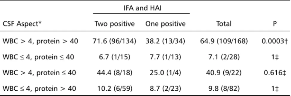
Table 5. Association between the presence of eosinophils and combined CSF aspects with the positiveness of IFA and HAI to S. mansoni in CSF.
IFA and HAI
CSF Aspect* Two positive One positive Total P WBC > 4, protein > 40 71.6 (96/134) 38.2 (13/34) 64.9 (109/168) 0.0003† WBC ≤ 4, protein ≤ 40 6.7 (1/15) 7.7 (1/13) 7.1 (2/28) 1‡ WBC > 4, protein ≤ 40 44.4 (8/18) 25.0 (1/4) 40.9 (9/22) 0.616‡ WBC ≤ 4, protein > 40 10.2 (6/59) 8.7 (2/23) 9.8 (8/82) 1‡
WBC = white blood cell. *Result in % (n/N), †X2, ‡Fisher’s exact test
Table 4. Comparison between means of isolated CSF aspects with the positiveness of IFA and HAI to S. mansoni in CSF. †
IFA and HAI
CSF Aspect Two positive One positive p 95% CI mean difference
WBC count 90 ± 142 46 ± 75 0.001 10, 77
Presence of Eosinophils 8 + 10 6 ± 8 0.069 -2, 7 Protein > 40 mg/dl 216 ± 624 84 ± 64 0.001 42, 222
abdominal and of the intra-spinal pressures con-current with the maintenance of negative pressure in the epidural space predisposes to the migration of eggs or worms to the spinal cord by Batson’s plexus24. It is important to highlight the little
importance of portal hypertension in the pathoge-nesis of SCS4, and the age in which the incidence of
this illness is higher, compromising young adults, during the most productive phase of life, factors that emphasize the importance of the study of SCS.
The most frequent clinical findings, paraparesis, lower limb pain and urinary retention, and the fre-quency of paraplegia (13.5%), (Table 2) are conse-quences of the commonest location of the lesion of SCS, that is, conus medullaris and equine cauda4,20.
The functional disturbances may be restrictive or disa-bling. In the present study, the clinical findings varied from 1 day up to 21 years, being the predominant duration ≤ 45 days at the time of the lumbar tap (Table 2). The SCS is an inflammatory disease, where the delayed-type hypersensitivity in response to an-tigens (eggs, worms) is the responsible for the neu-rological damage in the spinal cord18. Therefore, it is
important to ensure an early diagnosis and treat-ment, in order to warrant a better prognosis and the preservation or recuperation of neurological functions4,10,18,20-22,25.
The gold standard for the diagnosis of SCS is the demonstration of the pathologic process after a biopsy or a necropsy10. Nevertheless, the surgery to
collect clinical specimen for biopsy is an invasive pro-cedure that may compromise by itself the neuro-logical function of the patient, and must be kept for doubtful or unresponsive cases4,18,26. The
standardi-zation and availability of tests to identify S. mansoni antigens in the CSF, such as the Polymerase Chain Reaction, will be an advance in the SCS diagnosis. Nevertheless, in the meantime, it is recommended to establish criteria for presumptive diagnosis and to know their reliability. The most recommended study would consist in determining the predictive value of a positive and of a negative test which depends on the presence or absence of the disease as determined with the gold standard27. Such a study
was not feasible because of the difficulties mentio-ned previously4,18,26.
The CSF reflects the inflammatory process that compromises the spinal cord very reliably18. In 1985,
Livramento et al. described the syndrome of CSF in NS28, when the immunologic reactions to S. mansoni
in the CSF were standardized. This syndrome included lymphomononuclear hypercytosis associa-ted with the presence of eosinophil cells, protein con-centration increase and the presence of antibodies to S. mansoni28. In the present investigation, the
pre-sumptive diagnosis was strengthened by the presence of IgG antibodies to S. mansoni in two dif-ferent tests. From the data shown in Tables 3 and 4, it is possible to infer that the intensity of hypercytosis and of the increase in protein concentration influence the association of those CSF aspects and the positi-veness of the immunologic tests to S. mansoni, and that this is not the case of presence of eosinophil cells. For this latter CSF aspect, the association re-mains on the presence of eosinophil cells per si, wi-thout any influence of the intensity of its presence. The permanence of the association of concomitant hypercytosis, protein concentration increase and pre-sence of eosinophil cells, with the positiveness of IFA and HAI to S. mansoni in the CSF (Table 5) de-monstrates the importance of all these aspects of the syndrome of CSF for the presumptive diagnosis of SCS. From the foregoing data, a classification for the CSF diagnosis of presumptive NS is proposed in Figure 1. The difference among those levels (Table 6) is based on the analysis of the CSF abnormal fin-dings intensity that may represent different pro-babilities of SCS. The specific treatment to SCS must be considered at any of those levels (Table 6) because the prognosis is extremely favorable, if the disease is diagnosed and treated at an early stage12-17. It is
important to emphasize that two negative immu-nologic reactions do not necessarily exclude the diagnosis of NS.
Every patient included in this study had mansonic schistosomiasis and neurological complaints that
Table 6. Classification for the CSF diagnosis of NS. Gold standard diagnosis
Gold standard diagnosis Gold standard diagnosis Gold standard diagnosis
Gold standard diagnosis depends on the development and standardization of a test that identify the antigens of S. mansoni in the CSF;
Presumptive diagnosis Presumptive diagnosis Presumptive diagnosis Presumptive diagnosis Presumptive diagnosis
highly probable highly probablehighly probable
highly probablehighly probable when there are 2 positive immunologic reactions plus hypercytosis plus presence of eosinophil cells plus protein concentration increase;
probable probableprobable
probableprobable when there are 2 positive immunologic reactions plus either hypercytosis and presence of eosinophil or hypercytosis and protein concentration > 60mg/dl or presence of eosinophil and protein concentration > 60mg/dl; possible
possible possible
may not have been due to SCS. CSF antibodies may have crossed the blood-CSF barrier and the neurolo-gical findings may have been due to another disease. The cause-effect relationship could have been esta-blished if the serum/CSF index of specific antibodies had been studied along with the investigation of the integrity of the blood-CSF barrier29. These
pro-cedures were not performed. Information about other complementary exams was not computed be-cause such information was not regularly registered on the charts of the CSF Lab. Therefore, a prospective study is highly recommended in order to confirm our results.
For every patient with history of previous contact with schistosome-infected water, with compromise of spinal cord, it is important to consider the SCS as a possible diagnosis. The definitive diagnosis relies on the histopathological demonstration of the inflammatory process around the eggs or the worm in the spinal cord. The presumptive diagnosis of SCS must rely on history of exposure to schistosome-infected water, clinical findings, demonstration of schistosome eggs in feces or in a biopsy specimen from the rectum, CSF findings and exclusion of other illnesses that can cause the same symptoms.
REFERENCES
1. CEGET-CNRS/OMS. Atlas de la repartition mondial des schistosomia-ses. Geneva: WHO, 1987.
2. Lambertucci JR, Rocha RS, Carvalho OS, Katz N. Esquistossomose mansoni em Minas Gerais. Rev Soc Bras Med Trop 1987;20:47-52. 3. Andrade NA, Bastos Cl. Esquistossomose mansônica cerebral. Arq
Neuropsiquiatr 1989; 47: 100-104.
4. Peregrino AJP, Oliveira SP, Porto CA et al. Meningomielorradiculite por Schistosoma mansoni. Arq Neuropsiquiatr 1988; 46: 49-60. 5. Spina-França A, Salum PNB, Limongi JCP, Berger A, Losso ER.
Mielopatias: aspectos diagnósticos. Arq Neuropsiquiatr 1980;38:360-366. 6. Scrimgeour EM. Non-traumatic paraplegia in Northern Tanzânia. Br
Med J 1981;283:975-978.
7. Andrade AN. Neuroesquistossomose. Arq Neuropsiquiatr 1986;44:275-279.
8. Corrêa RLB, Lima JMB, Alencar A, Bastos ICC, Duro LA. Comprometimento neurológico na esquistossomose mansônica. Rev Bras Neurol 1983;19:101-104. 9. Galvão ACR. Radiculomielopatias esquistossomóticas. Arq Bras
Neurocirurg 1985;4:133-139.
10. Santos EC, Campos GB, Diniz AC, Leal JC, Rocha MOC. Perfil clínico e critérios diagnósticos da mielorradiculopatia esquistossomótica. Arq Neuropsiquiatr 2001;59:772-777.
11. Peregrino AJP. Neuroesquistossomose. In: Machado LR, Livramento JA, Nóbrega JPS, Gomes HR, Spina-França A (eds). Neuroinfecção 98. São Paulo: Academia Brasileira de Neurologia, 1998:45-50.
12. Gama C, Sá JM. Esquistossomose medular: granulomas produzidos por ovos de Schistosoma mansoni comprimindo a medula, epicone, cone
e cauda eqüina. Arq Neuropsiquiatr 1945;3:334-336.
13. Ross GL, Norcross JW, Horrax G. Spinal cord involvement in Schistosomiasis mansoni. N Engl J Med 1952;246:823-826.
14. Bird AV. Acute spinal schistosomiasis. Neurology 1964;14:647-656. 15. Rosebaum RM, Ishii M, Tanowitz H, et al. Schistosomiasis mansoni of
spinal cord. Am J Trop Hyg 1972;21:182-184.
16. Leads from the MMWR. Acute schistosomiasis with transverse myelitis in American students returning from Kenya. JAMA 1984;252:1116-1123. 17. Case Records of the Massachusetts General Hospital. A 40-year-old woman with the rapid onset of flacid paraplegia. N Engl J Med 1996;334: 382-389. 18. Peregrino AJP, Puglia PMK, Nóbrega JPS, Livramento JA,
Marques-Dias MJ, Scaff M. Esquistossomose medular: análise de 80 casos. Arq Neuropsiquiatr 2002;60:603-608.
19. Prata A, Bina JC, Barreto AC, Alecrim MG. Attempt to control the schistosomiasis transmission by oxamniquine, in an hyperendemic locality. Rev Inst Med Trop 1980; 22 (Suppl 4):65-72,182-189. 20. Brito JCF, Silva JAG, Silva EB, Viana NO. Neuroesquistossomose medular:
avaliação clínico-laboratorial de 5 anos. Arq Neuropsiquiatr 1992; 50:207-211. 21. Andrade AS Filho, Reis MG, Souza AL, et al. Neuroesquistossomose mansônica: aspectos clínicos, laboratoriais e terapêuticos. Arq Neuropsiquiatr 1996; 54:232-237.
22. Nobre V, Silva LC, Ribas JG, et al. Schistosomal myeloradiculopathy due to Schistosoma mansoni: report on 23 cases. Mem Inst Oswaldo Cruz
2001;96(Suppl):137-141.
23. Scrimgeour EM, Gajdusek DC. Involvement of the central nervous system in Schistosoma mansoni and S. haematobium infection: a review.
Brain 1985;108:1023-1038.
24. Budzilovich GN, Most H, Feigin I. Pathogenesis and latency of spinal cord schistosomiasis. Arch Path 1964;77:383–386.
25. Junker J, Eckardt L, Husstedt I. Cervical intramedullar schistosomiais as a rare cause of acute tetraparesis. Clin Neurol Neurosurg 2001;103:39-42. 26. Peregrino AJP, Puglia PMK, Bacheschi LA, et al. Diagnóstico da
esquistossomose medular: contribuição da ressonância magnética e eletroneuromiografia. Arq Neuropsiquiatr 2002;60:597-602. 27. Browner WS, Newman TB, Cummings SR. Designing a new study: III.
Diagnostic tests. In Hulley SB, Cummings SR (EDS). Designing clinical research. Baltimore: Williams & Wilkins, 1988:87-92.
28. Livramento JA, Machado LR, Silva CL, et al. Síndrome do líquido cefalorraqueano na neuroesquistossomose. Arq Neuropsiquiatr 1985;43: 372-377.