Laboratory of Neuropathology, Department of Pathology and Forensic Medicine1and Department of Neurology and Psychiatry2, School of Medicine, Federal University of M inas Gerais, M inas Gerais, Belo Horizonte M G Brazil:1M . D., Professor,2M . D., Associate Professor. This study was supported by grant 302036/76 (Dr. JEH Pittella) from Conselho Nacional de Desenvolvimento Científico e Tecnológico (CNPq) and by Fundação de Amparo a Pesquisa do Estado de Minas Gerais (FAPEMIG). Received 10 September 2003, received in final form 27 Novembro 2003. Accepted 8 January 2004.
Dr. José Eymard Pittella - Rua dos Otoni 712/304 - 30150-270 Belo Horizonte M G - Brasil. E-mail: [email protected]
THE CONFORM ATION OF THE BRAIN PLAYS AN IM PORTANT
ROLE IN THE DISTRIBUTION OF DIFFUSE AXONAL INJURY IN
FATAL ROAD TRAFFIC ACCIDENT
José Eymard Homem Pittella
1, Sebastião Nataniel da Silva Gusmão
2ABSTRACT - Objective: A study was made of the brain lesions in 120 random victims of fatal road traffic accidents to determine the frequency and topographic distribution of diffuse axonal damage (DAI) in relation to the midline brain structures. Method: The identification of axons was carried out with a mouse antibody anti-neurofilament proteins 70-, 160-, and 210-kD. Results: DAI was identified in 96 (80%) brains and classified as Grade 1 in 21.9%, as Grade 2 in 51%, and as Grade 3 in 27.1% of the patients. In spite of the diffuse distribution that is characteristic of DAI, damage occurred preferentially in the interhemispheric for-mations (corpus callosum and fornix) and rostral portion of the brainstem, usually to one side of the midline. Conclusion: From a mechanical point of view, the interhemispheric formations and the rostral portion of the brainstem act as fixating structures for the cerebral hemispheres during rotational acceleration of the head. It is known that the motion of the cerebral hemispheres is delayed at the points of fixation, where greater stress would be produced, particularly on the side subjected to greater displace-ment. The frequent involvement by DAI of deep, center-medial brain structures, usually to one side of the midline, supports the mechanism proposed above.
KEY WORDS: diffuse axonal injury, head injury, road traffic accident, biomechanics.
A conformação do encéfalo é um fat or import ant e na dist ribuição da lesão axonal difusa no acident e de t rân-sit o fat al
RESUMO - Objetivo: Foram estudadas as lesões encefálicas de 120 vítimas fatais de acidentes de trânsito, selecionadas aleatoria-mente, com a finalidade de determinar a freqüência e distribuição topográfica da lesão axonal difusa (LAD) em relação com as estruturas encefálicas da linha média. Método: A identificação dos axônios foi efetuada com antisoro anti-proteínas do neuro-filamento 70-, 160- e 210-kD obtido em camundongo. Resultados: A LAD foi observada em 96 (80%) dos encéfalos examinados, tendo sido classificada em Grau 1 em 21,9%, Grau 2 em 51% e Grau 3 em 27,1% dos pacientes. A despeito da distribuição difusa que é característica da LAD, a lesão afetou preferencialmente as formações inter-hemisféricas (corpo caloso e fórnix) e a porção rostral do tronco encefálico, usualmente em um dos lados da linha média. Conclusão: As formações inter-hemisféricas e a porção rostral do tronco encefálico funcionam, do ponto de vista mecânico, como estruturas de fixação dos hemisférios cerebrais durante a aceleração rotacional da cabeça. Sabe-se que a movimentação dos hemisférios cerebrais é retardada nas áreas de fixação, geran-do aí maior estresse, particularmente no lageran-do submetigeran-do ao maior deslocamento. O frequente envolvimento pela LAD das estru-turas encefálicas centro-mediais profundas, usualmente em um dos lados da linha média, favorece o mecanismo acima proposto. PALAVRAS-CHAVE: lesão axonal difusa, trauma crânio-encefálico, acidente de trânsito, biomecânica.
Diffuse axonal injury (DAI) is a consistent feature of trau-matic brain injury following long duration, high speed acceler-ation or deceleracceler-ation, as occurs in road traffic accidents, falls and some sports injuries1-3. DAI is currently considered as the
most important factor in determining the morbi-mortality in non-missile head trauma. Not only is it the most common cause of post-traumatic coma in the absence of an intracranial
expand-ing lesion and of vegetative state, but also of disability after head injury1,4-7.
ros-fin-embedding, cut into 7 µm sections and stained for the identifi-cation of axons w ith a monoclonal mouse antibody anti-neurofila-ment proteins 70-, 160-, and 210-kD (Dianova-Immunotech, Hamburg, Germany) at a dilution of 1:200. The sections were incubated for two hours at 4oC. Following primary antibody incubation, the sections were
incubated for one hour in goat anti-mouse antibody (Sigma Chemical Company, St Louis, M O, USA) at a dilution of 1:100. Then, the sec-tions w ere incubated for one hour in mouse peroxidase-antiperoxi-dase (Sigma Chemical Company, St Louis, M O, USA) at a dilution of 1:100. For the visualization of the reaction product, the sections w ere reacted in 0.05% 3,3 diaminobenzidine tetrahydrochloride (DAB) and H2O2. The histological sections w ere counterstained w ith hematoxylin. For positive controls histological sections of normal brains w ere used. For negative controls, the phosphate buffer solution or normal mouse serum w ere used instead of the primary antibody.
Identification and grading of DAI - Axonal injury was considered as evidence of DAI when axonal swellings and bulbs were diffusely distributed throughout the brain, although preferentially located in the corpus callosum, the fornix, the internal capsule, the cerebral white matter and the rostral brainstem8,12. Axonal injury around
fo-cal lesions (hemorrhages, contusions, infarcts) were not considered as DAI.
DAI was graded according to the criteria proposed by Adams et al.1: grade 1 - presence of axonal swellings and bulbs; grade 2 -
pres-ence of hemorrhagic lesion in the corpus callosum; grade 3 - prespres-ence of primary hemorrhagic lesion in the dorsolateral quadrant of the ros-tral brainstem. In those patients that died at the site of the accident or before reaching hospital or that had died immediately after being admitted to the hospital, the presence of DAI was assigned by the find-ing of hemorrhagic lesion (macro or microscopically) in the corpus cal-losum and/or in the dorsolateral quadrant of the rostral brainstem. Isolated or sparse focal hemorrhages, anywhere, were not considered as DAI. Also, focal hemorrhagic lesions in the midline of the tegmen-tum of the midbrain and pons secondary to increased intracranial pressure (Duret’s hemorrhage) were not considered as DAI19-21.
RESULTS
Clinical findings - The age range of the 120 patients was from 2 to 88 years and the mean age was 37.5 ± 18.3 years. There were 90 (75.0%) males and 30 (25.0%) females. Eighty-three patients (69.2%) survived for less than one day after the accident: 41 patients (34.2%) died at the site of the accident and 42 (35% ) after hospital admission; the remaining 37 (30.8%) survived betw een 1 and 28 days at the hospital.
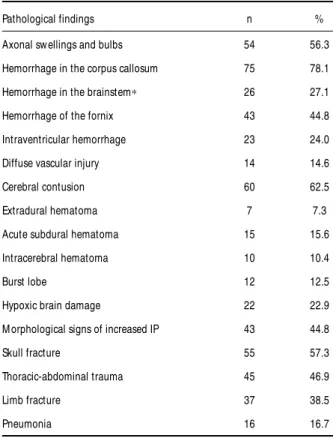
Neuropathological findings - DAI was seen in 96 (80.0%) patients. Table 1 show s the frequency of the various head and extracranial lesions found in the 96 patients with DAI. Cerebral contusion, skull fracture and morphological signs of increased intracranial pressure w ere the most common head lesions, w hereas thoracic-abdominal trauma and limb fracture w ere the most common extracranial lesions found. In those patients that died at the site of the accident or very early after hospi-tal admission, the cause of death was attributed to head tra-uma and/or increased intracranial pressure, internal hemor-tral brainstem2,5,8-14. The amount and topographic distribution
of DAI has been explained based on the centripetal progres-sion of strains to the core of brain15, and on the
concentra-tion of tensile and shear strains on the central brain and bra-instem determined in part by the compartmentalization of the intracranial cavity by the falx cerebri and tentorium cerebel-li7,16,17.
In this study, a morphological analysis is made of the macro and microscopic brain lesions in 120 individuals that died due to road traffic accidents, regardless of their period of survival after the head injury. The purpose of this study is to determi-ne the frequency and topographic distribution of diffuse axon-al damage in relation to the midline brain structures and to pro-pose a mechanism that would explain the correlation between the topography of DAI and the distribution of predicted defor-mations as related with the conformation of the brain, when the head is submitted to rotational acceleration.
M ETHOD
Patients - One hundred and twenty random victims of road traf-fic accidents, who had sustained either a motor vehicle accident (51 patients), or an auto-pedestrian injury (69 patients), autopsied in the period between 1989 and 1993, in the Institute of Legal Medicine, Belo Horizonte, Minas Gerais, Brazil, were studied. Both the victims that had died after being admitted to the hospital and those whose death had occurred at the site of the accident or before admission were includ-ed. The state of consciousness on hospital admission was evaluated by the Glasgow coma scale (GCS) established by Teasdale & Jennett18.
The autopsies were performed within 24 hours of death. Complete post-mortem examinations were performed in every case. After the trunk and limbs had been examined, the external lesions on the head and neck were described, followed by removal of the brain and descrip-tion of the bone and intracranial lesions found.
Neuropathology - In all 120 patients the neuropathological examination was performed by the same pathologist (JEH Pittella). The brains w ere suspended in 10% formalin solution for a minimum period of three w eeks. After the external brain surface had been described, frontal sections through the cerebral hemispheres, hori-zontal sections through the brainstem, sagittal sections through the left cerebellar hemisphere, and oblique sections through the right cere-bellar hemisphere were made.The sections were separated by 10 mm intervals at cerebral hemispheres level and by 5 mm intervals at brain-stem and cerebellum levels.
paraf-rhage, and multiple fractures. On the other hand, head trau-ma, increased intracranial pressure, hypoxic brain damage and pneumonia, either isolated or in association, were the cause of death in the patients that survived betw een 1 and 28 days. Grading and topographic distribution of DAI - DAI was grad-ed 1 in 21 (21.9%) patients, 2 in 49 (51,0%) patients and 3 in 26 (27.1%) patients.Axonal swellings and bulbs (Fig 1) were found in 54 (56.3%) patients (Table 1). Hemorrhagic lesions in the corpus callosum and in the dorsolateral quadrant of the rostral brainstem (Figs 2 and 3) w ere identified in 75 (78.1%) and 26 (27.1%) patients, respectively (Table 1). In the corpus callosum there w ere gross and microscopic hemorrhages in
39.2% and 60.8% of the patients, w hereas in the dorsolate-ral quadrant of the rostdorsolate-ral brainstem gross and microscopic hemorrhages were found in 57.6% and 42.4% of the patients, respectively.Axonal swellings and bulbs were diffusely distrib-uted throughout the brain, but w ere preferentially located in the corpus callosum and rostral brainstem (Table 2). The dis-tribution of focal hemorrhagic lesions and of the damage to axons in the corpus callosum and the rostral brainstem was markedly one-sided (Tables 3 and 4). Also, the distribution of the axonal injury in the cerebral white matter, the internal cap-sule, the fornix and the anterior commissure was markedly one-sided or asymmetrical. Of the 64 hospitalized patients with DAI,
Fig 1 Diffuse axonal injury. Axonal swellings and bulbs in the corpus callosum. PAP, antineurofilament proteins and hematoxylin counterstaining, X450.
Fig 2. Diffuse axonal injury. There is multiple small hemorrhage foci in the
56 (87.5%) w ere in a state of coma (Glasgow coma scores of 3 to 8) and eight (12.5%) had Glasgow coma scores of 9 to 15. All these eight patients had less severe DAI (grade I: five patients; grade II: three patients).
DISCUSSION
Identification of axonal injury - The identification of axon-al injury can be made by hematoxylin-eosin staining and sil-ver impregnation or by immunohistochemical techniques that use antibodies to the β-amyloid precursor protein2,12,13,22,23,
68-, 170- and 200-kDa neurofilament proteins10,24, and other
axon-ally transported proteins. The immunohistochemical tech-niques are comparatively much more sensitive than the con-ventional silver impregnation to demonstrate axonal injury10,13.
Table 1. Head and extracranial lesions in 96 patients with diffuse axonal injury.
Pathological findings n %
Axonal sw ellings and bulbs 54 56.3
Hemorrhage in the corpus callosum 75 78.1
Hemorrhage in the brainstem∗ 26 27.1
Hemorrhage of the fornix 43 44.8
Intraventricular hemorrhage 23 24.0
Diffuse vascular injury 14 14.6
Cerebral contusion 60 62.5
Extradural hematoma 7 7.3
Acute subdural hematoma 15 15.6
Intracerebral hematoma 10 10.4
Burst lobe 12 12.5
Hypoxic brain damage 22 22.9
M orphological signs of increased IP 43 44.8
Skull fracture 55 57.3
Thoracic-abdominal trauma 45 46.9
Limb fracture 37 38.5
Pneumonia 16 16.7
∗dorsolateral quadrant of the rostral brainstem.
IP, intracranial pressure.
Table 2. Topographic distribution of axonal swellings and bulbs in 96 patients with diffuse axonal injury.
Location n %
Cerebral w hite matter 26 27.1
Internal capsule 32 33.3
Corpus callosum 49 51.0
Fornix 22 22.9
Anterior commissure 19 19.8
Rostral brainstem 50 52.1
Table 3. Distribution of hemorrhagic lesions in the corpus callosum and dorsolateral quadrant of the rostral brain-stem in relation to the midline in 96 patients with diffuse axonal injury.
Distribution Corpus callosum Dorsolateral rostral brainstem
n % n %
Bilateral 12 16.0 8 30.8
One-sided 61 81.3 18 69.2
M edian 2 2.7 -
-Total 75 100 26 100
Table 4. Distribution of axonal swellings and bulbs in the corpus callosum and the rostral brain-stem in relation to the midline in 96 patients with diffuse axonal injury.
Distribution Corpus callosum Rostral brainstem
n % n %
Bilateral 3 6.1 4 8.00
One-sided 45 91.9 46 92.0
M edian 1 2.0 -
These studies indicated that the frequency of axonal injury has probably been underestimated using silver impregnation and that axonal injury may in fact be an almost universal conse-quence of fatal head injury7,13. In addition, neurofilament
pro-teins and the β-amyloid precursor protein have been show n to be effective markers of axonal damage in the form of focal axonal swellings within the first 1-2 hours after head injury9,12,
whereas by the conventional histological techniques of hema-toxylin-eosin and silver impregnation it is not possible to iden-tify w ith certainty axonal bulbs until about 15 h after injury14.
In a comparative study using antibodies to nine different anti-gens (including β-amyloid precursor protein and neurofilament protein), immunostaining for β-amyloid precursor protein pro-duced the most sensitive and reliable staining of axonal injury23,
although the authors used an antibody targeted exclusively to the 68-kDa neurofilament subunit.
Topographic distribution of DAI - In immunohistochemi-cal studies, the regions most frequently affected and contain-ing the largest number of axonal swellcontain-ings and bulbs were the corpus callosum, the internal capsule, the fornix, the midbrain and the pons9-13, such findings being very similar to those
reported by Adams et al.8and Adams25,26and by those of the
patients in our series.
DAI, in the form of both hemorrhagic lesions and injured axons, show ed a distinct tendency for one-sided concentra-tion in the interhemispheric structures and rostral porconcentra-tion of
the brainstem. According to Adams et al.1 and Graham et
al.14, the focal lesion in the corpus callosum is typically
hem-orrhagic, generally occurring in the inferior part of the corpus callosum and on one or other side of the midline, although it may extend to the midline and involve the interventricular sep-tum and the pillars of the fornix. Intraventricular hemorrhage can be found w hen there is disruption of the interventricular septum6. The focal hemorrhagic lesion in the dorsolateral
quadrant of the rostral brainstem is usually identified in the dorsolateral part of the midbrain and the rostral pons14. When
bilateral, one lesion is usually larger than the other. As in our series, in many patients the focal lesion in the corpus callo-sum and dorsolateral rostral brainstem is identifiable only microscopically1,10. Following the same pattern of the focal
hem-orrhagic lesion, the distribution of the damage to axons in the corpus callosum, the cerebral white matter, the cerebellum and the brainstem is not uniform or symmetrical14. Also w orth
noting is the high frequency of center-medial lesions associ-ated with DAI in our series: hemorrhage in the fornix, and intra-ventricular hemorrhage.
Biomechanics of DAI - Since DAI is a primary lesion25,27
caused by shearing forces, it can be admitted that its distri-bution is explainable by the application of physical law s28,29.
The biomechanics of DAI has been discussed for nearly three decades7,14-17(see, too, review of the literature in Goldsmith30
and M eythaler et al.3). Based on the experimental results
obtained by Holbourn28,29w ith a spheroid brain model and on
their ow n experiments31-33, Ommaya & Gennarelli15proposed
that the inertial strains produced by angular acceleration affect the brain in a centripetal progression, i.e., the dama-ging force w ould decrease as it is transmitted from the sur-face to the center of the brain. Based on that hypothesis, the authors concluded that center brain lesions are rare because centripetal force of sufficient intensity to reach that region is infrequent and, w hen present, w ould be associated w ith lesions in the periphery of the brain.
However, the results obtained by Tomlinson19, Crompton20,
Rosenblum et al.34, Grcevic16, Adams et al.1, and from our own
series confirm that lesions in the brainstem and center-medi-an brain structures (corpus callosum center-medi-and fornix) are quite fre-quent in fatal head trauma. Such high frequency was not ex-pected taking in consideration the decrease in the traumatic strains as they progress from the surface to the center of the brain.
The hypothesis advanced by Ommaya & Gennarelli15was
developed based on a spheroid brain model. The brain is bet-ter reproduced, however, not by a homogeneous spheroid, but by two large semi-spheroids (the cerebral hemispheres) inter-connected at the central portion of their medial faces by nar-row interhemispheric formations (corpus callosum, fornix and septum pellucidum) and connected to the brainstem inferior-ly by the cerebral peduncles. Relative fixation of the brainstem is provided anteriorly by the perforating vessels that originate from the basilar artery, and posterolaterally by the cerebellum. Besides, the anterior surface of the pons and the basilar artery rest on the basilar portion of the occipital bone and the dor-sum sellae of the sphenoid (clivus). The cerebral hemispheres, on the contrary, are relatively more subject to rotational move-ment35. Thus, from a mechanical point of view, the
interhemi-spheric formations and the rostral portion of the brainstem act as fixating structures for the hemispheres during movement of the brain w ithin the skull.
The shape and points of fixation of a body subjected to rotational acceleration are fundamental in the distribution of shearing stresses36. On the shape will depend the axis of
rota-tion and, therefore, the distriburota-tion of torque. At the point of fixation, rotational movement w ill be delayed (due to the torque produced by the reaction forces acting at these points), w ith consequent stress concentration and deformation at these points. Because of the conformation of the brain, inde-pendent and complementary displacements of the hemisphe-res occur during rotation of the head35. It is know n that such
areas, particularly on the side subjected to greater displace-ment. On the other hand, acceleration of the brain in the coronal plan w ould lead to greater stress in the rostral and lateral portion of the brainstem, much in the same manner as the acceleration of the crow n of a tree w ould cause greater stress, and possibly deformation, at the level of the tree’s trunk (Fig 4).
According to Grcevic16, Graham & Gennarelli7and Graham
et al.14, the direction in which the head moves plays an
impor-tant role in the amount and distribution of vascular and axon-al damage in a given situation. For equivaxon-alent levels of angu-lar acceleration, the brain is most vulnerable if it is moved la-terally. When the entire head undergoes rotational accelera-tion, in certain locations in the central brain and brainstem, portions of the brain move in opposite directions, thus gen-erating shear and tensile strains. The maximum deformation is in midline structures determined in part by the compartmen-talization of the intracranial cavity by the falx and tentorium7,16.
It is these regions that show the greatest and most consistent amounts of tissue tear hemorrhages and axonal injury. Similar conclusions were reported by Nishimoto & M urakami17based
on head models submitted to direct impact w ith translation-al acceleration.
It is possible, how ever, that the ow n conformation of the brain, as above explained, and not the compartmentalization of the intracranial cavity, can be the main responsible for the concentration of tensile and shear strains on the central brain and rostral brainstem during rotational acceleration of the head. In fact, a distinct pattern of lesions, influenced by the falx and
by the tentorium, follow to the presence of a supratentorial expanding mass lesion accompanied by increased intracranial pressure, deformation and shift of the brain and the appear-ance of internal herniae37,38. Expansion of a mass in the frontal
or parietal lobe lead to depression of the corpus callosum on the same side and herniation of w hole or part of the cin-gulate gyrus under the the free edge of the falx. The latter pro-duces an indendation or, occasionally, a w edge of pressure necrosis along the dorsal border of the herniated gyrus37-39.
On the other hand, herniation of part or w hole of the parahippocampal gyrus medially and dow nward through the tentorial opening results in deformation and compression of the midbrain37-39.The contralateral cerebral peduncle is pushed
against the rigid, free tentorial edge, leading to infarction w ith or w ithout hemorrhage in the dorsal part of the pedun-cle40. Hemorrhage and infarction occur adjacent to the
mid-line in the tegmentum of the midbrain and in the tegmental and basal parts of the pons (Duret’s hemorrhage). These lat-ter lesions are more likely the result of dow nwards displace-ment and anterior-posterior elongation of the rostral brain-stem caused by side-to-side compression by the tentorial her-nia, coupled w ith relative immobility of the basilar artery. In these situations of deformity, compression and shift of the brain, nerve tissue injury induced by contact w ith the falx and the tentorium does not produce focal hemorrhagic lesions in the corpus callosum and in the dorsolateral sector of the rostral brainstem.
CONCLUSION
The frequent involvement of deep, center-median brain structures (corpus callosum, fornix, and rostral brainstem), usually to one side of the midline, cannot be explained mere-ly either by the centripetal distribution of shearing stress dur-ing trauma or the concentration of tensile and shear strains on the central brain and brainstem determined in part by the compartmentalization of the intracranial cavity by the falx cere-bri and tentorium cerebelli. Our results show that it is the own conformation of the brain that makes these structures partic-ularly vulnerable to deformation by shear strains during rota-tional acceleration of the head.
REFERENCES
1. Adams JH, Doyle D, Ford I, Gennarelli TA, Graham DI, McLellan DR. Diffuse axonal injury in head injury: definition, diagnosis and grading. Histopathology 1989;15:49-59.
2. Abou-Hamden A, Blumbergs PC, Scott G, et al. Axonal injury in falls. J Neurotrauma 1997;4:699-713.
3. Meythaler JM, Peduzzi JD, Eleftheriou E, Novack TA. Current concepts: diffuse axonal injury-associated traumatic brain injury. Arch Phys Med Rehabil 2001;82:1461-1471.
4. Jennett B, Plum F. Persistent vegetative state after brain damage. Lancet 1972;1:734-737.
5. Adams JH, Graham DI, Murray LS, Scott G. Diffuse axonal injury due to non-missile head injury in humans: an analysis of 45 cases. Ann Neurol 1982;12:557-563.
6. Graham DI, Adams JH, Nicoll JAR, Maxwell WL, Gennarelli TA. The nature, distribution and causes of traumatic brain injury. Brain Pathol 1995;5:397-406.
7. Graham DI, Gennarelli TA. Trauma. In Graham DI, Lantos PL (eds). Greenfield’s neuropathology. 6 Ed. London: Arnold, 1997:197-262. 8. Adams JH, Graham DI, Scott G, Parker L, Doyle D. Brain damage in
fatal non-missile head injury. J Clin Pathol 1980;33:1132-1145. 9. Blumbergs PC, Jones NR, North JB. Diffuse axonal injury in head
trau-ma. J Neurol Neurosurg Psychiatry 1989;52:838-841.
10. Ng HK, Mahaliyana RD, Poon WS. The pathological spectrum of dif-fuse axonal injury in blunt head trauma: assessment with axon and myelin stains. Clin Neurol Neurosurg 1994;96:24-31.
11. Sherriff FE, Bridges LR, Sivaloganathan S. Early detection of axonal injury after human head trauma using immunocytochemistry for b-amyloid precursor protein. Acta Neuropathol 1994;87:55-62.
12. Blumbergs PC, Scott G, Manavis J, et al. Topography of axonal injury as defined by amyloid precursor protein and the sector scoring method in mild and severe closed head injury. J Neurotrauma 1995;12:565-572. 13. Gentleman SM, Roberts GW, Gennarelli TA, et al. Axonal injury: a uni-versal consequence of fatal closed head injury? Acta Neuropathol 1995;89:537-543.
14. Graham DI, Gennarelli TA, McIntosh TK. Trauma. In Graham DI, Lantos PL (eds). Greenfield’s neuropathology. 7. Ed. London: Arnold, 2002:823-898.
15. Ommaya AK, Gennarelli TA. Cerebral concussion and traumatic uncon-sciousness: correlation of experimental and clinical observations on blunt head injuries. Brain 1974;97:633-654.
16. Grcevic N. I. Head injury: the concept of inner cerebral trauma. Scand J Rehab Med Suppl 1988;17:25-31.
17. Nishimoto T, Murakami S. Relation between diffuse axonal injury and internal head structures on blunt impact. J Biomech Eng 1998;120:140-147. 18. Teasdale G, Jennett B. Assessment of coma and impaired consciousness:
a practical scale. Lancet 1974;2:81-84.
19. Tomlinson BE. Brain-stem lesions after head injury. J Clin Pathol 1970;23 (Suppl 4):154-165.
20. Crompton R. Brainstem lesions due to closed head injury. Lancet 1971;3:669-673.
21. Pittellla JEH, Gusmão SNS. Diffuse vascular injury in fatal road traffic accident victims: its relationship to diffuse axonal injury. J Forensic Sci 2003;48:626-630.
22. Gentleman SM, Nash MJ, Sweeting CJ, Graham DI, Roberts GW. b-Amyloid precursor protein (b-APP) as a marker of axonal injury after
head injury. Neurosci Lett 1993;160:139-144.
23. Sherriff FE, Bridges LR, Gentleman SM, Sivaloganathan S, Wilson S. Markers of axonal injury in post mortem human brain. Acta Neuropathol 1994;88:433-439.
24. Grady MS, McLaughlin MR, Christman CW, Valadka AB, Fligner CL, Povlishock JT. The use of antibodies targeted against the neurofilament subunits for the detection of diffuse axonal injury in humans. J Neuropathol Exp Neurol 1993;52:143-152.
25. Adams JH. Head injury. In Adams JH, Corsellis JAN, Duchen LW (eds). Greenfield’s Neuropathology. 4 Ed. London: Edward Arnold, 1984:85-124. 26. Adams JH. Head injury. In Adams JH, Duchen LW (eds). Greenfiels’s
neuropathology, 5 Ed. New York: Oxford, 1992:106-152.
27. Strich SJ. Shearing of nerve fibers as a cause of brain damage due to head injury: a pathological study of twenty cases. Lancet 1961;2:443-448. 28. Holbourn AHS. Mechanics of head injuries. Lancet 1943;2:438-441. 29. Holbourn AHS. The mechanics of brain injuries. Br Med Bull
1945;3:147-149.
30. Goldsmith W. The state of head injury biomechanics: past, present, and future. Part 1. Crit Rev Biomed Eng 2001;29:441-600.
31. Ommaya AK. Experimental head injury in the monkey. In Caveness HF, Walker AE (eds). Head injury. Philadelphia: Lippincott, 1966:260-275. 32. Ommaya AK. Trauma to the nervous system. Ann R Coll Surg Engl
1966;39:317-347.
33. Ommaya AK, Hirsch AE. Tolerances for cerebral concussion from head impact and whiplash in primates. J Biomech 1971;4:13-21.
34. Rosenblum WI, Greenberg RP, Seelig JM, Becker DP. Midbrain lesions: frequent and significant prognostic features in closed head injury. Neurosurgery 1981;9:613-620.
35. Pudenz RH, Shelden CH. The lucite calvarium: a method for direct obser-vation of the brain. II. Cranial trauma and brain movement. J Neurosurg 1946;3:487-505.
36. Holbourn AHS. The mechanics of trauma with special reference to herniation of cerebral tissue. J Neurosurg 1944;1:190-200.
37. Miller JD, Adams JH. The pathophysiology of raised intracranial pres-sure. In Adams JH, Corsellis JAN, Duchen LW (eds). Greenfield’s neu-ropathology. 4. Ed. London: Edward Arnold, 1984:53-84.
38. Ironside JW, Pickard JD. Raised intracranial pressure, oedema and hydrocephalus. In Graham DI, Lantos PL (eds). Greenfield’s neu-ropathology. 7. Ed. London: Arnold, 2002:193-231.
39. Russell DS, Rubinstein LJ. Pathology of tumours of the nervous sys-tem. 3. Ed. London: Edward Arnold, 1971:273-283.