Brazilian M inistry of Health Data Bank:1M D, PhD, Head of Neurology Unit, Hospital de Base do Distrito Federal, Brasilia DF, Brazil;2M D, Neurologist of Hospital Anchieta, Brasília DF, Brazil.
Received 29 September 2003, received in final form 10 January 2004. Accepted 5 February 2004.
Dra.Elza Dias Tosta - SHIS QL 16 Conj 5 casa 17 Lago Sul 71640-255 Brasília DF - Brasil. E-mail: [email protected]
NEUROLOGICAL M ORBIDITY IN VACCINE-ASSOCIATED
PARA-LYTIC POLIOM YELITIS IN BRAZIL
From 1989 up to 1995
Elza Dias-Tosta
1, Carlos Santos Kückelhaus
2ABSTRACT - We collected 30 cases of vaccine associated paralytic poliomyelitis (VAPP) from 4081 cases of acute flaccid palsies cases notified from 1989 to 1995 to the Brazilian Ministry of Health. There were 30 VAPP cases with 56% of children younger than 1 year old, 56.7% of female. 46% of cases were reported in the Northeast. Ten P2 vaccine virus, 8 P3 and 2 P1 and associations amongst them were isolated. The clinical pattern in 60 days was: monoplegia (16), paraplegia (6), tetraplegia (5), hemiplegia (2) and triplegia (1). There was no strong relationship between fever, before or after the prodrome period, or the use of intramuscular medication to morbidity. Conclusion: if the anti-poliomyelitis strategy adopted in Brazil has lead to the eradication of the poliomyelitis with wild virus infection, the existence of a minimum risk of vaccine-associated poliomyelitis is a matter of concern because there will be a permanent neurological deficit.
KEY WORDS: oral polimyelitis vaccine, paralytic poliomyelitis, neurological deficits.
M orbidade neurológica em poliomielit e paralít ica pós vacinal no Brasil de 1989 a 1995
RESUMO - Trinta casos de poliomielite associada à vacinação oral (Sabin) foram estudados a partir de 4081 notificações de par-alisias agudas e flácidas feitas ao Ministério da Saúde no período de 1989 a 1995, com o objetivo de avaliar a gravidade do quadro neurológico. Dezesseis pacientes tiveram monoplegia, 6 paraplegia, 5 tetraplegia , 2 hemiplegia e 1 triplegia. Foram 56% em menores de 1 ano, 56,7% no sexo feminino, 46% dos casos provenientes do nordeste. Em 10 pacientes foi isolado o vírus vacinal P2, em oito o P3 e dois o P1.Os demais tinham associações de mais de um tipo de vírus. Febre antes ou após o período prodrômi-co e o uso de medicação intramuscular não se relacionaram a maior morbidade. A política antipoliomielite adotada no Brasil lev-ou à erradicação da poliomielite pelo vírus selvagem com um risco mínimo do ponto de vista epidemiológico, porém ainda com custos individuais não desprezíveis.
PALAVRAS-CHAVE: vacina oral, poliomielite paralítica, seqüelas neurológicas.
The introduction of mass vaccination w ith the live atten-uated oral poliovirus vaccine has dramatically modified the epi-demic poliomyelitis, causing a sharp decline of the disease w orldw ide. The oral poliomyelitis vaccine (OPV) was success-fully used to control paralytic poliomyelitis in some regions of the w orld, including China, the former USSR, Japan, the Americas and Oceania, and many smaller European coun-tries, w hich all together have a population of almost 2 billion. The elimination of w ild-virus associated poliomyelitis in the Western Hemisphere in 1991 and the rapid progress in glob-al poliomyelitis eradication efforts changed the risk-benefit ratio associated w ith the exclusive use of oral poliovirus vaccine1.
Since 1961, shortly after the introduction of the OPV, cases of paralytic poliomyelitis caused by the vaccine in the United
Sta-tes w ere reported. Terry cited by Strebel, says that betw een 1961 and 1984, a total of 229 cases of vaccine-associated par-alytic poliomyelitis (VAPP) were reported in the United States, w ith a range of 14 cases annually after 19652.
In Brazil, the epidemiological surveillance system of the poliomyelitis eradication program of the M inistry of Health, keep in their records a total of 1290 cases of poliomyelitis in t he year of 1980.Wit h t he beginning of t he Nat ional Poliomyelitis Eradication campaign, there was a decline in the incidence of the disease and in 1989 the last case of po-liomyelitis in Brazil was reported3. However, in the last decade
reintro-duction of the w ild poliovirus in the population, since the dis-ease is still endemic in some neighboring continents. Moreover, in these days the risk of importing the virus is higher due to the facilities of traveling by airplane as already happened in Canada w here there was an outbreak of poliomyelitis ori-ginated from the Netherlands3-5.
The aim of the present study is to determine the actual neu-rological morbidity of vaccine associated paralytic poliomyelitis in the period of 1989 to 1995 in Brazil and their risk factors, to contribute to the discussion about a better way to vaccinate people against poliomyelitis: oral (Sabin) x intramuscular (Salk). It is important to keep in mind that the eradication of the dis-ease in South America through vaccination was a major achieve-ment. Nevertheless, isolated cases of VAPP have been report-ed in Brazil and other South American countries. After the first enthusiasm, it is timely to study the adverse events and to dis-cuss the vaccines costs/benefit ratio and the ongoing health strategies developed to maintain the eradication of poliomyelitis.
M ETHOD
The Brazilian poliomyelitis surveillance program data was searched for all cases diagnosed as poliomyelitis from 1989 to 1995. Cases were classified as: a) vaccine-associated poliomyelitis: those with acute flaccid paralysis (AFP) who persisted with residual neurological deficit after 60 days and the vaccine poliovirus was identified from the patients stools, collected during the first 15 days of paralysis, or from a sample of the cerebrospinal fluid: b) compatible poliomyelitis: AFP w ith residual neurological deficit after 60 days, or death, or had an unknow n follow up, but could not have the stool samples to isolate the poliovirus, c) not poliomyelitis: all subjects from whom poliovirus was not isolated in stool or other sample culture. According to the way to acquire the vaccine associated poliomyelitis they were: 1) VAPP recipients: cases with paralysis that received the OPV in a period bet-w een 4 to 30 days; 2) acquired cases from contact bet-with vaccinated children and presented with AFP in the period of 4 to 75 days after the vaccination day; 3) if neither 1 or 2 w ere identified, it was con-cluded that the case was acquired from the community.
The patients w ere studied in relation to some possible risk fac-tors such as: intramuscular injection, previous illness, the type of iso-lated virus strain (P1, P2, P3), age and gender. The vaccine-associat-ed poliomyelitis cases were distributvaccine-associat-ed according to the Brazilian geo-graphic regions in order to know its frequency and distribution.
All patients had stool samples culture.The isolated virus was clas-sified according to the strains as vaccine poliovirus type 1 (P1), vac-cine poliovirus type 2 (P2), and vacvac-cine poliovirus type 3 (P3) or w ild poliovirus. The analysis was performed by specialized laboratory ref-erence to the poliomyelitis program, and the stool had to be collect-ed in tw o samples w ith an interval of 24 hours until the fourteenth day. If the local condition permitted the patients had their cere-brospinal fluid analyzed and w ere submitted to an electromyogra-phy examination according to the judgement of the assistant electromyogra- physi-cian.The severity of the cases was shown by the intensity of the aggres-sion exemplified by the number of limbs affected: from tetraplegia, the most severe form, to monoplegia, the less severe one.
RESULTS
From a total of 4081 notifications of acute flaccid paraly-sis, 62 not ificat ions w ere classified as compat ible w it h poliomyelitis, and 30 cases (0.74%) as vaccine-associated poliomyelitis (age 2 to 100 months, mean 18.9 months, medi-an 10.5, stmedi-andard deviation SD = 10.7) there was 13 males (age 3 to 52 months, mean 14.8 months, median 7, SD = 14.8) and 17 females (age 2 to 100 months, mean 22.1, medi-an 16, SD = 22.1).
From these 30 cases, 14 w ere classified as recipients, as they developed the disease less than 30 days after the vacci-nation. Ten cases broke up betw een 31 to 60 days after the vaccine campaign and were considered as acquired from com-municants. It was not possible to identify the incubation peri-od in six cases. As long as these people had not been vacci-nated in the previous 60 days, the disease was considered as having been acquired in the community.
In 1989 ten cases of vaccine-associated cases w ere diag-nosed. The follow ing years show ed that there was decrease in the incidence. Only tw o cases w ere registered in 1993 and 1994, raising again in 1995 (Fig 1).
The year of 1989 was studied separately due to the fact that until that year all cases of acute flaccid palsies w ere investigated independently of the patient‘s age. Since 1990, only the acute palsies that affects children less than 15 years old w ere investigated. In 1989, from a total of 917 notifica-tions of acute flaccid paralysis, there w ere 10 cases of vac-cination-associated poliomyelitis. Out of these, three w ere identified as having received the vaccine. Tw o cases w ere acquired from communicants and five cases acquired the infection in the community. There was confirmed participation in the vaccination campaign of all theses cases. From 1990 to 1995, 20 cases of vaccine-associated poliomyelitis were iden-tified; 11 had received the vaccine, one was acquired from com-municants and the others from the community.
When the cases reported were plotted by regions, the vac-cine-associated paralysis predominated in the Northeast region (46%), w ith a minor frequency in the M idw est and North regions (Fig 2).
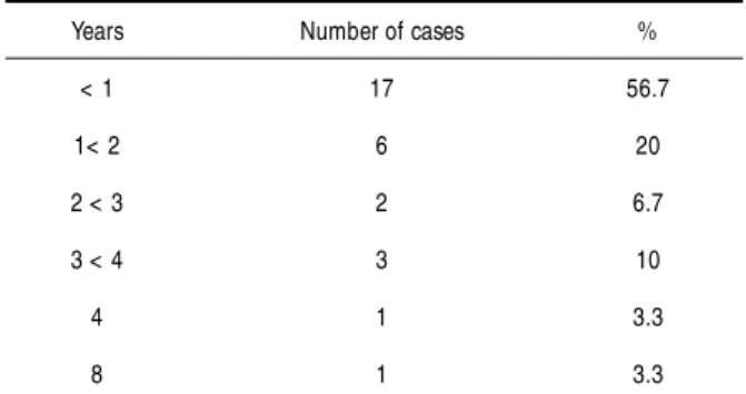
Gender and age - There was predominance of females, with 17 cases (56.7%). The great majority of cases were in children less than five years old with 96.6% of VAPP cases, and 17 cas-es (56.7%) lcas-ess than one year old, decreasing progrcas-essively in frequency according to the age. Betw een five and nine years old the frequency was only 4% (Table 1).
time as the paralysis, so they w ere interpreted as a compo-nent of the clinical picture and not as an indicative of immune deficiency or a sign of complication.
Diagnosis and morbidity - Table 2 shows the distribution of all vaccine-associated poliomyelitis cases according to the clinic, type of virus identified and the presence of risk factors. Table 3 correlates the illness severity to the type of virus.
In five cases the disease had affected all four limbs. In only one case it had affected three limbs. In eight cases tw o limbs
w ere affected. From those, tw o cases presented hemiplegia and six paraplegia. Finally, the last 16 patients had paralysis in only one limb (Table 2).
In only one case association w ith cranial nerve palsy was noticed. There w ere tw o cases of respiratory tract complica-tions. The inferior limbs w ere affected more severely and in the w hole the predominance was of the less severe forms of the disease. (Table 2).
In the follow -up at 60 days all cases w ith monoplegia remained as so but some became less severe. One case of
Fig 1. Vaccine-associated poliomyelitis in Brazil from 1989 to 1995.
tetraplegia turned to paraplegia and another with residual neu-rological deficit in the right arm and the left leg. Another case remained with triplegia.Two cases of tetraplegia remained un-changed at the evaluation w ithin 60 days, one of them w ith tracheotomy. How ever, in one of those, a later reevaluation in ten months show ed only monoplegia.
The cerebrospinal fluid was examined in 10 patients. It showed mononuclear pleocytosis in five cases and in three cas-es there was high protein content, w hich was compatible w ith viral meningitis. There w ere tw o cases w ith dissociation betw een protein and cytology content, but that proved not to be Guillain-Barré syndrome by the clinical and other labora-tory features. The electromyography test was performed in 25 of these 30 cases, being compatible w ith poliomyelitis in 22 cases.
The isolation of the virus in the stool was possible in 29 cases. In one case (case 12) identification of the virus was pos-sible only in the cerebrospinal fluid. From the three different viruses presented in the oral vaccine, the P2 type was present in 10 cases (representing 33% of the cases), the P3 type in eight cases and the P1 type in tw o cases. In the other 10 cas-es there was an association of poliovirus prcas-esent in the vac-cine. In four cases the P1P2P3 w ere identified, in four cases P2P3 and in one case P1P3 (Table 2).
DISCUSSION
The definition of VAPP cases differs according to the author6-8. In this study, it was considered as
vaccine-associat-ed poliomyelitis recipient only those cases in w hich the dis-ease started betw een four and thirty days after the vaccina-tion. The cases acquired from communicants w ere those w ho presented the disease between 4 and 75 days. Since 1997, the Brazilian Health M inistry changed and adopted the Pan American Health Organization definitions to classify the recip-ient cases as those cases occurring betw een 4 up to 40 days and cases acquired from communicants those in w hich the symptoms began between 4 to 85 days after the vaccine cam-paign, w ith the need to still have residual neurological deficit after 60 days of the disease. Other studies may use other cri-teria or even simplify them as in the United States, w here all cases of flaccid palsies with residual deficit after sixty days and that have had contact with vaccinated persons are considered VAPP, but it is not necessary to identify the virus7.
The risk rate and incidence of VAPP has already been cal-culated by Oliveira and Struchiner6, when discussing the same
cases presented here, finding a low er risk rate and incidence of VAPP than those found in other studies. This data differs from Andrus and col.7that found fifty four cases in a period
between 1989 and 1991, but in this study all patients that had received the vaccine between 4 and 40 days before the palsies manifestation w ere considered as recipients cases and they not included the virus isolation in stools as a criterion for the
case definition.To be more precise, the present study combined the isolation of the virus with the clinical and laboratorial data.
Our data agree w ith the literature concerning the age incidence and the monoplegia as the prevalent clinical pat-tern. Authors in Romania that reported poliomyelitis palsies related to vaccine and w ild poliovirus did the same observa-tion9. In that paper, it was observed a greater risk of VAPP than
the usual, which was attributed by the authors to elevated risk factors in the region, pointing the intramuscular injections as risk factor, similar to what occurs in paralytic poliomyelitis. But further studies did not confirm this hypothesis8,10. The present
study has found that 8 of the 30 cases (26.6%) w ere exposed to intramuscular injections in the period from oral vaccine expo-sition to the beginning of the poliomyelitis. Nevertheless, before taking any definite conclusion about this possible risk factor, w e agree w ith other authors that it is necessary to car-ry out more specific analysis11.
It was not clear that the severity of infection was related to a special virus strain either alone or in combination, prob-ably due to the great predominance of monoplegia, but the odds rates show ed that the more severe forms had 1.67 more probability to be related to P3 than to P1, failling to demon-strate differences betw een P1 and P2 or P1 and those w ith more than one virus.
Fever has been present in 24 cases, but only in tw o cases there was a febrile illness before the beginning of the prodrome period and these cases w ere not related to any clinical evi-dence of a congenital or acquired immunodeficiency.Although the immunology and nutritional aspects of these patients w ere not studied, the higher concentration of VAPP cases in the states w ith less favorable social and economic situation is notorious. The Northeast region of Brazil was responsible for 46% of the reported vaccine-associated poliomyelitis in con-trast to the North and the M idw est regions, each one repre-senting only 7%. As long as there was no report of problems concerning the cold chain and the vaccines are distributed in the same day and from the same source, w e cannot explain those diversities. Further studies are demanded.
Table 1. Distribution of vaccine-associated cases by age 1989 to 1995.
Years Number of cases %
< 1 17 56.7
1< 2 6 20
2 < 3 2 6.7
3 < 4 3 10
4 1 3.3
8 1 3.3
Table 2. Distribution of the vaccine-associated poliomyelitis Brazilian cases according to age, risk factors and etiologic agent. Period 1989 to 1995.
Risk factors Isolated virus
Case Age Presence of intramuscular from patient from M orbidity
feverish illness injection communicants
3a 1y 6m Prodrome - P2 P2 Tetraplegia
1 2y - - P3 P3 Tetraplegia
9 2m - - P3 P3 Tetraplegia
15 4y4m Prodrome - P2 P2 Tetraplegia
20 7m Prodrome - P3 P3 Tetraplegia
6 4m Before and - P3 - Triplegia
prodrome
4 2y - - P2 * P2 Paraplegia
P1,P2,P3 * *
5 1y4m - - P3 * - Paraplegia
P1,P2,P3 * *
10 2y5m Prodrome - P1,P2,P3 - Paraplegia
16 3y9m Prodrome - P1,P2,P3 - Paraplegia
17 3y11m Prodrome Same day OPV P1 - Paraplegia
18 3m Prodrome - P2,P3 - Paraplegia
8 7m Varicella-Zoster # - P3 - Hemiplegia
14 8m Prodrome Gentamicine P2 P2 Hemiplegia
2 2y Prodrome - P3 - M onoplegia
3 1y10m Prodrome - P2 - M onoplegia
7 8m Prodrome Peniciline P1 - M onoplegia
11 10m Prodrome - P1, P3 No polio M onoplegia
12 8y4m Prodrome M easle vaccine P3 P3 M onoplegia
13 6m Prodrome TDW P2 - M onoplegia
19 4m Prodrome TDW P2, P3 * - M onoplegia
P2 * *
1 a 8m Paralysis - P2,P3 - M onoplegia
2 a 6m Prodrome - P2 P2 M onoplegia
4 a 11m Paralysis - P2 - M onoplegia
5 a 3y Prodrome - P2,P3 - M onoplegia
6 a 8m Prodrome - P1,P2,P3 - M onoplegia
7 a 3m Prodrome - P3 - M onoplegia
8 a 4m Prodrome - P2,P3 - M onoplegia
9 a 1y 8m Prodrome First day P2 - M onoplegia
prodrome
10 a 1y Prodrome - P2 P2 M onoplegia
The electromyography performed in 25 cases showed den-ervation signals pointing to lesion in the anterior spinal cord. But it is not possible to distinguish betw een all the peripher-al neuropathies presenting as AFP w ith such test. The cere-brospinal fluid turns out to be very important in the differen-tial diagnosis as it show s septic meningitis and in one case permitted the isolation of virus.There was one case in the pres-ent study in w hich no virus was isolated in the stool, only in the cerebrospinal fluid, and the same virus was isolated in one communicant. This child began w ith a paralysis that persist-ed after 60 days, fulfilling the diagnostic criterion of a vaccine-associated poliomyelitis case. This is rarely reported in the lit-erature12.
The importance of the virus virulence versus the patient immunological condition has been discussed in the literature. The presence of the same frequency of P2 and P3 strains is in agreement with other authors8and it is suggested that the P2
and P3 strains have a great probability of reversing the viru-lence compared to the P1 strain.The viruviru-lence attenuation and in the same way the virulence reversion can be attributed to genetic mutations and is probably one of the factors that is needed to development of VAPP, in combination w ith some other host factors13-15. De Filipps and cols.16 published the
poliovirus differentiation types in acute flaccid paralysis in Brazil from 1990 to 1993, pointing out the predominance of P3 (45%), follow ed by the P1 (30%) and the P2 (24%). The pre-dominance of P3 and P2 is also observed when studied in rela-tion to VAPP, confirming their major probability of virulence reversion16.
Another factor that could be implicated as a major risk to VAPP could be the vaccine utilized in the immunization pro-gram. In Brazil the oral vaccine composed with a combination of three alive poliovirus (P1P2P3), has been used.The poliovirus type 1 (P1) is present in greater proportion, follow ed by the P3 and P2. In the United States the same oral vaccine (Sabin) and the intramuscular vaccine (Salk) composed by three inac-tive virus in equal doses are used5,17.
Comparing the capacity of inducing a protective immuno-logic response, both vaccines have proved to be equally
effi-cient, but some authors consider that the oral vaccine has the advantage of protection against the intestinal infection caused by the w ild poliovirus3,18and the immunity induced appears
to last longer compared to the inactivated intramuscular virus vaccine19. Therefore, the oral vaccine has a greater impact in
developing countries3,18. Furthermore, the oral vaccine by
selective pressure elevates the circulation of vaccine poliovirus, increasing the chance of infection by a vaccine poliovirus and reducing the w ild poliovirus circulation19.
If one considers the oral vaccine’s great contribution to lower the incidence of paralytic poliomyelitis, the number of cas-es of VAPP here reported is of minor significance when com-pared to the risk of introducing the wild virus in the country. This affirmation is valid even comparing with data of other co-untries where the estimated risk is more elevated.
CONCLUDING REM ARKS
Although the anti poliomyelitis strategies adopted in Brazil have lead to the eradication of the poliomyelitis wild virus infec-tion with a minimum risk of complicainfec-tion, the existence of mor-bidity of the vaccine associated paralytic disease is here re-ported. From the epidemiological view the low risk of VAPP has been proved, but in an individual basis it is a matter of concern because the injured person presents a great individ-ual, familial and social problem. Therefore, the risk of vaccine-associated poliomyelitis must be subject to further reflection and studies in search of safer solutions.
Aknow ledgm ent s- The autors thanks the poliomyelitis group from Brazilian Ministry of Health which allow us to have acces to theirs data bank, and also to Lena Dias Tosta for the careful revision of the English manuscript.
REFERENCES
1. Wattigney WA, Mootrey GT, Braun MM, Chen RT. Surveillance for poliovirus vaccine adverse events, 1991 to 1998: impact of a sequential vaccination schedule of inactivated poliovirus vaccine followed by oral poliovirus vaccine. pediatrics 2001;107:(e83):1-7.
2. Strebel PM, Sutter RW, Cochi SL, et al. Epidemiology of poliomyelitis in the United States one decade after the last reported case of indige-nous wild virus-associated disease. Clin Infect Dis 1992;14:568-579. 3. Ministerio da Saúde. Dossiê do programa de erradicação da
transmis-são autóctone do poliovirus selvagem no Brasil. Brasília, 1994;1. 4. Quadros CA, Hersh BS, Olivé JM, Andrus JK, Silveira CM, Carrasco
PA. Eradication of wild poliovirus from the Americas: acute flaccid paral-ysis surveillance, 1988-1995. J Infec Dis 1997;175 (Suppl. 1):S37-S42. 5. Robbins FC. Polio historical. In: Plotkin SA, Mortimer EA Jr (eds).
Vaccines, Philadelphia: Saunders, 1988;98-114.
6. Oliveira LH, Struchiner CJ. Vaccine-associated paralytic poliomyelitis: a retrospective cohort study of acute flaccid paralyses in Brazil. Int J Epidemiol 2000;29:757-763.
7. Andrus JK, Strebel PM, Quadros CA, Olive JM. Risk of vaccine-asso-ciated paralytic poliomyelitis in Latin America, 1989-1991. Bull World Health Org 1995;73:33-40.
8. Izurieta HS, Sutter RW, Baughman AL, Strebel PM, Stevenson JM, Wharton M. Vacinne-associated paralytic poliomyelitis in the United States: no evidence of elevated risk after simultaneous intramuscular injection of vaccine. Pediatr Infect Dis J 1995;14:840-846.
9. Strebel PM, Aubert-Combiescu A, Ion-Nedelcu N, et al. Paralytic poliomyelitis in Romenia 1984-1992: evidence for a high risk of vaccine-associated disease and reintroduction of wild-virus infection. Am J
Table 3. Association between virus strains and illness severity. Less severe M ore severe
P1 1 1
P2 6 3
P3 3 5
Px 6 5
Epidemiol 1994;140:1111-1124.
10. Strebel PM, Ion-Nedelcu N, Baughman AL, Sutter RW, Cochi SL. Intramuscular injections within 30 days of immunization with oral poliovirus vaccine-associated paralytic poliomyelitis. N Engl J Med 1995;332:529-530.
11. Oliveira LH, Struchiner C J. Vaccine-associated paralytic poliomyelitis in Brazil, 1989-1995. Rev Panam Salud Publica/Pan Am J Public Health 2000;7:219–224.
12. Gregory MJ, Malone JL, Ross EV. Poliovirus in cerebrospinal fluid from an infant with adenovirus infection. Clin Infect Dis 1993;16:342-343. 13. Friedrich F, Filippis AMB, Ferreira FC, Schatzmayr HG, Da-Silva EE.
Genomic characterization of type 2 polioviruses isolated from vaccine-associated cases in Brazil. Braz J Med Biol Res 1995;28:733-742. 14. Friedrich F, Filipis AMB, Ferreira FC, Schatzmayr HG, Da-Silva EE.
Genomic characterization of type 1 Sabin-related polioviruses isolat-ed in Brazil. Acta Virol 1995;39:23-29.
15. Friedrich F, Filippis AMB, Ferreira FC, Oliveira MJC, Schatzmayr HG, Da-Silva EE. Polioviruses type 1 isolated from vaccine-associated case of paralytic poliomyelitis in Brazil. Braz J Med Biol Res 1996;29:15-18. 16. De Filippis AMB, Schatzmayr HG, Ferreira FC, et al. Intratypic differ-entiation of poliovirus isolated from suspected cases of poliomyelitis in Brazil during the period of 1990 to 1993. Mem Inst Oswaldo Cruz 1994;89:513-518.
17. Nkowane BM, Wassilak SG, Orenstein WA, et al. Vaccine-associated par-alytic poliomyelitis, United States: 1973-1984. JAMA 1987;257:1335-1340. 18. Peter G, Halsey NA, Marcuse EK, Pickering LK. Infecciones por poliovírus. In Red Book. Enfermidades infecciosas en pediatria. 23.Ed. Buenos Aires: Editorial Médica Panamericana, 1996;390-397. 19. Salk J, Drucker JA, Malvy D. Non infectious poliovirus vaccine. In Vaccines,