Departments of Endocrinology1and Neurosurgery2, Hospital Brigadeiro, Sao Paulo SP, Brazil. Received 18 August 2003, received in final form 10 November 2003. Accepted 16 December 2003.
Dr. Arthur Cukiert - Rua Dr. Alceu Campos Rodrigues, 247/121 - 04544-000 São Paulo SP - Brasil. E-mail: [email protected]
M AGNETIC RESONANCE IM AGING OF CAVERNOUS SINUS
INVASION BY PITUITARY ADENOM A
Diagnostic criteria and surgical findings
Joaquim O. Vieira Jr.
2, Arthur Cukiert
2, Bernardo Liberman
1ABSTRACT - This study used MRI to define preoperative imaging criteria for cavernous sinus invasion (CSI) by pituitary adenoma (PA). MR images of 103 patients with PA submitted to surgery (48 with CSI) were retrospectively reviewed. The following MR signs were studied and compared to intraoperative findings (the latter were considered the gold standard for CSI detection): presence of normal pituitary gland between the adenoma and CS, status of the CS venous compartments, CS size, CS lateral wall bulging, displacement of the intracavernous internal carotid artery (ICA) by adenoma, grade of parasellar extension (Knosp-Steiner classi-fication) and percentage of intracavernous ICA encased by the tumor. Statistical analysis was performed using qui-square testing and sensitivity, specificity, positive predictive value (PPV) and negative predictive value (NPV) were obtained for each MR finding. The following signs have been found to represent accurate criteria for non-invasion of the CS: 1- normal pituitary gland interposed between the adenoma and the CS (PPV, 100%); 2- intact medial venous compartment (PPV, 100%); 3- percentage of encasement of the intracavernous ICA lower than 25% (NPV, 100%) and 4- medial intercarotid line not crossed by the tumor (NPV, 100%). Criteria for CSI were: 1- percentage of encasement of the intracavernous ICA higher than 45%; 2- occlusion of three or more CS venous compartments and 3- occlusion of the CS lateral venous compartment. The CS was very likely to be invaded if the inferior venous compartment was not detected (PPV. 92,8%), if the lateral intercarotid line was crossed (PPV. 96,1%) or if a bulging later-al durlater-al wlater-all of the CS was seen (PPV, 92,3%). The preoperative diagnosis of CSI by PA is extremely important since endocrinolog-ical remission is rarely obtained after microsurgery alone in patients with invasive tumors. The above mentioned MR imaging cri-teria may be useful in advising most of the patients preoperatively on the potential need for complimentary therapy after surgery.
KEY WORDS: magnetic resonance imaging, pituitary adenoma, cavernous sinus, invasion, microneurosurgery.
Ressonância magnét ica da invasão do seio cavernoso por adenomas hipofisários, crit érios diagnóst icos e acha-dos cirúrgicos
RESUMO - Este estudo utilizou exames de RM para definir critérios pré-operatórios de imagem para invasão do seio cavernoso (ISC) em adenomas hipofisários (AH). As imagens de RM de 103 pacientes com AH tratados cirurgicamente (48 com ISC) foram revisadas retrospectivamente. Os seguintes sinais de RM foram estudados e comparados aos achados intraoperatórios (consider-ados o padrão-ouro para invasão do seio cavernoso): presença de glândula hipofisária normal interposta entre o adenoma e o SC, situação dos compartimentos venosos do SC, tamanho do SC, abaulamento da parede lateral do SC, deslocamento da artéria caróti-da interna (ACI) intracavernosa pelo adenoma, grau de extensão paraselar (classificação de Knosp-Steiner) e porcentagem de envolvi-mento da ACI intracavernosa pelo tumor. A análise estatística foi realizada utilizando o teste de qui-quadrado e a sensibilidade, especificidade, valor preditivo positivo (VPP) e valor preditivo negativo (VPN) foram obtidos para cada critério de imagem. Os seguintes sinais representaram critérios precisos de ausência de invasão do SC: 1- presença de glândula hipofisária normal interposta (VPP de 100%); 2-compartimento venoso medial visível (VPP de 100%); 3-porcentagem de envolvimento da ACI intracavernosa inferi-or a 25% (VPN de 100%); 4-não cruzamento da linha intercarotídea medial pelo tuminferi-or (VPN de 100%). Os critérios definidos para invasão do SC foram: 1-porcentagem de envolvimento da ACI intracavernosa maior que 45%; 2-não visualização de 3 ou mais com-partimentos venosos do SC; 3-não visualização do compartimento venoso lateral do SC. A presença de invasão do SC era muito sugestiva quando o compartimento venoso inferior não era visível (VPP de 92,8%), a linha intercarotídea lateral era cruzada (VPP de 96,1%) ou quando a parede lateral do seio cavernoso estava abaulada (VPP de 92,3%). O diagnóstico pré-operatório de ISC por adenomas hipofisários é extremamente importante, pois a remissão endócrina é raramente obtida em pacientes com tumores invasivos tratados apenas por microcirurgia. Os critérios de imagem acima mencionados podem ser úteis para alertar a maioria dos pacientes no pré-operatório da necessidade potencial de tratamento complementar adjuvante após a cirurgia.
Pituitary adenomas (PA) are benign tumors, w hich usual-ly grow causing compression of adjacent anatomical structures and sellar enlargement. However, some PA may infiltrate adja-cent tissues, such as the sphenoid sinus, diaphragma sellae and cavernous sinus (CS). These adenomas have a more aggres-sive biological behavior and are considered to be invaaggres-sive. Ten percent of PA invades the CS1,2.The surgical morbidity and
mor-tality might be increased in these patients and tumor resec-tion is usually partial, yielding a low rate of endocrinological remission. Adjuvant treatment (radiotherapy or medication) is often necessary in these patients. The preoperative diagnosis of cavernous sinus invasion (CSI) is important in the planning of surgical and adjuvant treatment strategies.
The clinical signs of CSI occur only late in time, so that its precocious diagnosis can only be performed through imaging. M RI is the best technique to evaluate the sellar region, but it is not always accurate to precisely demonstrate the CS/pitu-itary interface. Total encasement of the internal carotid artery (ICA) by the tumor has been classically defined as a sign of invasion on M RI, but this also occur only late during tumor progression. The diagnosis of CSI is usually performed at sur-gery. Experienced neurosurgeons can easily distinguish between normal and abnormal dural walls. Some authors1-6have tried
to define imaging criteria for CSI using anatomical parame-ters, such as the intracavernous ICA and the CS venous com-partments; they have also compared imaging and surgical findings.
The purpose of this paper was to investigate M RI criteria that could potentially provide reliable preoperative informa-tion on the presence of CSI.
M ETHOD
Clinical data related to the endocrinological syndrome, the pres-ence or not of headache and surgical outcome w ere available for all patients. M RI findings of 103 patients (62 w omen, 41 man; age ranging from 13 to 75 years) w ith PA w ho underw ent surgical treat-ment at Hospital Brigadeiro from M arch 1992 to August 2002 w ere retrospectively review ed.
There were 47(45.5%) patients with acromegaly, 36 (35.0%) with Cushing’s disease, 14(13.5%) w ith nonsecreting tumors, 3(3.0%) w ith prolactinoma, 2(2.0%) w ith gigantism and 1(1.0%) w ith TSH secreting adenoma.
All patients have M R examinations performed on 1.5T units. We analyzed gadolinium enhanced coronal images, obtained through T1-w eighted spin-echo sequences T1-w ith TR= 500 and TE= 20, a 192x256 acquisition matrix, a 20 cm field of view and 3 mm thick slices.
Tw o observers w ho had no know ledge of the surgical findings jointly analyzed the M R images. PA w ere classified by size in micro (< 10mm) or macroadenomas (> 10mm). CS spaces were divided into 4 venous compartments as related to the ICA, as previously described7:
the medial compartment, w hich is located betw een the ICA and pituitary fossa; the superior compartment, w hich is above the ICA; the lateral compartment, w hich is lateral to the ICA and the inferior compartment, which is under the ICA. The parasellar extension of the tumors was classified according the Knosp-Steiner classification1: grade
0, w hen the adenoma did not cross the medial intercarotid line; grade 1, w hen the tumor passed the medial intercarotid line but did not cross the median intercarotid line; grade 2, w hen the tumor passed the median intercarotid line but did not cross the lateral intercarotid line; grade 3, w hen the adenoma passed the lateral intercarotid line and grade 4, w hen the ICA was totally encased by the tumor.The observers evaluated the following MRI features: 1. pres-ence of normal pituitary gland betw een the adenoma and CS; 2. sta-tus of the CS venous compartments; 3. CS size; 4. CS lateral wall bul-ging; 5. displacement of the intracavernous ICA by the adenoma; 6. grade of parasellar extension (Knosp-Steiner classification1); 7.
per-centage of the intracavernous ICA encased by the tumor, w hich was calculated measuring the angle of the artery’s perimeter invaded by the tumor, using digital tools (Fig 1).
All patients underw ent surgical treatment (99 patients through a transesphenoidal approach and 4 through craniotomy) by experi-enced neurosurgeons. The surgical findings were considered the gold standard for CSI. At surgery, invasion was defined by the direct obser-vation through the surgical microscope of perforation of the medial wall of the CS or clear-cut CS dural involvement by the tumor. The presence of invasion of the duramater of the sellar floor and the extent of tumor resection (partial or total) w ere also documented. The majority of the procedures w ere recorded on tape.
MR and surgical findings were compared usingχ2testing. P< 0.05
values w ere considered statistically significant. Sensitivity, specifici-ty, positive predictive value (PPV) and negative predictive value (NPV) w ere calculated for each criterion. The criteria w ith best results in χ2
testing w ere evaluated together by forward stepw ise selection and the significant criteria (p< 0.01) w ere used to elaborate a predictive score of CS invasion. The probabilities of CSI w ere obtained w ith the significant criteria by the multiple logistic regression equation (Y=β0 + β1.X1 + β2.X2).
The results of surgical morbidity and outcome w ere compared in patients w ith or w ithout CSI or sellar floor’s duramater involve-ment. The median follow -up time was 46 months (range 4 to 149
months).
This study w as approved by the Hospital Brigadeiro Ethics Committee.
RESULTS
Fifty-one (25%) of the 206 CS (103 patients) were unequiv-ocally invaded by PA at surgery. The invasion was unilateral in 45 patients (23 at right side and 22 at left side) and bilat-eral in 3.
Among the 48 adenomas w ith CSI, 8 w ere microadeno-mas (16.5%) and 40 macroadenomicroadeno-mas (83.5%). Twenty-two of the 55 adenomas w ithout invasion w ere microadenomas (40.0%) and 33 macroadenomas (60.0%).This association was statistically significant (p< 0.009).
Complete surgical removal of the tumor was performed in 13 (27.0%) of the 48 patients w ith CSI and partial resec-tion in 35 cases (73.0%). In noninvasive adenoma, total tumor resection was obtained in 47 patients (85.5%) and partial removal in 8 (15.5%) (p< 0.001).
Follow -up data w ere available in 98 patients (55 w ith CS and sellar floor dural invasion and 43 w ithout invasion). Ten patients (18.0%) have been cured by surgical treatment with-in the with-invasive group; the remawith-inwith-ing 45 patients (82.0% ) required adjuvant treatment (radiotherapy in 37 patients and suppressive drug therapy in 24). In the group w ith non-inva-sive tumors, 27 patients (63.0%) have been cured by surgery
alone and 16 (37.0%) required adjuvant therapy (radiother-apy in 11 patients, suppressive drug ther(radiother-apy in 8 and adrena-lectomy in 1) (p< 0.001).
Headache was present in 25 patients (41,5%) w ith dural invasion (CS and sellar floor) and in 24 (56.0%) patients w ith non-invasive tumors (not statistically significant).
All the CS in w hich the medial venous compartment was visualized or there was normal pituitary gland interposed between the tumor and the CS were not invaded by the lesion (PPV, 100.0%) (p< 0.001).
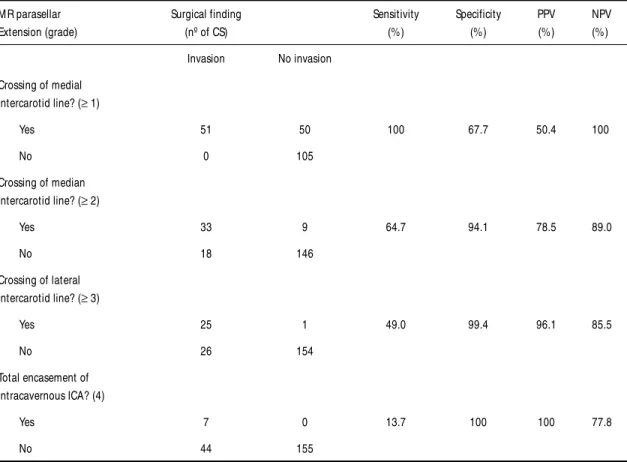
None of the patients with parasellar extension grade 0 had CSI. On the other hand, all patients w ith parasellar extension grade 4 had CSI (PPV.100% ). The summary of the results regarding parasellar extension can be seen in Table 1.
Displacement of the intracavernous ICA, bulging of the CS lateral wall and an increase in size of the CS w ere found to be associated w ith invasion (p< 0.001), although only CS lat-eral wall bulging demonstrated a good PPV (92.3%).
The medial venous compartment was not depicted in all invaded CS (100.0% sensitivity and NPV, p< 0.001) and in 54 CS w ithout invasion. This compartment was depicted in the remaining 101 CS w ithout invasion. Non-visualization of the inferior and lateral venous compartments had the highest positive predictive values (92.8% and 100.0%, respectively; p< 0.001). The results regarding visualization of the different CS venous compartments are summarized in Table 2.
Table 1. M R-defined parasellar extension correlated with CSI. (p< 0.001).
M R parasellar Surgical finding Sensitivity Specificity PPV NPV
Extension (grade) (nº of CS) (%) (%) (%) (%)
Invasion No invasion Crossing of medial
intercarotid line? (≥1)
Yes 51 50 100 67.7 50.4 100
No 0 105
Crossing of median intercarotid line? (≥2)
Yes 33 9 64.7 94.1 78.5 89.0
No 18 146
Crossing of lateral intercarotid line? (≥3)
Yes 25 1 49.0 99.4 96.1 85.5
No 26 154
Total encasement of intracavernous ICA? (4)
Yes 7 0 13.7 100 100 77.8
The CS was always invaded w hen 3 or more venous com-partments w ere not visualized (PPV 100%, p< 0.001).
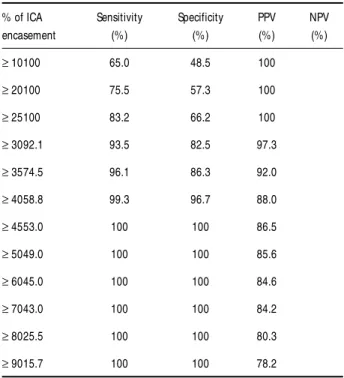
CS was never invaded when the percentage of encasement of the ICA was lower than 25% (NPV 100%, p< 0.001). On the other hand, w hen more then 45% of the circumference of the intracavernous ICA was involved by the adenoma, all CS w e-re invaded (PPV 100%, p< 0.001) (Table 3).
The criteria w ith stronger association w ith CSI by χ2
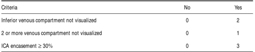
test-ing w ere: a) increase of CS size, b) bulgtest-ing of CS lateral wall, c) displacement of the intracavernous ICA, d) parasellar exten-sion grade 2 or more, e) non depiction of the superior or infe-rior venous compartment, f) 2 or more venous compartment not visualized and g) ICA encasement > 30%. Comparing all these criteria together, only 3 w ere statistically significant alone: 2 or more venous compartment not visualized, non depic-tion of the inferior venous compartment and ICA encasement > 30% (Table 4).
For each of these 3 selected criteria a value w eighted by its significance w ere chosen to make the predictive score of CSI (Table 5). This predictive score ranged from 0 to 6. A total score greater than 2 had the best diagnostic value for CSI (sen-sitivity 94.1%, specificity 92.9%).
DISCUSSION
The presence of CSI by PA is clinically relevant. Surgical procedures might be more difficult and tumor resection is usually partial in patients with invasive tumors.This yields poor-er endocrinological results and adjuvant treatment (radio-therapy, medical suppressive drugs or adrenalectomy) is often necessary. Only 18.0% of the patients in our series w ith CS and sellar floor dural invasion have been cured by surgery alone. Radiotherapy was performed in 67.0% of the patients w ith invasive adenomas and drug therapy in 43.5%. The surgical resection was complete in 85.5% of the patients w ith non-invasive tumors. Sixty-three percent of them have been cured by surgery alone; 25.5% of the patients needed radiotherapy and 18.5% drug therapy. Although preoperative diagnosis of CSI has a major impact on surgical outcome, only a few pa-pers have specifically discussed the issue.
The absence of a bony interface in the lateral limits of the
pituitary fossa might explain why tumor extension to the parasel-lar region and invasion of the CS are relatively common. A dur-al bag limited laterdur-ally by venous spaces within the CS surrounds the pituitary gland. There is only a thin dural layer between the gland and CS, which is called the medial wall. In about 30% of normal individuals, the pituitary gland has a lateral expansion reaching the intracavernous ICA, but the encasement of the ICA by the gland is always less than 25%8. Laterally invading tumors
have to be distinguished from these lateral normal pituitary expansions.
Intraoperative findings w ere considered the gold stan-dard for CSI in our study. This is possibly the best single objec-tive available criterion, but it is highly dependent on the sur-geon’s personal experience and it has to be recognized that in some patients it is very difficult to adequately visualize the lateral aspects of the pituitary fossa.
Table 2. Sensitivity, specificity, PPV and NPV of M R findings regarding the different CS venous com-partments.
Venous compartment Sensitivity Specificity PPV NPV
not visualized (%) (%) (%) (%)
M edial 100 65.0 48.5 100
Superior 76.5 94.8 83.0 92.4
Inferior 51.0 98.7 92.8 86.0
Lateral 45.0 100 100 84.7
Table 3. Sensitivity, specificity, PPV and NPV for CSI according to the amount of ICA encasement.
% of ICA Sensitivity Specificity PPV NPV
encasement (%) (%) (%) (%)
≥10100 65.0 48.5 100
≥20100 75.5 57.3 100
≥25100 83.2 66.2 100
≥3092.1 93.5 82.5 97.3
≥3574.5 96.1 86.3 92.0
≥4058.8 99.3 96.7 88.0
≥4553.0 100 100 86.5
≥5049.0 100 100 85.6
≥6045.0 100 100 84.6
≥7043.0 100 100 84.2
≥8025.5 100 100 80.3
MRI is superior to CT scanning in defining the pituitary gland and the sellar region and its boundaries. On the other hand, both techniques do not adequately depict the medial wall of the CS4,9-13.
The prevalence of CSI in our series is probably overesti-mated (46.5% of the patients). The present series did not include all the patients w ith PA treated at our institution. Intervening patients w ere excluded due to non-availability of adequate M RI images for analysis, lack of full clinical or fol-low-up data or lack of unequivocal intraoperative information regarding invasion or not by the tumor. CSI occurs more often unilaterally. In Scotti et al.4and Cottier et al.2series, invasion
by PA occurred only unilaterally. In our series, unilateral inva-sion by PA was observed in 93.7% of the patients and bilat-eral invasion occurred in 3 patients (all w ith GH-secreting adenoma). Knosp et al.1 observed bilateral invasion in 3
patients from their series (all w ith nonfunctioning PA).
We observed a significantly increased prevalence of inva-sion in macroadenomas (83.5% of the patients w ith invasive tumors).This higher prevalence of invasive tumors has also been observed by others1-3,5, although Scotti et al.4have not noticed
such difference. Knosp et al.1have also noted that the grade
of parasellar extension was directly related to the tumor’s size. Selman et al.14 have demonstrated histologically that
dural invasion in adenoma was w ell correlated to the tumor’s size.
Headache is a frequent symptom in PA, but its pathogen-esis is not w ell understood. Arafah et al.15 observed that
patients with PA and headache had increased intrasellar pres-sure compared to those w ithout headache. In our series, w e found no correlation betw een the presence of dural invasion by the tumor and headache.
We w ere able to define 4 M R criteria for the absence of CSI in patients w ith PA: 1- normal pituitary gland interposed
between the tumor and the CS; 2-intact CS medial venous com-partment; 3- tumor not crossing the medial intercarotid line and 4- percentage of ICA encasement by the lesion lower than 25%.
Cottier et al.2also considered the presence of normal
pitu-itary gland interposed between the tumor and the CS and intact CS medial venous compartment as seen on MRI as cri-teria for absence of CSI. Moreau et al.5 also observed that
there was no actual tumor invasion when the lesion did not cross the medial intercarotid line. Despite high specificity (100,0%), the 3 criteria above for absence of CSI had a very low sensitivity. ICA encasement lower then 25% disclosed the highest specificity (100,0%) and sensitivity (83,0%) for absence of CSI.
Other criteria highly suggestive of absence of CSI in our study were intact superior venous compartment (NPV, 92.4%) and the visualization of at least 3 CS venous compartments (NPV, 96%). Cottier et al.2noted that besides the
visualiza-tion of an intact CS superior venous compartment, the absence of size asymmetry betw een the 2 CS, absence of bulging of lateral wall of CS and grade 2 or less of parasellar extension were also good signs of absence of CSI. In the study of Moreau et al5, the signs highly suggestive of absence of invasion w ere
lack of size asymmetry, lack of bulging of the lateral wall of the CS, visualization of at least two venous compartments and non-displaced ICA.
The displacement of ICA and CS size asymmetry w ere show n to be criteria w ith good specificity but low sensitivity and PPV for CSI in our series. Cottier et al.2obtained similar
results for these criteria.
Bulging of the lateral wall of the CS had a good specifici-ty and PPV but low sensitivispecifici-ty in our series.These findings were in disagreement w ith Cottier’s et al.2data w hich disclosed a
very low PPV.
Table 5. Score values of each significant criteria.
Criteria No Yes
Inferior venous compartment not visualized 0 2
2 or more venous compartment not visualized 0 1
ICA encasement ≥30% 0 3
Table 4. Odds ratio and significance of the criteria selected by the forward stepwise analysis.
Criteria Significance Odds ratio
Inferior venous compartment not visualized P< 0.011 18.5 2 or more venous compartment not visualized P< 0.089 4.0
In our study, w e observed that in patients w ith Knosp parasellar extension grade 3 the CS was probably invaded (VPP, 96.1%) and w ith grade 4 (total ICA encasement), it was cer-tainly invaded. How ever, the sensitivity for these criteria was very low. Cottier et al.2 and M oreau et al.5also observed that
CSI was very likely w hen the lateral intercarotid line was crossed by the tumor. How ever, Knosp et al.1noted that
ade-noma with grade 2 or higher always had CSI. Their series eval-uated only patients w ith CSI.
The non-visualization of the inferior or lateral CS venous compartments was very suggestive of CSI (PPV, 92.8% and 100% respectively). CSI was always present w hen 3 or more CS venous compartments w ere not depicted on M RI (PPV, 100%). Cottier et al2further divided the CS inferior venous
com-partment into carotid sulcus and inferolateral comcom-partments, and observed that non-visualization of the carotid sulcus venous compartment had a high PPV (95.0%) for CSI. However, contrary to our results, these authors did not observe good PPV for invasion when the CS lateral venous compartment was not seen.
Total encasement of ICA by adenoma (grade 4 of Knosp1)
definitely indicated CSI (PPV and specificity of 100%) in our series, but it had low sensitivity and occurred late in time. In our series, only 13.7% of the CSI show ed total encasement of ICA. Knosp et al.1observed this sign in 32.0% of the invaded
CS and Cottier et al.2in 24.0%.
Adenoma grow s progressively around the ICA after infil-trating the CS medial wall. M oreau et al.5observed that in
patients in w hom there was 25% or more of ICA encasement by the tumor CSI was very likely. In our study, we observed that ICA encasement higher then 45% was always associated with CSI (PPV, 100%). Cottier et al.2suggested that at least 67%
of ICA encasement w ould be needed to assure CSI.
Comparing statistically the significant criteria all togeth-er, only 3 had separately association w ith CSI: 2 or more venous compartment not visualized, non depiction of the infe-rior venous compartment and ICA encasement > 30%. With these criteria, w e proposed a practical predictive score for CSI to make its preoperative diagnosis easier.
CONCLUSION
Our data show ed that accurate analysis of M R findings is useful in the preoperative diagnosis of CSI by PA and is able to generate relevant information regarding the microsurgical outcome in this patient’s population.The most specific and pre-cocious sign of invasion w as the encasement of the in-tracavernous ICA greater than 45%; when ICA encasement was low er than 25% the CS was never invaded. CS was definite-ly invaded w hen the lateral venous compartment was not visualized or w hen three or more venous compartments w ere not depicted. CS was never invaded w hen the medial venous
compartment was intact or normal pituitary gland was inter-posed betw een the adenoma and the CS.
The most useful sign to predict CSI was the encasement of ICA greater than 30%. This precocious finding had the best sensitivity (92.1%), specificity (93.5%) and PPV (82.5%).
REFERENCES
1. Knosp E, Steiner E, Kitz K, Matula C. Pituitary adenomas with inva-sion of the CS space: a magnetic resonance imaging classification com-pared with surgical findings. Neurosurgery 1993;33:610-618. 2. Cottier JP, Destrieux C, Brunereau L, et al. CSI by pituitary adenoma:
MR imaging. Radiology 2000;215:463-469.
3. Cukiert A, Andrioli M, Goldman J, et al. CSI by pituitary macroadeno-mas: neuroradiological, clinical and surgical correlation. Arq Neuropsiquiatr 1998;56:107-110.
4. Scotti G, Yu CY, Dillon WP, et al. MR imaging of CS involvement by pituitary adenomas. Am J Roentgenol 1988;151:799-806.
5 Moreau L, Cottier JP, Bertrand P, et al. MRI diagnosis of sinus cavernous invasion by pituitary adenomas. J Radiol 1998;79:241-246.
6. Roux FX, Obreja C, Moussa R, et al. L’extesion intra-caverneuse des macro-adénomes hypophysaires: infiltration ou invagination ? Neurochirurgie 1998;44:344-351.
7. Inoue T, Rhoton AL Jr, Theele D, Barry ME. Surgical approaches to the CS: a microsurgical study. Neurosurgery 1990;26:903-932.
8. Destrieux C, Kakou MK, Velut S, LeFranco T, Jan M. Microanatomy of the hypophyseal fossa boundaries. J. Neurosurg. 1998;88:743-752. 9. Wu W, Thuomas KA. Pituitary microadenoma: MR appearance and
cor-relation with CT. Acta Radiolog 1995;36:529-535.
10. Davis PC, Hoffman Jr. JC, Spencer T, Tindall GT, Braun IF. MR imag-ing of pituitary adenoma: CT, clinical, and surgical correlation. AJNR 1987;8:107-112.
11. Buchfelder M, Nistor R, Fahlbusch R, Huk WJ. The accuracy of the CT and MR evaluation of the sella turcica for detection of adrenocorticotropic hor-mone-secreting adenomas in Cushing disease. AJNR 1993;14:1183-1190. 12. Kulkarni MV, Lee KF, Mcardle CB, Yeakley JW, Haar FL. 1.5-T MR
imaging of pituitary microadenomas: technical considerations and CT correlation. AJNR 1988;9:5-11.
13. Nichols DA, Laws ER Jr, Houser OW, Abboud CF. Comparison of mag-netic resonance imaging and computed tomography in the preopera-tive evaluation of pituitary adenomas. Neurosurgery 1988;22:380-385. 14. Selman WR, Laws ER Jr, Scheithauer BW, Carpenter SM. The ocurrence