Nauplius
THE JOURNAL OF THE BRAZILIAN CRUSTACEAN SOCIETY
e-ISSN 2358-2936 www.scielo.br/nau
www.crustacea.org.br
First zoeal stage of
Macrocoeloma
subparallelum
(Stimpson, 1860)
(Decapoda: Brachyura: Majoidea)
described from laboratory hatched
material
Jéssica Colavite
1,2, Regulo López
3, Jesús Hernández
3,
Juan Bolaños
3,*and William Santana
1,21 Laboratório de Sistemática Zoológica, Pró-reitoria de Pesquisa e Pós-Graduação, Universidade Sagrado Coração, Rua Irmã Arminda, 10-50, Jd. Brasil. 17011-160 Bauru, São Paulo, Brazil.
2 Programa de Pós-Graduação em Zoologia, Universidade Estadual Paulista “Júlio de Mesquita Filho”. 18618-970 Botucatu, São Paulo, Brazil.
3 Grupo de Investigación en Carcinología, Universidad de Oriente, Escuela de Ciencias Aplicadas del Mar, Boca del Río, Isla de Margarita, Venezuela.
* In memoriam
ZOOBANK htt p://zoobank.org/urn:lsid:zoobank.org:pub:F59AB0D7-7044-4273-B91F-EDB0B2349E8B
ABSTRACT
Th e morphology of the fi rst zoeal stage of the decorator crab Macrocoeloma subparallelum (Stimpson, 1860) from Cuba is described and compared with the available descriptions for the genus. Among Macrocoeloma Miers, 1879 species, the fi rst zoea of M. subparallelum can be diff erentiated only by the number of aesthetascs and setae of the antennule (4 and 2, respectively). Recently, Macrocoeloma has been placed as an incertae sedis genus in Majoidea, closely allied to other Epialtidae and Pisinae members. Th e distinct spine in the distal segment of the endopod of the fi rst maxilliped in the zoeal stages of Macrocoeloma is similar to what we observe in other Epialtidae genera, a character that supports the recent phylogenetic fi ndings.
KEY WORDS
Cuba, Pisinae, Epialtidae, Mithracidae, larval development.
ORIGINAL ARTICLE
by the Brazilian Crustacean Society
in memoriam of Michael Türkay for his
outstanding contribution to Carcinology
CORRESPONDING AUTHOR
William Santana
SUBMITTED 13 June 2016
ACCEPTED 25 July 2016
PUBLISHED 20 October 2016
Guest Editor
Célio Magalhães
INTRODUCTION
Majoid crabs, commonly known as decorator or spider crabs, form one of the most diverse groups among brachyurans including nowadays six families with a total of 907 valid species (Davie et al., 2015). It is a superfamily that is distributed throughout nearly all oceans and is well represented, especially in warm regions, with about 239 species in the Americas, 124 species in the western Atlantic, and 115 species in the eastern Pacific (Griffin, 1966; Boschi, 2000).
Among majoids, the genus Macrocoeloma Miers, 1879 has been traditionally considered a member of the family Mithracidae; however, recent phylogenetic studies have suggested the exclusion of Macrocoeloma
from this family, being now an incertae sedis group closely allied to other Epialtidae genera (Windsor and Felder, 2014). Currently, there are 14 described species of Macrocoeloma (see Ng et al., 2008), of which 10 are distributed in the western Atlantic from North Carolina to Brazil (Melo, 1996; Coelho et al., 2008). The larval development of only two Caribbean species belonging to this genus is known: that of Macrocoeloma camptocerum (Stimpson, 1871) and Macrocoeloma diplacanthum (Stimpson, 1860). Both have been partially described in the thesis of Yang (1967), and the latter has been re-described by Marques et al. (2003).
Macrocoeloma subparallelum (Stimpson, 1860) is a western Atlantic species found in the Gulf of Mexico, the Antilles, Venezuela, and in Brazil as far south as the Espírito Santo State, from shallow waters to 25 m deep (Rodriguez, 1980; Melo, 1996; Felder et al., 2009), and appears to prefer coral substrates (Rathbun, 1925). Despite being widely distributed, M. subparallelum
is rarely collected, with only very few records in the literature (e.g. Rathbun, 1925; Rodriguez 1980; Scelzo and Varela, 1988).
We describe herein the first larval stage of M. subparallelum from Cuba and compare it with the available descriptions for the genus.
MATERIAL AND METHODS
Two ovigerous females of M. subparallelum were
collected in November 1994 in the beach of Jaimanitas, Havana, Cuba, between 1–2 meters of depth. The ovigerous females were held in an aquarium in a temperature–controlled room (24 ± 2°C) until hatching, which occurred at night. After hatching, the
zoeas were fixed in 70% ethanol. Twenty specimens of the first zoeal stage were stained using methylene blue, acid fuchsin and/or chlorazol black, and then dissected in a light stereomicroscope Leica Wild M8 for morphological descriptions. For slide preparations polyvinyl lactophenol or Canada balsam were used as mounting mediums. The larval description follows the pattern used in previous works (e.g. Pohle and Marques, 2000; Santana et al., 2003; 2004; Colavite et al., 2014) and the description of setae follows Pohle and Telford (1981), but here included only analysis by light microscopy (LM), using an Olympus CHA equipped with camera lucida. Some of the setae designated as plumose herein may be plumodenticulate due to the lower resolution limits of LM as compared to scanning electron microscopy (SEM). Description guidelines of Clark et al. (1998) were generally followed. A minimum of five zoeae were measured for carapace length (from the basis of the rostrum to the posterior margin of the carapace in lateral view) and total length (from the tip of the rostrum to the furcal shaft in lateral view). The adult females were measured for carapace width (CW) including branchiostegal spines.
Specimens of larval stages and the two spent female crabs (CW 24.1 mm and CW 23.95 mm) have been deposited at the GICUDONE – Grupo de Investigación in Carcinologia de la Universidad de Oriente, Escuela de Ciencias Aplicadas del Mar, Margarita Island, Venezuela (accession number # GIC-1001).
RESULTS
Morphological description
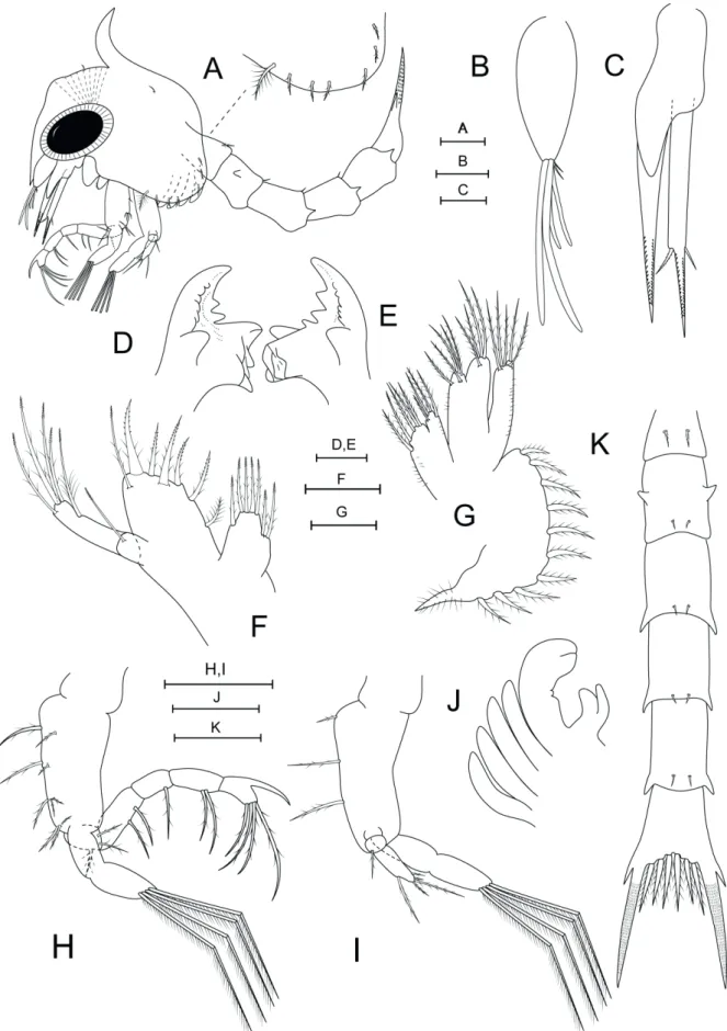
Carapace length. 0.63 mm ± 0.15 (0.61– 0.65 mm). Total length. 2.02 mm ± 0.05 (1.97–2.07 mm). Carapace (Fig. 1A). With dorsal spine, curved posteriorly, rostral spine smooth, ventrally directed, almost reaching antenna endopod bud, lateral spines absent. Pair of short simple setae anteriorly just above eyes, simple seta dorsolaterally to dorsal spine. Ventral margin with densely plumose ‘anterior setae’ (Clark et al., 1998) posterior to scaphognatite notch, followed by 5–6 smaller plumose setae. Eyes sessile. Small indistinct prominence frontally between dorsal spine and rostrum bearing cuticular dorsal organ (sensu Martin and Laverack, 1992; Lerosey-Aubrill and Meyer, 2013).
conical. Terminally with 2 long, 2 shorter aesthetascs, 2 short simple setae.
Antenna(Fig. 1C). Birramous, protopod long,
pointed, bearing two rows of sharp spinules; endopod bud approximately less than half of protopod size; exopod unsegmented, slightly longer than protopod, with long spinulated distal process; pair of setae about one third from tip, uneven, one small simple, one larger serrulate.
Mandible (Fig. 1D, E). Asymmetrical, with
medial toothed molar processes uneven; enlarged lateral incisor processes; 2–3 marginal teeth between molar and incisor processes. Internal margin of incisor processes with a series of teeth, ending in a larger tooth in both mandibles. Palp absent.
Maxillule (Fig. 1F). Coxal endite with seven setae: four terminal graded plumodenticulate, 3 plumodenticulate subterminally. Basial endite with seven setae: five terminal plumodenticulate cuspidate, one subterminal plumodenticulate, one plumose proximally. Two-segmented endopod, proximal segment with long plumodenticulate seta, distal segment with five long plumodenticulate setae: one subapical, four apical. Exopod seta absent.
Maxilla (Fig. 1G). Coxal endite bilobed, proximal lobe with 4–5 setae: 3–4 plumose, 1 plumodenticulate; distal lobe with five plumodenticulate setae. Microtrichia present on both endites. Basial endite bilobed, proximal and distal lobes with five and four plumodenticulate setae, respectively. Endopod unsegmented, unilobed, with five plumodenticulate apical setae; microtrichia on lateral margin. Scaphognatite marginally with 10–11 densely plumose setae, including distal process.
Maxilliped I (Fig. 1A, H). Coxa without seta. Basis with 10 plumodenticulate setae arranged 2+2+3+3. Endopod 5-segmented with 3,2,1,2,4 plumodenticulate setae, with strong accessory spine on distal segment. Exopod incompletely bisegmented with four plumose terminal natatory setae.
Maxilliped II (Fig. 1A, I). Coxa without seta. Basis with three plumodenticulate setae. Endopod three-segmented, with 0,1,4 sparsely plumodenticulate setae, distal segment with three subapical, two apical setae. Exopod incompletely bisegmented with four plumose terminal natatory setae.
Maxilliped III (Fig. 1A, J). Present as a small bud. Endo and exopod distinct.
Pereopods (Fig. 1A, J). Present as small buds, chela distinct, without apparent segmentation.
Pleon (Fig. 1A, K). Five pleonites. First pleonite with pair of middorsal, long, plumodenticulate setae, pleonites 2–5 with pair of shorter posterodorsal simple setae each. Posterolateral margin of second pleonite with blunt process, pleonites 3–5 with spines; second pleonite with pair of dorsolateral processes. Pleopod buds rudimentary.
Telson(Fig. 1K). Bifurcated, indistinct median notch, three pairs of plumodenticulate setae (Type 13-viii sensu Pohle and Telford, 1981) on inner margin; each furcal shaft proximally bearing a distinct lateral spine, furcal shafts covered with rows of spinules to just below the tips.
DISCUSSION
Macrocoeloma subparallelum closely resembles congeners for which the first larval stage has been described. Macrocoeloma subparallelum, M. diplacanthum and M. camptocerum differ only in the number of aesthetascs and setae of the antennule; while the former has 4 aesthetascs and 2 setae, the latter two have 3 and 1, respectively. Another minor difference can be observed in the number of setae in the ventral margin of the carapace: M. subparallelum
and M. diplacanthum have six to seven setae, while
M. camptocerum has only six (Yang, 1967; Marques
et al., 2003).
The larvae of the genus Macrocoeloma can be easily recognized through a combination of characteristics including the presence of a distended forehead with strong underlying muscle bands, a dorsal spine, and a distinct, strong spine in the distal segment of the endopod of the first maxilliped (Yang, 1967; Marques
et al., 2003). All of these characteristics are present in
M. subparallelum. As noted by Marques et al. (2003), this distinct spine can also be found in other Epialtidae species, such as Menaethius monoceros (Latreille, 1825),
Pugettia Dana, 1851 and Huenia De Haan, 1837 (see Ko, 1998; Kornienko and Korn, 2004; Marques et al., 2003; Colavite et al., 2014).
and larval characters could be the best way to find diagnostic characteristics for some groups. Despite the placement of Macrocoeloma as an incertae sedis group by Windsor and Felder (2014), this genus nested close to other Epialtidae and Pisinae members in their analyses. This is what we observe in the first zoeal stage of this genus, with the distinct spine in the distal segment of the endopod of the first maxilliped. This character is similar to what we see in other Epialtidae genera, which supports the recent phylogenetic studies (Windsor and Felder, 2014). However, Epialtidae is the most speciose family of Majoidea, with 452 recognized species (Davie
et al., 2015), and shows a high degree of variability in several larval characters (see Clark and Cuesta, 2015). This could indicate that Epialtidae is not monophyletic in its present composition suggesting the taxonomic reevaluation and additional phylogenetic studies to better define Epialtidae in general and the position of
Macrocoeloma in particular.
ACKNOWLEDGEMENTS
We thank the Universidad de Oriente - UDO, Venezuela, and the Universidade do Sagrado Coração – USC, Brazil for supporting studies on the systematics of decapod crustaceans. This work was supported by the Fundação de Amparo à Pesquisa do Estado de São Paulo – FAPESP (WS grant number 2013/01201-0 and JC fellowship number 2016/02775-8).
REFERENCES
Boschi, E.E. 2000. Biodiversity of marine decapod brachyurans of the Americas. Journal of Crustacean Biology, 20(3): 337–342. Clark, P.F.; Calazans, D.K. and Pohle, G. 1998. Accuracy and
standardization of brachyuran larval descriptions. Invertebrate Reproduction and Development, 33(2–3): 127–144.
Clark, P.F. and Cuesta, J.A. 2015. Larval systematics of Brachyura. p. 981–1048. In: P. Castro; P.J.F. Davie; D. Guinot; F.R. Schram and J.C. von Vaupel Klein (eds), Treatise on Zoology – Anatomy, Taxonomy, Biology. The Crustacea, Vol. 9, Part C (2 vol.). Leiden, Brill.
Coelho, P.A., Almeida, A.O. and Bezerra, L.E.A. 2008. Checklist of the marine and estuarine Brachyura (Crustacea: Decapoda) of the northern and northeastern Brazil. Zootaxa, 1956: 1–58. Colavite, J.; Santana, W. and Pohle, G. 2014. Larval development of the spider crab Menaethius monoceros (Latreille, 1825) (Crustacea: Decapoda: Brachyura: Epialtidae). Journal of Natural History, 48(37–38): 2273–2292.
Dana, J.D. 1851. On the classification of the maioid Crustacea or Oxyrhyncha. American Journal of Science, series 2, 11(33): 425–434.
Davie, P.J.; Guinot, D. and Ng, P.K. 2015. Systematics and
classification of Brachyura. p. 1049–1130. In: P. Castro; P.J.F. Davie; D. Guinot; F.R. Schram and J.C. von Vaupel Klein (eds), Treatise on Zoology – Anatomy, Taxonomy, Biology. The Crustacea, Vol. 9, Part C (2 vol.). Leiden, Brill. Felder, D.L; Álvarez, F.; Goy, J.W. and Lemaitre, R. 2009. Decapoda
(Crustacea) of the Gulf of Mexico, with comments on the Amphionidacea. p. 1019–1104. In: D.L. Felder and D.K. Camp (eds), Gulf of Mexico Origin, Waters, and Biota, Vol 1, Biodiversity. College Station, Texas, Texas A&M University Press.
Griffin, D.J.G. 1966. A review of the Australian majid spider crabs (Crustacea, Brachyura). Australian Zoologist, 13: 259–298, pls. 15–17.
Haan, W. de 1833–1850. Crustacea. In: P.F. von Siebold (ed) Fauna Japonica sive Descriptio Animalium, Quae in Itinere per Japoniam, Jussu et Auspiciis Superiorum, qui Summum in India Batava Imperium Tenent, Suscepto, Annis 1823– 1830 Collegit, Noitis, Observationibus et Adumbrationibus Illustravit. Leiden, Lugduni-Batavorum. p. i–xvii, i–xxxi, ix–xvi, 1–243, Pls A–J, L–Q, 1–55, circ. tab. 2
Ko, H.S. 1998. Zoeal development of three species of Pugettia (Decapoda: Majidae) with a key to the known zoeas of the subfamily Epialtinae. Journal of Crustacean Biology, 18(3): 499–510.
Kornienko, E.S. and Korn, O.M. 2004. Morphological features of the larvae of spider crab Pugettia quadridens (Decapoda: Majidae) from the northwestern Sea of Japan. Russian Journal of Marine Biology, 30(6): 402–413.
Latreille, P.A. 1825. Histoire Naturelle. Entomologie, ou Histoire naturelle des Crustacés, des Arachnides et des Insectes. Encyclopédie Méthodique, Vol. 10. Paris, Agasse Imprimeur-Libraire, 832p.
Lerosey-Aubrill, R. and Meyer, R. 2013. The sensory dorsal organs of crustaceans. Biological reviews of the Cambridge Philosophical Society, 88(2):406–426.
Marques, F.P.L.; Pohle, G.W. and Vrbova, L. 2003. On the larval stages of Macrocoeloma diplacanthum (Decapoda: Brachyura: Majidae), with a review of mithracine phylogenetic aspects. Journal of Crustacean Biology, 23(1): 187–200.
Martin, J.W. and Laverack, M.S. 1992. On the distribution of the crustacean dorsal organ. Acta Zoologica, 73(5): 357–368. Melo, G.A.S. 1996. Manual de identificação dos Brachyura
(caranguejos e siris) do litoral brasileiro. São Paulo, Plêiade/ FAPESP ed., 603p.
Miers, E.J. 1879. On the classification of the Maioid Crustacea or Oxyrhyncha, with a synopsis of the families, sub-families and genera. Journal of the Linnean Society of London, Zoology, 14(79): 634–673, pls 12–13.
Ng, P.K.L.; Guinot, D. and Davie, P.J.F. 2008. Systema brachyurorum: Part 1. An annotated checklist of extant brachyuran crabs of the world. The Raffles Bulletin of Zoology, 17: 1–286.
Pohle, G. and Marques, F.P.L. 2000. Larval stages of Paradasygyius depressus Bell, 1835 (Crustacea: Decapoda: Brachyura: Majidae) and a phylogenetic analysis for 21 genera of Majidae. Proceedings of the Biological Society of Washington, 113(3): 739–760.
microscope study of Dissodactylus crinitichelis Moreira, 1901 (Brachyura: Pinnotheridae). Bulletin of Marine Science, 31(3): 736–752.
Rathbun, M.J. 1925. The spider crabs of America. Bulletin of the United States National Museum,129: 1–613.
Rodriguez, G. 1980. Los Crustaceos Decapodos de Venezuela. Caracas, Instituto Venezolano de Investigaciones Cientificas, 494p.
Santana, W.; Marques, F. and Pohle, G. 2004. Larval stages of Stenocionops furcatus (Olivier, 1781) (Decapoda: Brachyura: Majoidea) and a reappraisal of larval morphological characters for Mithracidae. Journal of Plankton Research, 26(8): 859–874. Santana, W.; Pohle, G. and Marques, F. 2003. Zoeal stages and
megalopa of Mithrax hispidus (Herbst, 1790) (Decapoda: Brachyura: Majoidea: Mithracidae): a reappraisal of larval characters from laboratory cultured material and a review of larvae of the Mithrax-Mithraculus species complex. Invertebrate Reproduction and Development,44(1): 17–32.
Scelzo, M. A. and Varela, R. 1988. Crustáceos Decápodos Litorales
de la Isla La Blanquilla, Venezuela. Memoria de la Sociedad de Ciencias Naturales La Salle, 47(129): 33–54.
Stimpson, W. 1860. Notes on North American Crustacea, in the Museum of the Smithsonian Institution, No. II. Annals of the Lyceum of Natural History of New York, 7(1): 177–246, Plates II and V.
Stimpson, W. 1871. Preliminary report on the Crustacea dredged in the Gulf Stream in the Straits of Florida by L.F. de Pourtales, Assist. U. S. Coast Survey. Part I. Brachyura. Bulletin of the Museum of Comparative Zoology at Harvard College, 2(2): 109–160.
Windsor, A.M. and Felder, D.L. 2014. Molecular phylogenetics and taxonomic reanalysis of the family Mithracidae Macleay (Decapoda: Brachyura: Majoidea). Invertebrate Systematics, 28(2): 145–173.