Frequency of B cells in normal
mice which recognize self proteins
Departamento de Genética, Universidade Federal do Rio Grande do Sul, 91501-900 Porto Alegre, RS, Brasil
M.C. Carlan, A. Peres and N.B. Nardi
Abstract
The mechanism whereby the immune system avoids self-aggression is one of the central issues of Immunology. The discovery of natural autoantibodies, mainly of IgM isotype, and of idiotypic interactions between antibodies indicates that elements of the immune system interact with self constituents and with themselves. Results of studies with soluble antibodies have indicated that the pool of circulating IgM represents the end result of a highly selective process of B cell activation and differentiation by self proteins resulting in the forma-tion of a network. The objective of the present work was to determine the frequency of self-reacting B cells in normal mice. We were able to detect B cells that recognize self proteins present in extracts of different organs in normal adult, 2-3-month old, BALB/c and C57BL/ 6 mice with an ELISA spot assay. About 1% of total IgM-secreting cells among small, LPS-stimulated spleen cells reacted with organ extracts, whereas among large spleen cells the frequency was 5- to 10-fold lower. Immunization induced an increase in the frequency of IgM-secreting cells. The present results provide cellular evidence for the results of studies done at the serological level. The physiological role of these self-recognizing cells, as well as their participation in autoimmune processes, remain to be established.
Correspondence N.B. Nardi
Departamento de Genética, UFRGS Caixa Postal 15053
91501-970 Porto Alegre, RS Brasil
Fax: 55 (051) 316-2011 E-mail: [email protected]
Research supported by CNPq (No. 520936/95-7).
Received April 25, 1996 Accepted December 4, 1996
Key words
•B cells
•Self-recognition •Ig repertoire •BALB/c mice •C57BL/6 mice
•LPS-stimulated spleen cells
Introduction
The normal immune system is capable of self-recognition without triggering processes that characterize the immune response against conventional antigens. The mechanism(s) whereby the immune system avoids self-aggression is one of the central issues in Immunology.
The B cell repertoire is generated by ran-dom rearrangements of immunoglobulin genes in the absence of nominal antigens.
con-nectivity with each other and with other self components (5-7). Idiotypic connectivity and self-recognition, in agreement with the ideas of Jerne (8), lead to the formation of a net-work, where the elements are in equilibrium and therefore are not activated to react with self.
Recent studies with soluble antibodies have indicated that the pool of circulating IgM in normal mice represents the end result of a highly selective process; naturally acti-vated, immunocompetent lymphocytes do not constitute a random sample of the resting cell pool, but are rather the result of a posi-tive selection guided by self proteins and resulting in the formation of a network (9,10). These studies have provided remarkable ev-idence for the fundamental role of self com-ponents in the establishment and mainte-nance of the system. Thus, a pool of lympho-cytes would be selected by dominant self proteins which constitute the “immunologi-cal homunculus” as proposed by Cohen (11). As a complementation of these studies, done at the level of soluble antibodies, the objective of the present investigation was to analyze self-reacting B cell frequencies in normal mice. Using an ELISA spot assay we were able to detect B cells that recognize self proteins present in extracts of different or-gans in normal adult BALB/c and C57BL/6 mice. Our results show a relatively high frequency of IgM-secreting B cells that rec-ognize self proteins, supporting reports of the existence of a positive selection mech-anism on the initial development of B cells and a negative selection of mature B lym-phocytes.
Material and Methods
Animals
BALB/c and C57BL/6 inbred mice were maintained under standard conditions in our animal facilities. Adult animals, 2-3 months of age, were used in these studies.
Protein extracts and immunizations
Protein extracts were prepared from the following organs: heart, lung, brain, liver, gut, spleen, kidney and stomach. Mice were killed by cervical dislocation, and the organs were removed under sterile conditions and disrupted by ultrasound (7 pulses of 20 mA for 30 s) on ice-cold phosphate-buffered saline (PBS) with 35 µg/ml phenylmethyl-sulfonyl fluoride. After centrifugation at
1,000 g at 4oC, the supernatant was collected
and protein concentration was determined by absorbance at 260 and 280 nm. The
mate-rial was aliquoted and stored at -20oC until
use.
Groups of 3-4 mice were immunized on days 0 and 7 by intraperitoneal injection of 50 µg lung protein extract diluted in 100 µl PBS and emulsified in the same volume of complete Freund’s adjuvant (CFA, Sigma Chemical Co.). Control groups consisted of animals injected with 100 µl PBS or PBS/ CFA. On day 10 spleen cells were screened for the frequency of Ig-secreting cells recog-nizing the organ extracts by the ELISA spot assay (ESA).
Cells and cultures
Cells from spleen, peritoneal exudate and bone marrow were collected from mice un-der sterile conditions, washed with cold Hank’s solution, adjusted to the desired con-centration and kept in an ice bath until use. Large and small spleen cells were separated by centrifugation using a Percoll gradient (12).
For some experiments, cells were
sus-pended at 106 or 2 x 10⁶/ml in RPMI 1640
medium (Sigma Chemical Co.) comple-mented with 25 mM HEPES buffer (Sigma), pH 7.3, gentamycin (280 mg/l), 10% fetal calf serum (Cultilab, São Paulo, Brazil) and
25 µg/ml E. coli lipopolysaccharide (LPS,
Sigma) and cultured for 3 days at 37oC with
washed, counted and analyzed for the fre-quency of Ig-secreting cells by ESA.
ELISA spot assay
The ELISA spot assay was performed as described by Nardi et al. (12). Briefly, flat-bottom ELISA plates were coated with 50 µl/well of either anti-mouse Ig (Sigma Chem-ical Co.) or protein extracts at 1 mg/ml. After saturating the plates with PBS containing 1% non-fat milk and washing with PBS, cells were added in serial dilutions starting with the appropriate concentrations
(usu-ally, 103 to 106 cells in 100 µl). Plates were
incubated at 37oC for 3 h and the cells were
then lysed by washing with distilled water. Rabbit anti-mouse IgG, IgM or IgA coupled to biotin (Sigma) was added and after
stand-ing overnight at 4oC the plates were
incu-bated for 1 h at 37oC with a solution of
streptavidin coupled to alkaline phosphatase (Sigma). The substrate consisted of a solu-tion of 2.3 mM 5-bromo-4-chloro-3-indolyl-phosphate (Sigma) diluted in 2-amino-2-methyl-1-propanol buffer (Merck) contain-ing 0.75% low-meltcontain-ing agarose (type 1, low EEO; Sigma). After a 3-4-h incubation
pe-riod at 37oC, the Ig-secreting cells,
visual-ized as blue spots, were counted, and their frequency was determined in relation to the total number of cells added to the well.
SDS-PAGE
Protein electrophoresis was performed by SDS-PAGE in the presence of mercapto-ethanol with a 4% stacking gel and 10% separating acrylamide gel.
Results
As expected, complex protein patterns were observed after SDS-PAGE of the ex-tracts (data not shown), and protein integrity was periodically monitored with this assay. The ESA was highly reproducible, as
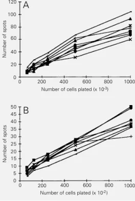
indicated by the same results after 5 to 8 repetitions. The results reported here were obtained in at least three different experi-ments. Initially, the frequency of BALB/c cells secreting anti-self antibodies was de-termined in total spleen with or without LPS stimulation. Figure 1 shows that the fre-quency of antibody-secreting cells recogniz-ing protein extracts is closely similar for all organs, and in LPS-stimulated spleen is about 5- to 10-fold higher than that of non-stimu-lated spleen cells. Only the IgM isotype was detected. Assays in which the plates were covered with irrelevant foreign proteins such as bovine serum albumin or casein showed that 10% of the cells were of the “sticky” type (data not shown).
Since extracts from all 8 organs presented the same results when assayed by ESA, 3 of them (gut, brain and lung) were selected for further study. Total IgM-secreting cells in this assay were detected at frequencies of 2.5% and 11.0% among large and LPS-stim-ulated small spleen cells, respectively. Anal-ysis of these cells in BALB/c mice showed very different frequencies of self-reacting B
Figure 1 - Frequency of BALB/c total spleen cells secreting anti-self IgM antibodies without (A) or with (B) LPS stimulation. Note that the ordinate of panel A
refers to thousands of cells plated and panel B refers to hundreds of cells plated. ¢, Kid-ney; £, stomach; à, gut; 5, heart; , spleen; +, liver;
·
, lung; , brain.Number of spots
120
100
80
60
40
20
0
A
0 200 400 600 800 1000 Number of cells plated (x 10-3)
Number of spots
50 45 40
30 25
10
0
0 200 400 600 800 1000 35
20 15
5
Number of cells plated (x 10-2)
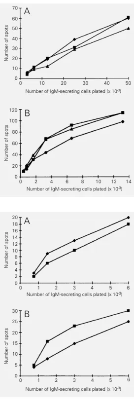
The spleen of immunized mice presented a 3-fold increase in the frequency of IgM-secreting B cells recognizing lung and intes-tine extract; the results were the same whether the animals received the protein extract in CFA or CFA alone (Figure 3).
Frequencies of antibody-secreting cells reactive with gut, brain and lung extracts were analyzed in C57BL/6 mice, and did not differ from those found for BALB/c mice (data not shown).
Discussion
Evidence has been accumulating which supports the hypothesis that differentiation in the immune system is driven by self as well as foreign antigens, and that this anti-self reactivity is under control to prevent autoimmunity (13). Whereas at the T cell level the presence of anti-self cells has been demonstrated in several studies (14,15), the B lymphocyte compartment has been stud-ied mainly at the antibody level. The pool of normal serum antibodies, as well as mono-clonal antibody collections prepared from normal mice, present extensive autoreactiv-ity (16,17).
The objective of the present study was to characterize the B cell compartment at the cellular level, and the frequency of B lym-phocytes reacting with self components was determined by ESA in normal BALB/c and C57BL/6 mice. In order to obtain a broader analysis of the reactivity repertoire, protein extracts from organs, instead of purified an-tigens were used in the assay. This approach imposes a limitation in terms of being able to identify the proteins towards which reactiv-ity is directed, and the results obtained must be interpreted in terms of experiments car-ried out with soluble antibodies of known specificity. Thus, the results obtained in the present study represent the cellular counter-part of studies done at the humoral level.
Closely similar levels of reactivity were found for all eight organ extracts employed
Number of spots
70
60
50
40
30
20
10
A
0 10 20 30 40 50
Number of IgM-secreting cells plated (x 10-3)
Number of spots
120
100
80
40
20
0 2 4 6 8 10
60
0
Number of IgM-secreting cells plated (x 10-3)
B
Number of spots
20 18 16 14 12 10
0
A
0 1 2 3 4 5
Number of IgM-secreting cells plated (x 10-3)
Number of spots
30
25
20
10
0
0 1 2 3 4 5
15
5
Number of IgM-secreting cells plated (x 10-3)
B
Figure 2 - Frequency of cellsse-creting IgM antibodies reactive with self components among IgM-secreting large (A) and LPS-stimulated small (B) spleen cells of BALB/c mice. Note that the ordinate scales of both panelsA
and B refer to thousands of cells plated. ¢, Gut; £, brain; à, lung.
Figure 3 - Frequency of cells se-creting IgM antibodies reactive with self components among IgM-secreting spleen cells ob-tained from mice immunized with lung extract plus CFA (A) or CFA alone (B). ¢, Gut; à, lung.
0
12 14
8 6 4 2
6
in this study. The possibility that this reactiv-ity could be due only to multispecific, “sticky” B cells which are present in normal individu-als (13) was eliminated by experiments which showed that about 10% of the B cells de-tected in the assay reacted with unrelated proteins. Actually, analysis of natural anti-bodies has shown that even polyreactive an-tibodies discriminate between the anti-gens they recognize (18), and that they pres-ent fine specificity towards a) defined sub-sets of antigens present in different organ extracts as well as b) different antigens within various strains (9). These results suggest that the basal frequencies of anti-self B cells detected in the present study, if analyzed for their fine specificities, should present dis-criminated profiles for different organ ex-tracts.
About 1% of total IgM-secreting cells among small, LPS-stimulated spleen cells reacted with organ extracts; among large spleen cells, this frequency was 5-10-fold lower. The fact that the frequency of self-recognizing B cells is higher among cells in the available repertoire than in the actual repertoire, though disagreeing with some re-ports (19), is in agreement with several oth-ers (9,20,21), and represents the result of
selection of the B cell repertoire by self antigens (13). An alternative explanation for the lower frequency of positive cells scored among naturally activated B cells is that multireactivity has been shown to decrease with the maturation stage of the B cell popu-lation analyzed (21).
As expected, immunization induced an increase in the frequency of IgM-secreting B cells; the effect is probably of a polyclonal, multispecific nature, and induced by myco-bacterial antigens, since the same results were observed when mice were immunized with organ extracts and CFA or CFA alone. Some other studies have been conducted to investigate the frequency of anti-self B lymphocytes. Thus, selection of self-react-ing B cells by proteins such as the F antigen (12) or heat shock proteins (22) has been reported in mice. This approach, however, is restricted to a few antigens. The study re-ported here, although not permitting the iden-tification of antigens towards which B lym-phocytes are directed, provides cellular evi-dence for the results obtained at the serologi-cal level. The physiologiserologi-cal role of these self-recognizing cells, as well as their par-ticipation in autoimmune processes (23), re-main to be established.
References
1. Hooijkaas H, Benner R, Pleasants JR & Wostmann BS (1984). Isotypes and speci-ficities of immunoglobulins produced by germ-free mice fed chemically defined ultrafiltered antigen-free diet. European
Journal of Immunology, 14: 1127-1130.
2. Pereira P, Forni L, Larsson EL, Cooper MD, Heusser C & Coutinho A (1986). Au-tonomous activation of T and B cells in antigen-free mice. European Journal of
Immunology, 16: 685-688.
3. Lundkvist I, Coutinho A, Varela F & Holmberg D (1989). Evidence for a func-tional idiotypic network among natural an-tibodies in normal mice. Proceedings of the National Academy of Sciences, USA,
86: 5074-5078.
4. Varela FJ, Anderson A, Dietrich G, Sundblad A, Holmberg BD, Kazatchkine M & Coutinho A (1991). Population dy-namics of natural antibodies in normal and autoimmune individuals. Proceedings of the National Academy of Sciences, USA,
88: 5917-5921.
5. Coutinho A (1989). Beyond clonal selec-tion and network. Immunological
Re-views, 110: 63-87.
6. Varela FJ & Coutinho A (1991). Second generation immune networks.
Immunol-ogy Today, 12: 159-166.
7. Coutinho A, Freitas AA, Holmberg D & Grandien A (1992). Expression and selec-tion of murine antibody repertoires.
Inter-national Review of Immunology, 8:
173-187.
8. Jerne NK (1974). Towards a network theory of the immune system. Annales
de lInstitute Pasteur, Immunologie,
125C: 373-389.
9. Nobrega A, Haury M, Grandien A, Malanchère E, Sundblad A & Coutinho A (1993). Global analysis of antibody reper-toires. II. Evidence for specificity, self-se-lection and the immunological homun-culus of antibodies in normal serum.
Eu-ropean Journal of Immunology, 23:
2851-2859.
11. Cohen IR & Young DB (1991). Autoimmu-nity, microbial immunity and the immuno-logical homunculus. Immunology Today,
12: 105-110.
12. Nardi NB, Freitas A & Coutinho A (1990). Selection of anti-F protein B-cell reper-toires in normal mice. Research in
Immu-nology, 141: 711-721.
13. Avrameas S (1991). Natural autoantibod-ies: from horror autotoxicus to gnothi seauton. Immunology Today, 12: 154-159.
14. Schild H, Rotzschke O, Kalbacher H & Rammensee HG (1990). Limit of T cell tolerance to self proteins by peptide pres-entation. Science, 247: 1587-1589. 15. Shoenfeld Y, Teplitzki HA, Mendlovic S,
Blank M, Mozes E & Isenberg DA (1989). The role of the human anti-DNA idiotype 16/6 in autoimmunity. Clinical
Immunol-ogy and ImmunopatholImmunol-ogy, 51: 313-325.
16. Dighiero G, Guilbert B & Avrameas S (1982). Naturally occurring antibodies against nine common antigens in human sera. High incidence of monoclonal Ig ex-hibiting antibody activity against actin and tubulin and sharing antibody specificities with natural antibodies. Journal of
Immu-nology, 128: 2788-2792.
17. Holmberg D, Freitas A, Portnoi D, Jacquemart F, Avrameas S & Coutinho A (1986). Antibody repertoires of normal BALB/c mice: B lymphocyte populations defined by state of activation.
Immuno-logical Reviews, 93: 147-169.
18. McHeyzer-Williams MG & Nossal GVJ (1988). Clonal analysis of autoantibody-producing cell precursors in preimmune B cell repertoire. Journal of Immunology,
141: 4118-4123.
19. Portnoi D, Freitas A, Bandeira A, Holmberg D & Coutinho A (1986). Immu-nocompetent autoreactive B lymphocytes are activated cycling cells in normal mice.
Journal of Experimental Medicine, 164:
25-35.
20. Ishigatsubo Y, Steinberg AD & Klinman DM (1988). Autoantibody production as-sociated with polyclonal B cell activation in autoimmune mice which express the lpr or gdl genes. European Journal of
Im-munology, 18: 1089-1093.
21. Grandien A, Fucs R, Nobrega A, Andersson J & Coutinho A (1994). Nega-tive selection of multireacNega-tive B cell clones in normal mice. European Journal
of Immunology, 24: 1345-1352.
22. Bonorino C, Folberg A, Silveira R & Nardi N (1993). Heat shock proteins and the shaping of the B cell compartment. Anais do III Congreso de la Associacion
Latinoamericana de Inmunologia: 171A.
23. Bockenstedt LK, Gee RJ & Mamula MJ (1995). Self-peptides in the initiation of lupus autoimmunity. Journal of