An Unexpected Mode Of Binding Defines
BMS948 as A Full Retinoic Acid Receptor
β
(RAR
β
, NR1B2) Selective Agonist
Eswarkumar Nadendla1,2,3☯, Catherine Teyssier1,2☯¤, Vanessa Delfosse1,2, Valérie Vivat4, Gunasekaran Krishnasamy3, Hinrich Gronemeyer5, William Bourguet1,2
*, Pierre Germain1,2*
1Inserm U1054, Centre de Biochimie Structurale, Montpellier, France,2CNRS UMR5048, Universités Montpellier 1 & 2, Montpellier, France,3CAS in Crystallography and Biophysics, University of Madras, Chennai, India,4Novalix, Illkirch, C.U. de Strasbourg, France,5IGBMC, CNRS/INSERM/UdS/CERBM, Illkirch-Cedex, C.U. de Strasbourg, France
☯These authors contributed equally to this work.
¤ Current address: IRCM, Inserm U896, Montpellier, France
*[email protected](WB);[email protected](PG)
Abstract
Retinoic acid is an important regulator of cell differentiation which plays major roles in em-bryonic development and tissue remodeling. The biological action of retinoic acid is mediat-ed by three nuclear receptors denotmediat-ed RARα,βandγ. Multiple studies support that RARβ
possesses functional characteristics of a tumor suppressor and indeed, its expression is fre-quently lost in neoplastic tissues. However, it has been recently reported that RARβcould also play a role in mammary gland tumorigenesis, thus demonstrating the important but yet incompletely understood function of this receptor in cancer development. As a conse-quence, there is a great need for RARβ-selective agonists and antagonists as tools to facili-tate the pharmacological analysis of this proteinin vitroandin vivoas well as for potential
therapeutic interventions. Here we provide experimental evidences that the novel synthetic retinoid BMS948 is an RARβ-selective ligand exhibiting a full transcriptional agonistic activi-ty and activating RARβas efficiently as the reference agonist TTNPB. In addition, we solved the crystal structures of the RARβligand-binding domain in complex with BMS948 and two related compounds, BMS641 and BMS411. These structures provided a rationale to explain how a single retinoid can be at the same time an RARαantagonist and an RARβfull agonist, and revealed the structural basis of partial agonism. Finally, in addition to revealing that a flip by 180° of the amide linker, that usually confers RARαselectivity, accounts for the RARβselectivity of BMS948, the structural analysis uncovers guidelines for the rational de-sign of RARβ-selective antagonists.
OPEN ACCESS
Citation:Nadendla E, Teyssier C, Delfosse V, Vivat V, Krishnasamy G, Gronemeyer H, et al. (2015) An Unexpected Mode Of Binding Defines BMS948 as A Full Retinoic Acid Receptorβ(RARβ, NR1B2) Selective Agonist. PLoS ONE 10(5): e0123195. doi:10.1371/journal.pone.0123195
Academic Editor:Makoto Makishima, Nihon University School of Medicine, JAPAN
Received:October 23, 2014
Accepted:February 19, 2015
Published:May 1, 2015
Copyright:© 2015 Nadendla et al. This is an open access article distributed under the terms of the
Creative Commons Attribution License, which permits unrestricted use, distribution, and reproduction in any medium, provided the original author and source are credited.
Data Availability Statement:Protein Data Bank: Atomic coordinates and structure factors for RARβ -BMS948-SRC-1 NR2, RARβ-BMS411-SRC-1 NR2, and RARβ-BMS641-SRC-1 NR2 complexes have been deposited under accession codes 4JYH, 4JYG, and 4JYI, respectively.
Introduction
Retinoic acids and their analogs, referred to as retinoids, exert their pleiotropic effects through three retinoic acid receptor subtypes [RARα(NR1B1), RARβ(NR1B2) and RARγ(NR1B3)] that originate from three distinct genes [1–3]. RARs are members of the nuclear receptor (NR) superfamily [4] and act as ligand-inducible transcription factors binding to DNA regulatory el-ements in the promoter regions of target genes by forming heterodimers with another NR, the retinoid X receptor (RXR) [5,6]. RARs are modular proteins composed of several domains, most notably the DNA-binding domain (DBD) and the ligand-binding domain (LBD). The well-established function of RARs is to regulate gene expression [5], and switching on RAR transcriptional activity relies on ligand-induced conformational changes provoking a dynamic series of coregulator exchanges. In the absence of agonists, RAR exerts a repressor activity by interacting with transcriptional corepressors (CoRs) which themselves serve as docking plat-forms for the recruitment of histone deacetylases that impose a higher order structure on chro-matin that is not permissive to gene transcription. Upon agonist (i.e. the natural ligand all-trans-retinoic acid, atRA, or synthetic compounds that mimic its action) binding, conforma-tional changes of the RAR LBD induce CoR release and favour the recruitment of coactivators (CoAs) such as CBP/p300, the p160 protein family, or CARM1 with histone acetylase activities allowing chromatin de-compaction and gene transcription [5,6].
Our understanding of how ligand binding leads to receptor regulation has been greatly ad-vanced by structural studies of RAR LBDs in complex with different pharmacological classes of retinoids as well as CoA- and CoR-derived fragments [7–12]. These structures revealed that RAR LBDs exhibit a common fold comprising 12α-helices (H1-H12) and a shortβ-turn (s1-s2) arranged in three layers to form an antiparallel“α-helical sandwich”. This particular ar-rangement generates a ligand binding pocket (LBP) in the lower part of the domain. Another key component of the LBD is the C-terminal helix (H12) which is repositioned upon agonist binding to complete a hydrophobic surface with residues from helices H3 and H4 that is specif-ically recognized by the LxxLL motifs (also called NR boxes) of CoAs. Hence the interactions between H12 or residues in its vicinity and the bound ligands are critical for the control of the transcriptional activity of RARs. In this regard, the structures of RARαin complex with Am580 [12], BMS614 [8] and BMS493 [12] have revealed the structural basis of agonist, antag-onist and inverse agantag-onist action, respectively. Briefly, while Am580 (Fig 1) stabilizes H12 in the so-called active conformation described above, thus inducing CoA recruitment, the bulky ex-tended 8”-quinolinyl group of the antagonist BMS614 (Fig 1) prevents CoA binding by redi-recting H12 into the CoA binding groove. On the other hand, the inverse agonist BMS493 (Fig 1) which also contains a bulky extension prevents CoA recruitment and, in addition, reinforces CoR interaction by stabilizing aβ-sheet interaction between the RAR LBD and specific CoR residues [12]. A fourth class of retinoids, referred to as partial agonists, consists in a group of molecules that fail to stabilize a particular conformation so their activity depends on the rela-tive concentrations of cellular CoAs and CoRs. In the past twenty years, a large panel of reti-noids with activities ranging from full agonists (any retinoid activating RAR as efficiently as atRA) to antagonists through partial agonists has been generated for potential therapeutic ap-plications [13].
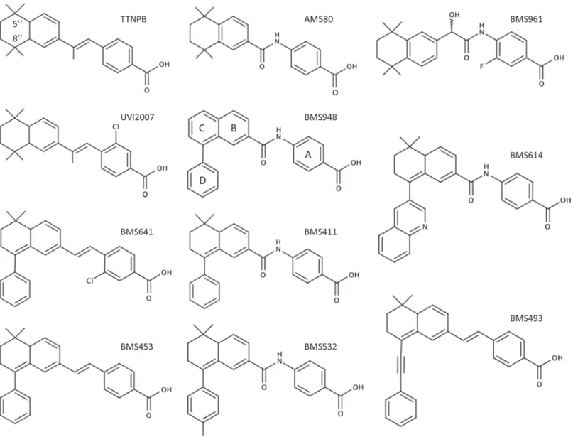
Despite their chemotherapeutical potential [14–18], the pharmacological use of retinoic acids is severely restricted because of their pleiotropic activities. The development of synthetic retinoids that specifically modulate only one RAR subtype might overcome these restrictions [17–19]. Note that a given ligand (agonist and antagonist) may be considered as selective when it exhibits an affinity difference greater than 100-fold between its primary target and other re-ceptors (see International Committee of Pharmacology Committee on Receptor Nomenclature Recherche Scientifique, Université de Montpellier 1 &
2. The funders had no role in study design, data collection and analysis, decision to publish, or preparation of the manuscript.
and Classification [4]). Under 100-fold difference the ligand may be defined as a compound that shows a preference for a given receptor. Comparison of RAR subtype sequences together with their 3D structures revealed only three divergent residues into their LBPs located in H3 (RARαSer232, RARβAla225, RARγAla234), H5 (RARαIle270, RARβIle263, RARγMet272) and H11 (RARαVal395, RARβVal388, RARγAla397) [20]. Ligands that display selectively or marked preferences for RARαor RARγhave been developed on the basis of specific hydrogen bonds formed with the polar Ser232(H3) [21] and the weakly polar Met272(H5) [9] in RARαand RARγ, respectively. However, no such discriminatory bond can be established in RARβLBP so the development of RARβ-selective ligands is more challenging [10] and requires alternative strategies. There is a great need for RARβ-selective agonists and antagonists as tools to facilitate the pharmacological analysis of this proteinin vitroandin vivoas well as for potential thera-peutic interventions. In humans, there are three major isoforms for RARβ(β1 initiated at the P1 promoter,β2 andβ4 initiated at the P2 promoter) harboring identical LBPs. The term RARβin the literature usually refers to the RARβ2 isoform. Importantly, many studies have
Fig 1. Chemical structures of the synthetic RAR ligands used in this study.
previously shown that RARβ2 possesses many of the functional characteristics of a tumor suppressor [22]. In some cellular models RARβ2 is required for the anti-proliferative effect of retinoic acid [23] and its expression is frequently lost in many neoplastic tissues [24–28]. Nev-ertheless, it has been recently reported that RARβ2 could also play a role in mammary gland tu-morigenesis [29], thus demonstrating the important but yet incompletely understood function of this nuclear receptor in cancer development. At the present time, it has been possible to generate molecules with complex activities such as ligands that are RARβagonists and RARα/ RARγantagonists [10,20,30,31] or which exhibit only a partial agonistic activity toward RARβsuch as BMS641 [10]. Here we report on the structural and functional characterization of BMS948 as RARβ-selective full agonist and provide rational guidelines for the development of selective antagonists.
Results
Despite a number of gene ablation [3] and gain-of-function studies, the mechanisms underly-ing the specific action of RARβhave remained poorly defined. These approaches can in no way replace receptor pharmacology. Hence development of more and effective RARβ-selective reti-noids is important to further dissect the function of this protein in various systems. For this purpose, novel retinoids inspired by well-characterized retinoids were prepared (Fig 1). Subse-quently, and because RARs are transcription factors, the biological activity of these new syn-thetic retinoids was characterized by the use of transactivation reporter assays with cultured cells. Ultimately, the transcriptional activity of these compounds led us to explore the structural basis of their behavior as RAR agonist and antagonist by defining the 3D structure of the RARβ LBD complexed with these retinoids. Overall, our approach allowed refining the structure-ac-tivity relationships in both retinoid agonists and antagonists.
BMS948 is a full RAR
β
-selective agonist
To evaluate the activity of a panel of synthetic retinoids (Fig 1) on RAR-mediated transactiva-tion, we used HeLa cells transiently transfected with human RARα1,-β2, or—γ2 and a (RARE)3x-tk-luciferase reporter gene. Well characterized full agonist and antagonist retinoids were also used as reference compounds (S2 Fig). The transcriptional activity obtained with the various compounds was compared with that observed with 10 nM of the pan-RAR agonist TTNPB (100%), a chemically stable compound activating all RAR subtypes as efficiently as atRA (S3 Fig). In these conditions, as expected, the RARα-selective agonist Am580 at 3 nM and the RARγ-selective agonist BMS961 at 100 nM exhibited full agonistic activity for RARα and RARγ, respectively (Figs2A–3C). Importantly, the novel compound BMS948 acted as a full RARβ-agonist at 1μM, whereas a very weak activity was seen with RARαand RARγ(Fig 2A–2C). In comparison with previously characterized compounds, BMS948 displayed an activ-ity profile similar to that of BMS411 [30] which also acted as an RARβ-selective full agonist whereas BMS641 [10] and BMS453 [30] acted as RARβ-selective partial agonists (Fig 2A–2C). To further characterize the activation profile of BMS compounds, transfected cells were ex-posed to a range of ligand concentrations to establish dose-response curves. As displayed inFig 2D–2F, BMS411, BMS641 and BMS948 activated RARβbut very weakly RARαor RARγ, and only at the highest ligand concentration used (1μM). Furthermore, the partial activity of
BMS641 toward RARβwas confirmed as the dose-response curve plateaued at about 50% of the TTNPB-induced activity (Fig 2E). BMS948 produced a concentration-dependent increase in transactivation for RARβwith an EC50 that was about 0.1μM and one order of magnitude
higher than those determined for BMS411 and BMS641 (EC5010 nM) and two order of
Fig 2. BMS948 acts as a full RARβagonist. (A to C) HeLa cells were transiently cotransfected with the reporter (RARE)3x-tk-Luc and RARα(A), RARβ(B) or RARγ(C), as indicated, to assess the RAR agonist potential of synthetic RAR ligands.Cells were incubated with ligands at selective concentrations (Am580 10–9M, BMS961 10–7M, BMS453 10–7M, BMS641 10–7M, BMS411 10–7M, BMS948 3.10–7M). 100% corresponds to reporter gene transcription induced in the presence of the full pan-RAR agonist TTNPB at 10–8M. (E to G) Dose-response curves to assess the binding affinity of synthetic RAR ligands towards RARα(E), RARβ(F), or RARγ(G). Cells were incubated with increasing concentrations of TTNPB (open squares), BMS948 (open circles), BMS411 (closed triangles), or BMS641 (closed diamonds), in a range of 10–10to 10–6M. All error bars are expressed as s.e.m.
Fig 3. BMS948 is an RARβ-selective ligands.(A to C) Transient transactivation assays as inFig 2to assess the antagonist potential of synthetic RAR ligands (RARα(A), RARβ(B) or RARγ(C)). The reporter was activated with 3nM TTNPB (100%) alone and plus the synthetic retinoids added at 1μM. (E to G) Dose-response curves (RARα(E), RARβ(F), or RARγ(G)) to assess the binding affinity of BMS493 (closed circles), BMS948 (open circles), BMS411 closed triangles), or BMS641 (closed diamonds) relative to TTNPB at 3 nM.
been shown to reflect its binding affinity for the target receptor [10,30]. For instance, BMS641 which binds to RARα, RARβand RARγwith dissociation constants (Kds) of 225 nM, 2.5 nM and 223 nM, respectively, was previously shown to activate RARβat a 100-times lower concen-tration than the two other RAR subtypes [10]. We thus concluded that BMS948 must display a somewhat lower affinity for RARβthan both BMS411 and BMS641.
Having shown that BMS948 was almost ineffective in inducing transactivation through both RARαand RARγ, we tested whether this compound displayed RAR antagonistic activi-ties. To this end, transient transactivation assays were conducted in order to assess the ability of the synthetic retinoids to affect TTNPB-induced RAR activity. In these experiments TTNPB at 3 nM was challenged with increasing amounts of the ligands and the powerful pRAR an-tagonist BMS493 at 1μM was used as positive control for transactivation inhibition (Fig 3A– 3C). As previously reported [32], BMS493 abolished the TTNPB-induced transactivation for all three RAR subtypes whereas the RARα-specific antagonist BMS614 [8,20] counteracted the TTNPB-induced transactivation on RARαonly. In the same vein, BMS453 [10,30] antago-nized almost entirely the TTNPB-induced RARαand RARγactivity and acted as a partial an-tagonist for RARβ, in line with its RARβpartial agonistic activity (Fig 2B). BMS411 appeared as a potent RARαantagonist with no or little antagonizing effect on RARβand RARx, respec-tively. In addition, dose-response curves were conducted to evaluate the potency of molecules relative to TTNPB (Fig 3D and 3F). Competition curves with increasing concentrations of BMS411 yielded an IC50 of roughly 30 nM for RARαin keeping with the high affinity of this compound for this receptor subtype [30] (Fig 3D). Similar experiments confirmed the partial RARβ-selective antagonistic activity of BMS641 as the competition curve plateaued at about 50% of the TTNPB-induced activity. Importantly, BMS948 showed no (RARβ) or very weak (RARαandγ) inhibiting activity, suggesting that this compound binds to RARαand RARγ only at very high concentration. Overall, our data lead to the general conclusion that BMS948 is a full RARβ-selective agonist.
Crystal structures of RAR
β
LBD in complex with BMS411, BMS641 and
BMS948
To gain structural insights into the specific binding and activity profiles of the newly character-ized synthetic ligands, we solved the crystal structures of RARβLBD in complex with BMS411, BMS641 and BMS948. To further stabilize the RARβLBD in its active form and in turn facili-tate crystallization, a peptide containing the second interaction motif of the coactivator SRC-1 was also added during the crystallization trials. Structure determination and refinement statis-tics are summarized inS1 Table. The structures solved at 1.9 Å (BMS641), 2.3 Å (BMS411), and 2.6 Å (BMS948) resolution display the canonical active conformation, with helix H12 cap-ping the ligand-binding pocket (LBP) and the SRC-1 peptide bound to the so-called“AF-2 sur-face”formed by helices H3, H4 and H12 (as shown for the RARβLBD–BMS641 complex in
Fig 4A), thereby reflecting the agonistic character of the ligands. The compounds could be pre-cisely placed in their respective electron density revealing a conserved binding mode. As shown inFig 4B–4D, the ligands occupy almost the same volume in the LBP and impose similar side chain conformations. They are stabilized in the RARβLBP through extensive van der Waals contacts and a network of ionic and hydrogen bonds between the carboxylate moiety of the ret-inoids, Arg269in H5, Ser280in theβ-turn and water molecules.
Structural features contributing to RAR
β
-selectivity
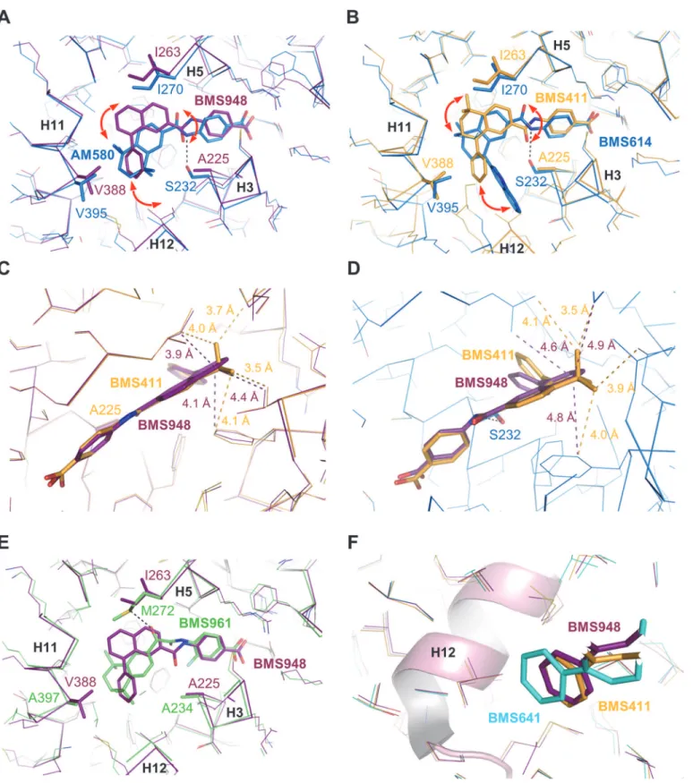
suggesting different recognition mechanisms. BMS641 that is closely related to both BMS453 with a chlorine atom at position C3 and UVI2007 with a phenyl group at position C8”(Fig 1), exhibited a marked RARβ-selectivity and acted as RARα/γantagonist at high concentration only (Fig 2D–2F). This is in contrast with BMS453 and UVI2007 which acted as potent RARα/ γantagonist and RARβpartial agonist or RARβ/γfull agonist, respectively [10]. Thus we con-cluded that, separately, the phenyl group at position C8”or the chlorine atom at position C3 do not allow discrimination between RAR subtypes and we suggested that the selectivity to-ward RARβdisplayed by BMS641 results from the combination of these two chemical groups. Superposition of the BMS641-bound RARβstructure onto that of RARαbound to the RARα -selective ligand Am580 revealed that the replacement of RARβAla225by Ser232in H3 of RARα generates steric hindrance that strongly reduces the binding of 3-chloro substituted com-pounds (Fig 5A). In the same line, comparison with the structure of RARγbound to the RARγ-selective ligand BMS961 showed that the position of BMS641 in the LBP is incompatible with the presence of Met272in RARγ(Fig 5B). Indeed, BMS641 lies in a portion of the LBP that is not occupied by the ligand in the RARα-Am580 and RARγ-BMS961 complexes. This shift in
Fig 4. Crystallographic analysis.(A) The whole structure of the RARβLBD in complex with BMS641 and SRC-1 coactivator peptide (in orange). The AF-2 surface formed by helices H3, H4 and H12 is highlighted in green. The lower part of the LBD, in blue, encloses the ligand-binding pocket (LBP). (B-D) The interaction networks of BMS compounds with LBD residues of RARβare also displayed. Oxygen, nitrogen and chlorine atoms are colored in red, blue, and green, respectively. Hydrogen bonds are indicated by black dashed lines and water molecules by red spheres. The electron density represents aFo-Fc
simulated annealing omit map contoured at 3σ.
the position of BMS641 relies on the presence of the bulky C8”-phenyl ring which pushes the ligand toward RARβIle263. Of note, comparison of the BMS641-bound RARβand
Fig 5. Structural features contributing to the RARβ-selectivity of BMS641.Structure superposition of BMS641-bound RARβ(cyan) with (A) Am580-bound RARα(blue), (B) BMS961-bound RARγ(green), or (C) BMS493-bound RARα(blue).
BMS493-bound RARα[12] structures reveals that the position of the two stilbene-based reti-noids adopt exactly the same position in RARαand RARβLBPs (Fig 5C). Overall these obser-vations provide a structural basis for the RARβselectivity of BMS641 and reveal that the presence of a 3-chloro and an 8”-phenyl prevent BMS641 from binding to RARαand RARγ while maintaining a partial agonistic activity on RARβ.
In contrast with BMS641, BMS948 harbors an amide linker (Fig 1). This is a rather unex-pected observation since previous studies have revealed that retinoids with such a connector display RARα-selectivity [20,21]. Indeed, the structures of RARα-LBD complexed with the RARα-selective agonist Am580 or the RARα-selective antagonist BMS614 (a quinolyl deriva-tive of Am580) revealed that the amide linker of both ligands adopts a particular constrained conformation by establishing a hydrogen bond with the RARα-specific Ser232through its amino moiety (Fig 6A and 6B), the carbonyl group pointing toward Ile270. Strikingly, the 3D structures of RARβLBD in complex with BMS411 or BMS948 showed an alternative confor-mation of the amide function. In contrast to RARα, and because of the presence of an alanine residue in RARβH3 instead of Ser232in RARα, the carbonyl moiety of the amide linker of BMS948 (Fig 6A) and BMS411 (Fig 6B) points toward RARβA225. This flipping by 180° of the amide bond induces a change in the position of the rings B and C of BMS948 and BMS411 as compared to that of Am580 and BMS614 in RARα. Interestingly, the only difference between BMS411, which retains a significant interaction capacity with RARα, and the RARβ-selective BMS948 resides in the replacement of a dimethyl-cyclohexenyl ring by a planar phenyl group (Fig 1). To better understand the drastic loss of affinity of BMS948 for RARαby comparison with BMS411, we used the BMS614-bound RARαstructure to dock BMS948 and BMS411 in RARαand compared their LBP environment to that observed in the crystal structures of BMS948 and BMS411 bound to RARβ. Whereas the two methyl groups of BMS411 are in-volved in strong van der Waals interactions with LBP residues in both receptor subtypes (Fig 6C and 6D), the lack of such chemical groups in BMS948 results respectively in a modest and strong loss of van der Waals contacts between this ligand and LBP residues in RARβand RARα. Indeed, in the absence of the two methyl groups, the carbon atom at position 5 of the BMS948 naphthalene ring remains involved in several contacts with RARβLBP residues (Fig 6C) whereas no interaction distance below 4.6 Å is observed in RARα(Fig 6D). Thus the diver-gent binding modes of BMS948 in the two receptor subtypes bring the naphthalene ring into a LBP environment that is more sensitive to the loss of the methyl groups in RARαthan in RARβ, in full agreement with the much higher affinity of BMS948 for RARβ. Finally, because RARγcontains an alanine residue in H3 (Ala234), we hypothesized that BMS948 adopts a posi-tion in this receptor subtype similar to that observed in RARβ. A superposiposi-tion of the
BMS948-bound RARβLBD structure with that of RARγbound to the selective ligand BMS961 shows that, as in the case of BMS641, the position of BMS948 is incompatible with the presence of Met272in RARγ(Fig 6E). A mutational analysis in which LBPs of both RARβand RARγ were interconverted confirmed that Met272is a most important discriminatory residue (S4
Fig). Together the above considerations provide a structural rationale accounting for the RARα antagonist/RARβagonist activities of BMS411.
Structural basis for the specific activity of BMS ligands in RAR subtypes
Fig 6. Structural features contributing to the RARβ-selectivity and full agonistic activity of BMS948.(A) Structure superposition of BMS948-bound RARβ(violet) with Am580-bound RARα(blue). (B) Structure superposition of BMS411-bound RARβ(yellow) with BMS614-bound RARα(blue). (C) Structure superposition of BMS411-bound RARβ(yellow) with BMS948-bound RARβ(violet). (D) Superposition of the docking models of BMS411 (yellow) and BMS948 (violet) in RARα. (E) Structure superposition of BMS948-bound RARβ(violet) with BMS961-bound RARγ(green). (F) Superposition of all BMS-bound RARβstructures highlighting the differential positioning of the phenyl extensions relative to helix H12. Superposition of the structures was done using the SSM function in Coot[33].
reasonably make the hypothesis that both ligands adopt a similar conformation in RARαLBP. Thus, as compared to the RARβsituation, the flipping by 180° of the amide bond of BMS411 in RARαis very likely to induce a complete reorientation of the rings B and C, and bring the phe-nyl extension D in a position similar to that observed for the quinolyl group of BMS614 (Fig 6B), thereby interfering unfavorably with the stable positioning of H12 in the active conforma-tion. This structural model is in full agreement with the dual RARαantagonist/RARβagonist activities of BMS411. Overall, this structural analysis shows that the mode of binding of BMS411 to RARβallows accommodation of the phenyl ring of the ligand within the confined environment of the active conformation. In contrast, in RARαthe conformational change of BMS411 induces the reorientation of the bulky extension which projects toward H12 and pre-vents the active conformation of RARαfrom being stably formed.
We subsequently looked for a rational explanation for the differential activation of RARβby the BMS compounds. Indeed, our transcriptional assays demonstrated that BMS641 acted as RARβpartial agonist whereas both BMS948 and BMS411 displayed full RARβagonistic activity (Fig 2B and 2E). The superposition of all three structures reveals that, although all BMS com-pounds are able to lock RARβin the active conformation which is consistent with their agonis-tic activity, the phenyl extension of BMS641 protrudes slightly more from the LBP toward helix H12 than that of BMS948 and BMS411 (Fig 6F). A likely consequence is that the RARβ -coactivator interaction may be less optimal in the presence of BMS641. Indeed BMS641 in-duced a rein-duced recruitment of the coactivator TIF2, accounting for the weakest agonistic ac-tivity of this compound (S5 Fig).
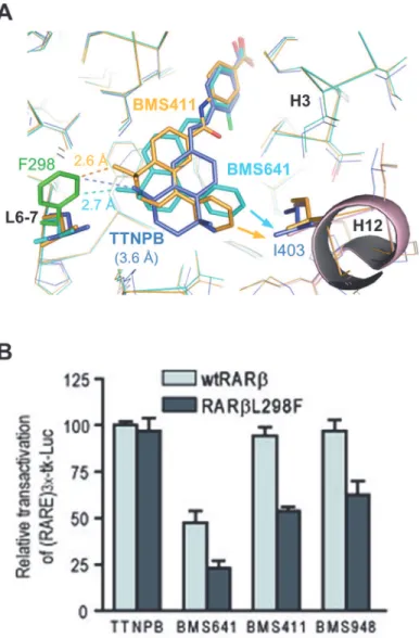
To validate experimentally our model of partial agonist action according to which the de-gree of agonistic/antagonistic activity varies as a function of the repulsive forces exerted by the ligand onto helix H12, we introduced a point mutation in the back of the RARβLBP where Leu298(loop between H6 and H7) was replaced by a bulkier phenylalanine residue (Fig 7A). Transient transfection assays revealed that the full agonistic activity of TTNPB was not affected by the mutation (Fig 7B). In contrast, this single mutation was detrimental for all compounds harboring a phenyl substituent at the C8”position. Whereas BMS411 and BMS948 acted as full agonists of wtRARβ, they induced only a 50–60% activity on RARβL298F as compared to TTNPB. Similarly, the partial agonistic activity of BMS641 was decreased from 50% (wtRARβ) to 20% (RARβL298F). Note that dose-response curves revealed unchanged EC50s as compared to those obtained with wtRARβ, suggesting that this reduction of the transcriptional activity is not due to a decrease of ligand affinities for the mutant (S6 Fig). Modeling of the RARβmutant reveals that Phe298generates steric clashes with rings C/D of C8”substituted compounds which is likely to provoke a displacement of the ligands toward H12 and a destabilization of the active conformation, and as a result a reduction of the binding affinity of CoA (Fig 7A). Comparison with the structure of RARβin complex with TTNPB [34] reveals that the agonist which does not contain a bulky extension adopts a position in the LBP that allows accommoda-tion of the phenylalanine residue without significant posiaccommoda-tional adaptaaccommoda-tion. These data substan-tiate the combined roles of the linker region and of the C8”substituents in the mixed agonistic/ antagonistic activity of BMS compounds and provides guidelines for the design of RARβ-selec-tive antagonists.
Discussion
transcriptional potential of the retinoids. In addition, BMS641, which contains atrans-olefin as a linker, acted as RARβ-selective partial agonist, presumably by provoking more disturbance of the H12 folding than full agonists. Finally, and strikingly, our study revealed the selectivity and the full agonistic activity of BMS948 for RARβas well as the structural basis of these features. BMS948 is then the first RARβ-selective full agonist identified. Of note, the BMS948 selectivity is surprising as its structure contains the identical amide function as a connector than that of known RARα-selective ligand such as Am580. The unexpected structural basis of BMS948 se-lectivity is based on the lack of hydrophobic gem-dimethyl group located at C5”position found in both RARαligands BMS411 (antagonist) and Am580 (agonist) which appears to be the de-terminant for the dramatic low affinity of BMS948 towards RARαand, as a consequence, for its RARβ-selectivity. To assess whether this absence of the two methyl groups suffices to impair
Fig 7. Structural model for full- and partial activity of BMS compounds.(A) Comparison of the steric clashes generated by the L298F mutation (green) in RARβcomplexed with TTNPB (blue), BMS411 (yellow)
and BMS641 (cyan). (B) Transient transactivation assays as inFig 2to assess the transcriptional activity of wild-type hRARβand mutant hRARβL298F in the presence of the indicated retinoids or vehicle (ethanol). The
reporter was activated with 10 nM TTNPB (100% with wild-type hRARβ) and the synthetic retinoids were added at 0.1μM.
the binding of a retinoid to RARαor whether the combination of this absence with the pres-ence of a linker harboring an amide function is required, one should generate stilbene-based retinoids, such as a derivative series of BMS453, lacking the gem-dimethyl group. On the other hand, and despite a great need, RARβantagonists have not been reported yet. Nevertheless, a diazepine fused to aryl rings, referred to as LE135 [35], has been reported as an antagonist showing some selectivity for RARβ. But, the affinity difference between RARβand the two other RAR subtypes makes that this compound may be defined as a retinoid that only shows preference for RARβ. In addition, this compound displayed some RARγpartial agonistic activi-ty ([35] and personal data not shown). Consequently, such a ligand is inadequate to properly investigate the function of this protein, mainly in the context ofin vivoanalyses. Hence the present challenge is to integrate our above molecular and structural information to provide po-tent and selective RARβantagonists. A possible strategy to achieve this goal is to start from the RARβ-selective ligands identified in this work, namely BMS641 and BMS948. Because the re-ported crystal structures of RARs [8,10,12] have showed that substitution at the naphthalene C8 position endow in general ligands with RAR antagonist activities, a further extension of the substituent at this position of both BMS641 and BMS948 could impact on H12 positioning and thus afford ligands with antagonist properties. In this respect, an entire series of C3-halogenat-ed derivatives and bulkier analogues at C8”position of the parent stilbene-based BMS641 has been generated and characterized in term of their RAR transcriptional potential [36]. Whereas most of these ligands acted as potent RARβantagonists, the RARβ-selectivity observed for BMS641 is lost in the presence of bulkier substituents. Then, one potential caveat is that an in-crease in bulk may change the rules defining the binding selectivity. More recently, inspired in the parent BMS948, a C5,C8-diphenylnaphtalene-2-yl linked to a benzoic acid via an amide connector has been reported [37]. Unfortunately, this compound did not exert an RAR antago-nistic activity, but exhibited a RARβagonistic activity at very high concentration (10μM).
Importantly, we have observed that replacement of the phenyl group at position C8”of the RARβagonist BMS411 by ap-tolyl group, yielding BMS532 (Fig 1), converted the ligand into a potent antagonist for RARβ, while BMS532 retained a high affinity for RAR03B1 (data not shown). Together with the fact that both BMS411 and BMS948 adopt the same conformation in the RARβLBP and that the absence of the gem-dimethyl group at C5”position is a crucial point to lose affinity towards RARα, these considerations led us to predict that increasing the bulkiness of the hydrophobic group of BMS948-like molecules, notably by introducing ap -tolyl group instead of the phenyl group, would generate retinoids that sterically interfere with the agonist positioning of H12 and thus turn into RARβ–selective antagonists.
In conclusion, the novel RARβ-selective full agonist presented here should represent a use-ful tool for both developing selective antagonists for RARβand pharmacologically addressing the specific RARβfunctionin vitroand in animal models, and possibly, therapeutic exploita-tion. In addition, our overall results demonstrate that subtle changes in the chemical structure of the ligand or in the residues lining the LBPs translate in very selective effects on the tran-scriptional potential of the receptor.
Materials and Methods
Ligands and plasmids
by PCR-assisted site-directed mutagenesis with Deep Vent DNA polymerase (New England Biolabs). The construct was verified by DNA sequencing.
Cell culture and transient transfections
HeLa cells were cultured in DMEM with Glutamax and 10% (v/v) FCS and transfected using JetPei transfectant (Ozyme). After 24 h, the medium was changed to a medium containing the indicated ligands or vehicle. Cells were lysed and assayed for reporter expression 48 h after transfection. The luciferase assay system was used according to the manufacturer’s instruction (Promega). In each case results were normalized to coexpressedβ–galactosidase. Each transfec-tion was carried out in duplicate and repeated each three to six times.
Protein production, purification and crystallization
The human wild-type RARβLBD (amino acids 169–414) was cloned into the pET-15b vector and expressed inEscherichia coliBL21(DE3) cells. Cells were grown at 37°C in LB medium supplemented with 50 mg.mL-1ampicillin until OD600reached about 0.6. Expression of T7 po-lymerase was induced by addition of isopropyl-β-D-thiogalactoside (IPTG) to a final concen-tration of 0.5 mM. After an additional incubation for 8 h at 20°C, cell cultures were harvested by centrifugation at 8,000 g for 20 min. Cell pellets from 2 L of culture were resuspended in 50 mL buffer-A (20 mM Tris-HCl pH 7.5, 500 mM NaCl, 1 mM DTT) supplemented with a protease inhibitor cocktail (cOmplete, Mini, EDTA-free; Roche Applied Science). The suspen-sion was lysed by sonication and centrifuged at 4°C for 45 min. The supernatant was loaded onto a nickel affinity column (HisTrap 5 mL; GE Healthcare) pre-equilibrated with buffer-A. The protein was eluted with buffer-B (20 mM Tris-HCl pH 7.5, 500 mM NaCl, 1 mM DTT, 500 mM Imidazole). The fractions containing hRARβLBD were pooled and further purified by size exclusion chromatography (Superdex 75 HR 26/60; GE Healthcare). Prior to crystalliza-tion the purified hRARβLBD was further complexed with 2 equimolar of ligand (BMS948, BMS641, and BMS411 provided by Bristol-Myers Squibb) and 3 equimolar of SRC-1 co-activa-tor peptide (the RHKILHRLLQEGS peptide corresponding to the NR box 2-binding motif was purchased from EZbiolab). The complexes were concentrated to 10 mg.ml-1in the gel filtration buffer (20 mM Tris pH 7.5, 150 mM NaCl, 5 mM DTT and 1 mM EDTA). The various RARβ-ligand-SRC-1 complex crystals were obtained in 200 mM Trisodium citrate pH 5.5 and 25% PEG 4000 using the hanging drop crystallization method.
Data collection and structure determination
Native data were collected from crystals cryoprotected with 30% glycerol on the BM30A and ID14-2 beamlines at the European Synchrotron Radiation Facility, Grenoble, France. Data were processed and scaled using XDS and XSCALE [38]. Crystals belong to space group P212121for all complexes. Structures were solved by molecular replacement method using PHENIX (phenix.automr) [39], and refinement and rebuilding were performed with COOT [33], PHENIX (phenix.refine) [39] and REFMAC [40] from the CCP4 suite [41]. Data collec-tion and refinement statistics are summarized in Supporting Informacollec-tion. Figures were pre-pared with PyMOL (http://pymol.org/).
Accession codes
Supporting Information
S1 Fig. Determination of the BMS948 purity by HPLC/LCMS analysis.The purity of BMS948 was determined by LCMS analysis, which was performed on a system consisting of an electrospray source on a Waters Micromasss ZQ mass spectrometer, a Waters 2996 diode array detector, a Waters alliance 2695 HPLC system with autosampler, and a Macherey-Nagel Nucleoshell RP18 plus HPLC column (5μm, 4 mm [1] 100 mm). The HPLC method
incorpo-rated UV detection on a range of 214–400 nm, a column temperature of 40°C, a flow rate of 1.0 mL/min, a 50μL injection volume, and a binary solvent system of 0.1% formic acid in
water (solvent A) and 0.1% formic acid in CH3CN (solvent B). The following gradient (10 min) was used: 0–1 min, 5% B; 1–7 min, 5–100% B; 7–10 min, 100% B. (A) BMS948 was isolat-ed to>98% purity (98.49%). (B) MS spectrum confirmed the presence of BMS948.
(EPS)
S2 Fig. Dose-response curves for reference retinoids.(A to C) Transient transactivation as-says as inFig 2(RARα(A), RARβ(B), or RARγ(C)). Cells were incubated with increasing con-centrations of TTNPB (open squares), Am580 (closed triangles), BMS961 (closed circles), or BMS453 (open circles), in a range of 10–10to 10–6M. All error bars are expressed as s.e.m. (E to G) Transient transactivation assays as inFig 3to assess the antagonist potential of synthetic retinoids (RARα(E), RARβ(F) or RARγ(G)). The reporter was activated with 3nM TTNPB (100%) alone and plus the synthetic retinoids (BMS614 (closed triangles), BMS453 (open cir-cles), BMS493 (closed circles) in a range of 10–10to 10–6M.
(EPS)
S3 Fig. TTNPB andall transretinoic acid exhibit a similar efficacy.HeLa cells were
tran-siently cotransfected with the reporter (RARE)3x-tk-Luc and RARα(black bars), RARβ(light grey) or RARγ(dark grey), as indicated, to assess the RAR agonist potential ofall transretinoic acid (atRA) and TTNPB at 10 nM.
(EPS)
S4 Fig. The methionine 272 of RARγLBP is a most important discriminatory element for the RARβ-specific response to BMS948.A sequence analysis of RARβ- and RARγ-LBPs showed that a leucine residue (βIle263) in RARβis replaced by a methionine residue (γMet272) in RARγ. To investigate the role of these residues in the RARβ–specific response to BMS948, we compared the agonistic and antagonistic potentials of this compound for RARβand RARγ, as well as chimeric RAR mutants in which LBPs were interconverted (denoted RARβ!γand
RARγ!β). Dose-response curves were established from transient transfection in HeLa cells of
these proteins and a (RARE)3x-tk-luciferase reporter gene as described in Figs2and3. When the LBP of RARγwas converted into that of RARβ, RARγ!βresponded to BMS948 like RARβ
did, that is BMS948 acted as a potent full agonist, whereas a very weak activation was seen only at high concentration for RARβ!γand the parental RARγ. In addition, BMS948 did not
re-duce TTNPB-inre-duced activity for RARβ!γwhich harbors a methionine residue. Overall these
results show that by changing the RARγLBP into that of RARβ, RARγ!βacquired the ability
to bind BMS948, demonstrating that the replacement ofγMet272with a leucine residue most likely accounts for the acquisition of BMS948-binding ability by the mutant RARγ!β. These
results underscore the importance of these divergent residues for the selectivity of BMS948 to-ward RARβand suggest thatγMet272is the most important discriminatory element, in full agreement with our structural analysis (Fig 5B). (A) HeLa cells were transiently cotransfected with the reporter (RARE)3x-tk-Luc and RARβ(open circles), RARγ(closed diamonds), RARγ!βin which the methionine residue 272 in RARγLBP is replaced by an isoleucine
by a methionine residue (closed triangles) to assess the agonist potential of BMS948. Cells were incubated with increasing concentrations of BMS948 in a range of 10–9to 10–6M. 100% corre-sponds to reporter gene transcription induced in the presence of the full agonist TTNPB at 10nM. (B) Transient transfection assays in HeLa cells with the reporter (RARE)3x-tk-Luc and RARβ!γas in (A) to assess the antagonist potential of BMS948 and BMS493. The reporter
was activated with 3 nM TTNPB (100%) alone and plus the BMS compounds at 1μM.
(EPS)
S5 Fig. Molecular basis of BMS641 partial activity.According to the current model of gene regulation by RARs, the agonistic property of a given retinoid depends on its ability to induce coregulator recruitment. RAR agonists dissociate corepressors (such as SMRT) and induce the recruitment coactivators (such as TIF2), resulting in transactivation. The transcriptional data reported in Figs2and3revealed that BMS641 acts as an RARβagonist with reduced efficacy (50%) when compared to the full agonist TTNPB (100%). BMS641 was therefore categorized as a partial RARβagonist. To further characterize the partial activity of BMS641, we used two-hybrid assays to monitor the interaction between RARβand transcriptional coregulators in the presence of various retinoids. In these assays, a chimeric luciferase-based reporter gene ((17 m)5x-βGlob-Luc) is transfected together with two expression vectors. One expresses a fusion protein (Gal-TIF2 or Gal-SMRT) that binds through the Gal DNA binding domain (Gal) to the pentamer of the“17m”DNA recognition site in the reporter gene and contains the nuclear receptor interacting domain of the coregulator. The other vector expresses a second fusion pro-tein composed of the VP16 acidic transcription activation domain, and the LBD of RARβ. If a ligand induces RARβ-coregulator interaction, this results in the indirect recruitment of the VP16 activation domain to the promoter of the luciferase reporter gene and to the luciferase protein synthesis which can be easily quantified by using a luminometer. Two-hybrid analyses using Gal-TIF2 indicated that the RARβ-TIF2 interaction was optimal in the presence of the full RARβagonists TTNPB, BMS411, and BMS948, while only a partial effect was seen with BMS641 (75%). As expected, BMS493 prevented the recruitment of TIF2 by RARβ. Regarding corepressor interaction, SMRT binding was decreased in the presence of all three full RARβ agonists and strongly enhanced in the presence of BMS493, in keeping with the inverse agonis-tic property of this ligand. Strikingly, the RARβ-SMRT interaction was maintained in the pres-ence of BMS641, in agreement with the partial agonistic activity of this compound. Overall these data indicated that ligands show distinct pattern of RARβ-coregulator interaction which apparently account for their transcriptional activity. Retinoids characterized as full agonists (TTNPB, BMS411, and BMS948) are able to dissociate the corepressor SMRT and to induce ef-ficient recruitment of the coactivator TIF2, consistent with their transcriptional efficacy. BMS641 induces reduced coactivator recruitment, but cannot dissociate corepressors. Conse-quently, as the binding of coactivators and corepressors are mutually exclusive, the resulting transcriptional activity led by BMS641 is partial. Thereby BMS641 can be classified as a potent partial agonist on the basis of its reduced efficacy relative to full agonists that originates from its pattern of coregulator interaction. Mammalian two-hybrid assays with (17m)5x-βGlob-Luc and Gal-TIF2 (A, 100% corresponds to reporter gene transcription induced by 10 nM TTNPB) or Gal-SMRT (B, 100% corresponds to reporter gene transcription in the absence of ligand) as bait and VP16-RARβLBD as prey were performed in HeLa cells to assess the influence of BMS compounds at 1μM (compared to TTNPB 10 nM) on interaction between RAR and both
core-gulators TIF2 and SMRT in a cellular context. (EPS)
was replaced by a bulkier phenylalanine residue (Fig 7A). Transient transfection assays re-vealed that, whereas the efficacy of TTNPB was not affected by the mutation (Fig 7B), this sin-gle mutation was detrimental for all BMS compounds harboring a phenyl substituent at the C8”position. Dose-response curves show a concentration-dependent increase in activation for RARβL298F with however a reduction of the efficacy of all BMS compounds (70%, 50%, and 25% for BMS948, BMS411, and BMS453, respectively) and, in contrast, unchanged EC50s when compared to those measured with the parental RARβ(Fig 2E), thus suggesting that the reduction of the transcriptional activity for RARβL298F is not due to a decrease of ligand affini-ties for this mutant. HeLa cells were transiently cotransfected with the reporter (RARE)3x -tk-Luc and the RARβL298F mutant (in which the phenylalanine residue 298 is replaced by a leu-cine residue in the RARβLBP) to assess the RAR agonist potential of synthetic RAR ligands. Cells were incubated with increasing concentrations of TTNPB (open squares), BMS948 (open circles), BMS411 (closed triangles), or BMS641 (closed diamonds). Gal-TIF2 [32], Gal-SMRT [6], and pSG5-based RAR [20] expression vectors were described previously.
(EPS)
S1 Table. Data collection and refinement statistics.
(DOCX)
Acknowledgments
We acknowledge the experimental assistance from the staff of ESRF (ID14-2 and BM30A beamlines, Grenoble, France) during data collection. We thank Patrick Balaguer for (RARE)3x -tk-Luc plasmid.
Author Contributions
Conceived and designed the experiments: VV WB PG. Performed the experiments: EN CT VD VV WB PG. Analyzed the data: GK HG WB PG. Contributed reagents/materials/analysis tools: HG. Wrote the paper: WB PG.
References
1. Chambon P. A decade of molecular biology of retinoic acid receptors. FASEB J 1996; 10(9):940–954.
PMID:8801176
2. Germain P, Chambon P, Eichele G, Evans RM, Lazar MA, Leid M, et al. International Union of Pharma-cology. LX. Retinoic acid receptors. Pharmacol Rev 2006; 58(4):712–25. PMID:17132850
3. Mark M, Ghyselinck NB, Chambon P. FUNCTION OF RETINOID NUCLEAR RECEPTORS: Lessons from Genetic and Pharmacological Dissections of the Retinoic Acid Signaling Pathway During Mouse Embryogenesis. Annu Rev Pharmacol Toxicol 2006; 46:451–480. PMID:16402912
4. Germain P, Staels B, Dacquet C, Spedding M, Laudet V. Overview of nomenclature of nuclear recep-tors. Pharmacol Rev 2006; 58(4):685–704. PMID:17132848
5. Rochette-Egly C, Germain P. Dynamic and combinatorial control of gene expression by nuclear retinoic acid receptors (RARs). Nucl Recept Signal 2009; 7:e005. doi:10.1621/nrs.07005PMID:19471584
6. Germain P, Iyer J, Zechel C, Gronemeyer H. Coregulator recruitment and the mechanism of retinoic acid receptor synergy. Nature 2002; 415:187–192. PMID:11805839
7. Renaud JP, Rochel N, Ruff M, Vivat V, Chambon P, Gronemeyer H, et al. Crystal structure of the RAR-gamma ligand-binding domain bound to all- trans retinoic acid. Nature 1995; 378(6558):681–9. PMID:
7501014
8. Bourguet W, Vivat V, Wurtz JM, Chambon P, Gronemeyer H, Moras D. Crystal structure of a heterodi-meric complex of RAR and RXR ligand-binding domains. Mol Cell 2000; 5(2):289–98. PMID:10882070
10. Germain P, Kammerer S, Perez E, Peluso-Iltis C, Tortolani D, Zusi FC, et al. Rational design of RAR-selective ligands revealed by RARbeta crystal stucture. EMBO Rep 2004; 5(9):877–82. PMID:
15319780
11. Pogenberg V, Guichou JF, Vivat-Hannah V, Kammerer S, Perez E, Germain P, et al. Characterization of the interaction between retinoic acid receptor/retinoid X receptor (RAR/RXR) heterodimers and tran-scriptional coactivators through structural and fluorescence anisotropy studies. J Biol Chem 2005; 280 (2):1625–33. PMID:15528208
12. le Maire A, Teyssier C, Erb C, Grimaldi M, Alvarez S, de Lera AR, et al. A unique secondary-structure switch controls constitutive gene repression by retinoic acid receptor. Nature Structural & Molecular Bi-ology 2010; 17(7):801–U43.
13. Alvarez R, Vaz B, Gronemeyer H, de Lera AR. Functions, therapeutic applications, and synthesis of ret-inoids and carotenoids. Chem Rev 2014; 114(1):1–125. doi:10.1021/cr400126uPMID:24266866
14. Altucci L, Gronemeyer H. The promise of retinoids to fight against cancer. Nat Rev Cancer 2001; 1 (3):181–93. PMID:11902573
15. Ortiz MA, Bayon Y, Lopez-Hernandez FJ, Piedrafita FJ. Retinoids in combination therapies for the treat-ment of cancer: mechanisms and perspectives. Drug Resist Updat 2002; 5(3–4):162–75. PMID:
12531180
16. Sun SY, Lotan R. Retinoids and their receptors in cancer development and chemoprevention. Crit Rev Oncol Hematol 2002; 41(1):41–55. PMID:11796231
17. Dawson MI. Synthetic retinoids and their nuclear receptors. Curr Med Chem Anticancer Agents 2004; 4 (3):199–230. PMID:15134501
18. Kagechika H, Shudo K. Synthetic retinoids: recent developments concerning structure and clinical utili-ty. J Med Chem 2005; 48(19):5875–83. PMID:16161990
19. le Maire A, Alvarez S, Shankaranarayanan P, Lera AR, Bourguet W, Gronemeyer H. Retinoid receptors and therapeutic applications of RAR/RXR modulators. Curr Top Med Chem 2012; 12(6):505–27. PMID:
22242853
20. Gehin M, Vivat V, Wurtz JM, Losson R, Chambon P, Moras D, et al. Structural basis for engineering of retinoic acid receptor isotype-selective agonists and antagonists. Chem Biol 1999; 6(8):519–29. PMID:
10421757
21. Kagechika H, Kawachi E, Hashimoto Y, Himi T, Shudo K. Retinobenzoic acids. 1. Structuactivity re-lationships of aromatic amides with retinoidal activity. J Med Chem 1988; 31(11):2182–92. PMID:
3184125
22. Alvarez S, Germain P, Alvarez R, Rodriguez-Barrios F, Gronemeyer H, de Lera AR. Structure, function and modulation of retinoic acid receptor beta, a tumor suppressor. Int J Biochem Cell Biol 2007; 39(7–
8):1406–15.
23. Faria TN, Mendelsohn C, Chambon P, Gudas LJ. The targeted disruption of both alleles of RARbeta(2) in F9 cells results in the loss of retinoic acid-associated growth arrest. J Biol Chem 1999; 274
(38):26783–8. PMID:10480883
24. Lotan R, Xu XC, Lippman SM, Ro JY, Lee JS, Lee JJ, et al. Suppression of retinoic acid receptor-beta in premalignant oral lesions and its up-regulation by isotretinoin. N Engl J Med 1995; 332(21):1405–10.
PMID:7723796
25. Castillo L, Milano G, Santini J, Demard F, Pierrefite V. Analysis of retinoic acid receptor beta expression in normal and malignant laryngeal mucosa by a sensitive and routine applicable reverse transcription-polymerase chain reaction enzyme-linked immunosorbent assay method. Clin Cancer Res 1997; 3 (11):2137–42. PMID:9815607
26. Shin DM, Xu XC, Lippman SM, Lee JJ, Lee JS, Batsakis JG, et al. Accumulation of p53 protein and reti-noic acid receptor beta in retinoid chemoprevention. Clin Cancer Res 1997; 3(6):875–80. PMID:
9815762
27. Widschwendter M, Berger J, Muller HM, Zeimet AG, Marth C. Epigenetic downregulation of the retinoic acid receptor-beta2 gene in breast cancer. J Mammary Gland Biol Neoplasia 2001; 6(2):193–201.
PMID:11501579
28. Sirchia SM, Ren M, Pili R, Sironi E, Somenzi G, Ghidoni R, et al. Endogenous reactivation of the RAR-beta2 tumor suppressor gene epigenetically silenced in breast cancer. Cancer Res 2002; 62(9):2455–
61. PMID:11980632
29. Liu X, Nugoli M, Laferriere J, Saleh SM, Rodrigue-Gervais IG, Saleh M, et al. Stromal retinoic acid re-ceptor beta promotes mammary gland tumorigenesis. Proc Natl Acad Sci U S A 2011; 108(2):774–9.
30. Chen JY, Penco S, Ostrowski J, Balaguer P, Pons M, Starrett JE, et al. RAR-specific agonist/antago-nists which dissociate transactivation and AP1 transrepression inhibit anchorage-independent cell pro-liferation. Embo J 1995; 14(6):1187–97. PMID:7720709
31. Klein ES, Pino ME, Johnson AT, Davies PJ, Nagpal S, Thacher SM, et al. Identification and functional separation of retinoic acid receptor neutral antagonists and inverse agonists. J Biol Chem 1996; 271 (37):22692–6. PMID:8798442
32. Germain P, Gaudon C, Pogenberg V, Sanglier S, Van Dorsselaer A, Royer CA, et al. Differential action on coregulator interaction defines inverse retinoid agonists and neutral antagonists. Chem Biol 2009; 16(5):479–89. doi:10.1016/j.chembiol.2009.03.008PMID:19477412
33. Osz J, Brelivet Y, Peluso-Iltis C, Cura V, Eiler S, Ruff M, et al. Structural basis for a molecular allosteric control mechanism of cofactor binding to nuclear receptors. Proc Natl Acad Sci U S A 2012; 109(10): E588–94. doi:10.1073/pnas.1118192109PMID:22355136
34. Li Y, Hashimoto Y, Agadir A, Kagechika H, Zhang X. Identification of a novel class of retinoic acid re-ceptor beta-selective retinoid antagonists and their inhibitory effects on AP-1 activity and retinoic acid-induced apoptosis in human breast cancer cells. J Biol Chem 1999; 274(22):15360–6. PMID:
10336422
35. Alvarez S, Khanwalkar H, Alvarez R, Erb C, Martinez C, Rodriguez-Barrios F, et al. C3 halogen and c8'' substituents on stilbene arotinoids modulate retinoic Acid receptor subtype function. ChemMedChem 2009; 4(10):1630–40. doi:10.1002/cmdc.200900214PMID:19670208
36. Martinez C, Lieb M, Alvarez S, Rodriguez-Barrios F, Alvarez R, Khanwalkar H, et al. Dual RXR Ago-nists and RAR AntagoAgo-nists Based on the Stilbene Retinoid Scaffold. ACS Med Chem Lett 2014; 5 (5):533–7. doi:10.1021/ml400521fPMID:24900875
37. Kabsch W. Integration, scaling, space-group assignment and post-refinement. Acta Crystallogr D Biol Crystallogr 2010; 66(Pt 2):133–44.
38. Adams PD, Afonine PV, Bunkoczi G, Chen VB, Davis IW, Echols N, et al. PHENIX: a comprehensive Python-based system for macromolecular structure solution. Acta Crystallogr D Biol Crystallogr 2010; 66(Pt 2):213–21.
39. Emsley P, Cowtan K. Coot: model-building tools for molecular graphics. Acta Crystallogr D Biol Crystal-logr 2004; 60(Pt 12 Pt 1):2126–32. PMID:15572765
40. Murshudov GN, Vagin AA, Dodson EJ. Refinement of macromolecular structures by the maximum-like-lihood method. Acta Crystallogr D Biol Crystallogr 1997; 53(Pt 3):240–55. PMID:15299926