Spectroscopy: An International Journal Volume 27 (2012), Issue 5-6, Pages 343–348
doi:10.1155/2012/502497
The V30M Amyloidogenic Mutation
Decreases the Rate of Refolding Kinetics of
the Tetrameric Protein Transthyretin
Catarina S. H. Jesus,1, 2Daniela C. Vaz,2Maria J. M. Saraiva,3 and Rui M. M. Brito1, 2
1Department of Chemistry, Faculty of Science and Technology, University of Coimbra,
3004-535 Coimbra, Portugal
2Center for Neuroscience and Cell Biology, University of Coimbra, 3004-517 Coimbra, Portugal
3Institute of Molecular and Cell Biology (IBMC), University of Porto, 4150-1801 Porto, Portugal
Correspondence should be addressed to Rui M. M. Brito,rbrito@ci.uc.pt
Copyright © 2012 Catarina S. H. Jesus et al. This is an open access article distributed under the Creative Commons Attribution License, which permits unrestricted use, distribution, and reproduction in any medium, provided the original work is properly cited.
Abstract. Transthyretin (TTR) is a homotetrameric protein implicated in several amyloid diseases. The mechanism by which
TTR is converted into elongated fibrillar assemblies has been extensively investigated, and numerous studies showed that dissociation of the native tetrameric structure into partially unfolded monomeric species precedes amyloid formation. The small differences observed in the crystal structures of different TTR variants, as well as the thermodynamics and kinetics of tetramer dissociation, do not seem to completely justify the amyloidogenic potential of different TTR variants. With this in mind, we have studied the refolding kinetics of WT-TTR and its most common amyloidogenic variant V30M-TTR, monitoring changes in intrinsic tryptophan fluorescence at different urea and protein concentrations. Our results demonstrate that the in vitro refolding mechanisms of WT- and V30M-TTR are similar, involving a dimeric intermediate. However, there are large differences in the refolding rate constants for the two variants, specially close to physiological conditions. Interestingly, tetramer formation occurs at a much slower rate in the amyloidogenic variant V30M-TTR than in WT-TTR, which in the in vivo setting may promote the accumulation of monomeric species in the extracellular environment, resulting in higher susceptibility for aggregation and amyloid formation instead of spontaneous refolding.
Keywords: Amyloidosis, FAP, folding kinetics, transthyretin, TTR
1. Introduction
Insoluble amyloid fibrils are found in patients with diseases such as systemic amyloidosis, spongiform encephalopathies and Alzheimer’s, among many others [1]. Pathologies such as senile systemic amyloidosis (SSA), familial amyloid polyneuropathy (FAP), and familial amyloid cardiomyopathy (FAC) are characterized by extracellular fibril deposits of transthyretin (TTR) [2]. In certain forms of FAP these deposits are mostly constituted by variants of the protein transthyretin, while in SSA the fibrils consist essentially of wild-type TTR. FAP is an autosomal dominant lethal disease affecting individuals from their twenties in many countries, particularly Portugal, Japan, Sweden, and the USA. The most common type of FAP is associated with the variant V30M-TTR.
Transthyretin is a homotetrameric plasma protein that transports thyroxine and retinol, the later in association with the retinol binding protein [2,3]. The concentration of TTR in serum ranges from 170 to 420 µg/mL and in the cerebrospinal fluid varies between 5 and 20 µg/mL [4]. The small structural differences observed in the crystal structures of TTR variants do not seem to justify their varying amyloidogenic potential [5,6], and a significant effort has been devoted to search for thermodynamic and kinetic factors that may play a critical role on TTR stability, in order to fully understand the molecular mechanism of amyloid formation by TTR. According to calorimetric studies, amyloidogenic and non-amyloidogenic TTR variants are highly stable to thermal unfolding [7]. Moreover, although suggested as rate limiting for amyloid formation [8, 9], tetramer dissociation kinetics do not seem to completely justify the observed spectrum of amyloidogenic potential among TTR variants [2]. Interestingly, other studies revealed that the more amyloidogenic TTR variants produce large amounts of partially unfolded monomeric species as a consequence of the marginal conformational stability of the non-native monomers resulting from tetramer dissociation, even in solution conditions close to physiological [10,11]. Thus, a combination of tetramer dissociation kinetics, monomer conformational stability, and aggregation kinetics may play a deciding role in amyloid formation by TTR [2].
In the present work, we investigate the potential role of refolding kinetics on amyloid formation by TTR. Physiologically relevant TTR concentrations were used, and the refolding mechanism for V30M- and WT-TTR is shown to involve the presence of a dimeric intermediate. The refolding kinetics data also show that, in the absence of denaturant, the refolding process from unfolded monomers to the corresponding native homotetramer is significantly more favourable for WT-TTR than for its amyloidogenic variant V30M-TTR.
2. Experimental Procedures
2.1. Materials
Recombinant WT- and V30M-TTR were expressed in Escherichia coli [12] and purified as described previously [13]. All chemicals were of the highest commercially available purity and were purchased from the Sigma Chemical Company. Fluorescence spectra were performed on a Varian Eclipse spectrofluorometer, with continuous stirring, at 25◦C.
2.2. TTR Denaturation
TTR concentrations were determined spectrophotometrically at 280 nm [14]. Protein samples were incubated in 2 M guanidinium thiocyanate (GdmSCN) for 12 hours, followed by dialysis against 6 M urea during 10 hours. Denaturant solutions were prepared in 20 mM sodium phosphate buffer, 150 mM sodium chloride, at pH 7.0. The concentration of stock solutions of GdmSCN and urea were checked by their refractive index.
2.3. Refolding Experiments
Protein refolding experiments were carried out at several urea and protein concentrations. WT- and V30M-TTR, denatured as described above, were refolded to the desired urea and protein concentrations, by dilution into 20 mM sodium phosphate buffer, 150 mM sodium chloride, pH 7.0, at 25◦C.
The refolding reaction was allowed to proceed for 12 hours and was monitored by fluorescence emission at 380 nm, upon excitation at 290 nm. In the case of WT-TTR, the urea concentration ranged between 1.8 and 2.6 M, while for V30M-TTR the refolding experiments were performed between 0.4 and 1.2 M urea. The refolding assays were repeated several times and found to be reproducible within experimental errors.
3. Results and Discussion
3.1. Transthyretin Denaturation and Refolding
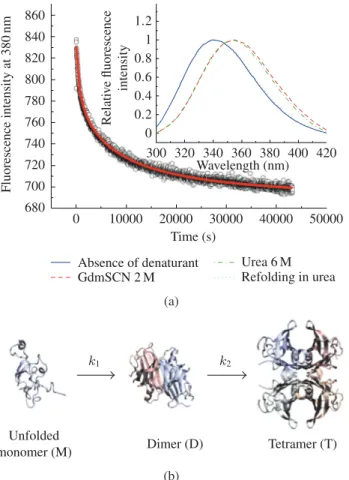
TTR unfolding was chemically induced in two steps as reported in the Experimental Procedures, because tetrameric TTR is highly stable and difficult to denature even in high urea concentrations [15]. The intrinsic fluorescence spectra of TTR (graph inset in Figure 1(a)) show a red shift in the emission maximum upon TTR unfolding, as a consequence of a more polar environment around the tryptophan residues due to higher solvent exposure in the denatured state [10]. Moreover, the fluorescence spectra of denatured TTR in 2 M GdmSCN and after dialysis against 6 M urea overlap, which demonstrates that urea is able to maintain the GdmSCN-induced denatured state of TTR. The fluorescence spectra (graph inset inFigure 1(a)) also show that the refolded species have emission spectra very similar to those of the native tetrameric TTR, with emission maxima of approximately 340 nm, indicating that protein refolding was achieved upon urea dilution. The refolded species of WT- and V30M-TTR were also characterized by size exclusion chromatography and thyroxine binding assays (data not shown), demonstrating that both TTR variants refold to their native tetrameric forms with a very high yield.
3.2. Transthyretin Refolding Mechanism
The refolding kinetic mechanisms of WT- and V30M-TTR were investigated following the changes in intrinsic tryptophan fluorescence at 380 nm, and using different urea and protein concentrations. Figure 1(a)shows an example of the fluorescence intensity changes accompanying TTR refolding as a function of time. Several kinetic models were tested, and the simplest refolding mechanism that better describes the folding and assembly of the TTR tetramers involves three states and one dimeric folding intermediate (Figure 1(b)). The dimeric nature of this folding intermediate may be postulated due to the observed dependence of both rate constants (k1and k2) on protein concentration, which indicates the
presence of two oligomerization steps. Thus, the simplest best-fitting mechanism to our kinetic data is a folding and assembly mechanism of the type monomer-dimer-tetramer (MDT), and the rate equations associated with this mechanism are listed below:
d[M] dt = −2k1[M] 2 , d[D] dt = k1[M] 2− 2k 2[D]2, d[T] dt = k2[D] 2 . (3.1)
860 840 820 800 780 760 740 720 700 680 0 10000 20000 30000 40000 50000 Fluorescence intensity at 380 nm Time (s) 1.2 1 0.8 0.6 0.4 0.2 0 300 320 340 360 380 400 420 Wavelength (nm) Absence of denaturant GdmSCN 2 M Urea 6 M Refolding in urea Relati v e fluorescence intensity (a) k1 k2 Unfolded
monomer (M) Dimer (D) Tetramer (T)
(b)
Figure 1: (a) Example of a refolding kinetics experiment for WT-TTR monitored by the variation over
time of the intrinsic fluorescence emission at 380 nm, pH 7.0, and 25◦C. The curve (solid line) is the fit
to the experimental data points using the simplest, best-fitting refolding model. The graph inset shows the spectral differences reflecting the conformational changes of TTR upon unfolding and refolding. (b) Schematic view of the TTR refolding mechanism.
Wiseman and collaborators performed refolding studies with WT-TTR, monitored by a small molecule binding assay, using protein concentrations in the range of 0.72 to 36 µM (in monomers), and, in order to globally fit the data for such a wide range of protein concentrations, the authors proposed an atypical monomer-dimer-trimer-tetramer (MDRT) assembly mechanism [16]. However, they suggested that, at lower protein concentrations, a simpler mechanism like the conventional MDT is bound to occur. Our kinetic data, at lower TTR concentrations and using a more direct monitoring method, fits best the MDT mechanism, in accordance to what has been postulated for various homotetrameric proteins [17].
3.3. Refolding Rates of the Amyloidogenic Mutant Protein V30M-TTR
The refolding mechanism of WT- and V30M-TTR, at low protein concentrations (1 µM to 0.1 µM), involves a single oligomeric intermediate, dimeric in nature. Both TTR variants refold through this MDT
Table 1: Apparent refolding kinetic constants of WT- and V30M-TTR at a protein concentration of
1 µM in the absence of denaturant, obtained by extrapolation of the refolding rates at different urea concentrations, using the equation:ln k = ln k0+ m[urea].
k01 (M−1s−1) k20 (M−1s−1)
WT-TTR 2.2× 10⁶ 8.1× 10³
V30M-TTR 1.8× 10³ 1.9× 10²
mechanism with a high yield. However in the case of V30M-TTR the folding and assembly process is much slower.Table 1shows the apparent kinetic constants k1and k2, at low protein concentration and
extrapolated to the absence of denaturant. Comparison of the individual kinetic constants for V30M-TTR with those obtained for WT-V30M-TTR reveals a noteworthy difference, with the lower rate constants corresponding to the amyloidogenic V30M-TTR. These lower rate constants, in particular k1, may
reflect the reduced conformational stability of the V30M-TTR monomers compared to WT-TTR [11].
4. Conclusions
In conclusion, we showed that the amyloidogenic variant V30M-TTR has much longer refolding times than WT-TTR, and this might be of critical importance to explain the increased amyloidogenicity of this variant. In vivo, the slower rate at which refolding and assembly of the native tetrameric form of the amyloidogenic variant V30M-TTR occurs may facilitate the accumulation of monomers of this variant in the extracellular environment, which could result in a higher susceptibility to aggregation and consequently amyloid formation, instead of refolding to the native tetramer.
Acknowledgments
The authors acknowledge the support of the “Fundac¸˜ao para a Ciˆencia e Tecnologia,” Portugal, through Grant PTDC/BIA-PRO/72838/2006 (to R. M. M. Brito) and doctoral fellowship SFRH/BD/43896/2008 (to C. S. H. Jesus). The authors also thank Elsa S. Henriques for critically reading the paper and Cˆandida G. Silva for advice on integration of differential equations.
References
[1] S. Ohnishi and K. Takano, “Amyloid fibrils from the viewpoint of protein folding,” Cellular and
Molecular Life Sciences, vol. 61, no. 5, pp. 511–524, 2004.
[2] R. M. M. Brito, A. M. Damas, and M. J. M Saraiva, “Amyloid formation by transthyretin: from protein stability to protein aggregation,” Current Medicinal Chemistry—Immunology, Endocrine
& Metabolic Agents, vol. 3, no. 4, pp. 349–360, 2003.
[3] D. R. Soprano, J. Herbert, K. J. Soprano, E. A. Schon, and D. S Goodman, “Demonstration of transthyretin mRNA in the brain and other extrahepatic tissues in the rat,” The Journal of Biological
Chemistry, vol. 260, no. 21, pp. 11793–11798, 1985.
[4] G. T. Vatassery, H. T. Quach, W. E. Smith, B. A. Benson, and J. H. Eckfeldt, “A sensitive assay of transthyretin (prealbumin) in human cerebrospinal fluid in nanogram amounts by ELISA,” Clinica
[5] J. A. Hamilton, L. K. Steinrauf, B. C. Braden et al., “The X-ray crystal structure refinements of normal human transthyretin and the amyloidogenic Val-30→ Met variant to 1.7- ˚A resolution,” The
Journal of Biological Chemistry, vol. 268, no. 4, pp. 2416–2424, 1993.
[6] C. J. Terry, A. M. Damas, P. Oliveira et al., “Structure of Met30 variant of transthyretin and its amyloidogenic implications,” The EMBO Journal, vol. 12, no. 2, pp. 735–741, 1993.
[7] V. L. Shnyrov, E. Villar, G. G. Zhadan et al., “Comparative calorimetric study of non-amyloi-dogenic and amyloinon-amyloi-dogenic variants of the homotetrameric protein transthyretin,” Biophysical
Chemistry, vol. 88, no. 1–3, pp. 61–67, 2000.
[8] P. Hammarstr¨om, X. Jiang, A. R. Hurshman, E. T. Powers, and J. W. Kelly, “Sequence-dependent denaturation energetics: a major determinant in amyloid disease diversity,” Proceedings of the
National Academy of Sciences of the United States of America, vol. 99, supplement 4, pp. 16427–
16432, 2002.
[9] A. R. H. Babbes, E. T. Powers, and J. W. Kelly, “Quantification of the thermodynamically linked quaternary and tertiary structural stabilities of transthyretin and its disease-associated variants: the relationship between stability and amyloidosis,” Biochemistry, vol. 47, no. 26, pp. 6969–6984, 2008.
[10] A. Quintas, M. J. M. Saraiva, and R. M. M. Brito, “The tetrameric protein transthyretin dissociates to a non-native monomer in solution. A novel model for amyloidogenesis,” The Journal of
Biological Chemistry, vol. 274, no. 46, pp. 32943–32949, 1999.
[11] A. Quintas, D. C. Vaz, I. Cardoso, M. J. M. Saraiva, and R. M. M. Brito, “Tetramer dissociation and monomer partial unfolding precedes protofibril formation in amyloidogenic transthyretin variants,”
The Journal of Biological Chemistry, vol. 276, no. 29, pp. 27207–27213, 2001.
[12] H. Furuya, M. J. Saraiva, M. A. Gawinowicz et al., “Production of recombinant human transthyretin with biological activities toward the understanding of the molecular basis of familial amyloidotic polyneuropathy (FAP),” Biochemistry, vol. 30, no. 9, pp. 2415–2421, 1991.
[13] M. R. Almeida, A. M. Damas, M. C. Lans, A. Brouwer, and M. J. M Saraiva, “Thyroxine binding to transthyretin Met 119: comparative studies of different heterozygotic carriers and structural analysis,” Endocrine, vol. 6, no. 3, pp. 309–315, 1997.
[14] A. Raz and D. S. Goodman, “The interaction of thyroxine with human plasma prealbumin and with the prealbumin-retinol-binding protein complex,” The Journal of Biological Chemistry, vol. 244, pp. 3230–3237, 1969.
[15] P. Hammarstr¨om, X. Jiang, S. Deechongkit, and J. W. Kelly, “Anion shielding of electrostatic repulsions in transthyretin modulates stability and amyloidosis: insight into the chaotrope unfolding dichotomy,” Biochemistry, vol. 40, no. 38, pp. 11453–11459, 2001.
[16] R. L. Wiseman, E. T. Powers, and J. W. Kelly, “Partitioning conformational intermediates between competing refolding and aggregation pathways: insights into transthyretin amyloid disease,”
Biochemistry, vol. 44, no. 50, pp. 16612–16623, 2005.
[17] E. T. Powers and D. L. Powers, “A perspective on mechanisms of protein tetramer formation,”
Submit your manuscripts at
http://www.hindawi.com
Hindawi Publishing Corporation
http://www.hindawi.com Volume 2014
Inorganic Chemistry International Journal of
Hindawi Publishing Corporation
http://www.hindawi.com Volume 2014
Photoenergy
Hindawi Publishing Corporation
http://www.hindawi.com Volume 2014
Carbohydrate Chemistry
International Journal of
Hindawi Publishing Corporation
http://www.hindawi.com Volume 2014 Journal of
Chemistry
Hindawi Publishing Corporation
http://www.hindawi.com Volume 2014
Physical Chemistry
Hindawi Publishing Corporation http://www.hindawi.com Analytical Methods in Chemistry Journal of Volume 2014 Bioinorganic Chemistry and Applications Hindawi Publishing Corporation
http://www.hindawi.com Volume 2014
Spectroscopy
International Journal of Hindawi Publishing Corporationhttp://www.hindawi.com Volume 2014 The Scientific
World Journal
Hindawi Publishing Corporation
http://www.hindawi.com Volume 2014 Medicinal Chemistry
Hindawi Publishing Corporation
http://www.hindawi.com Volume 2014
Chromatography Research International Hindawi Publishing Corporation
http://www.hindawi.com Volume 2014
Applied ChemistryJournal of
Hindawi Publishing Corporation
http://www.hindawi.com Volume 2014
Hindawi Publishing Corporation
http://www.hindawi.com Volume 2014
Theoretical Chemistry
Journal of
Hindawi Publishing Corporation
http://www.hindawi.com Volume 2014
Journal of
Spectroscopy
Analytical Chemistry
Hindawi Publishing Corporation
http://www.hindawi.com Volume 2014
Journal of Hindawi Publishing Corporation
http://www.hindawi.com Volume 2014 Quantum Chemistry Hindawi Publishing Corporation
http://www.hindawi.com Volume 2014 International
Electrochemistry
International Journal ofHindawi Publishing Corporation
http://www.hindawi.com Volume 2014
Hindawi Publishing Corporation
http://www.hindawi.com Volume 2014