BIOCOMPATIBLE AND BIODEGRADABLE FUNCTIONAL COATINGS WITH
NATURAL OCCURRING MATERIALS FOR THE CORROSION PROTECTION
OF MG ALLOYS
I. Sousa1 , C.S. Neves1, S. Fraga2,3, J.P. Teixeira2,3, N. Scharnagl4, M.L. Zheludkevich4, M.G.S. Ferreira1 and J.Tedim1
1. CICECO/DEMaC, Universidade de Aveiro, 3810-193 Aveiro, email: isabel.sa.correia@ua.pt. 2. EPIUnit Instituto de Saúde Pública, Universidade do Porto, 4050-091 Porto, Portugal.
3. Departamento de Saúde Ambiental, Instituto Nacional de Saúde Doutor Ricardo Jorge, 4000-053
Porto, Portugal.
4. Helholtz-Zentrum Geesthacht Zentrum für Material-und Küstenforschung, Max-Planck Straße 1, 21502 Geesthacht, Germany.
ABSTRACT
Magnesium alloys are amidst the most innovative materials for biomedical applications, as they show a set of unique properties, namely appropriate mechanical properties and biodegradability, when compared to other alloys. Although these properties make them suitable for medical implants, the main challenge is the uncontrolled corrosion. Mg degradation is fast, inhomogeneous, localized and often accompanied by hydrogen formation which can lead to complications in vivo. Here, we propose the development of a functional coating, containing natural-based capsules for the controlled release of biocompatible corrosion inhibitors and well known pharmaceutical agents. Empty and loaded capsules toxicity tests were performed as a first step for materials selection. Subsequently, they were incorporated into polyetherimide (PEI) coatings and tested using electrochemical impedance spectroscopy (EIS) under aggressive conditions. The obtained results showed a successful synthesis of natural-based microcapsules, constituting a fast, simple and environmentally friendly method. Additionally, the high cell proliferation observed in the presence of the aforementioned materials demonstrates their low toxicity. Preliminary results carried out with capsule-modified coatings show that the incorporation of Ca2+-loaded gelatin capsules in PEI coatings leads to barrier and active corrosion protection properties improvement and that anti-inflammatory agent ibuprofen may have a role in active corrosion protection as well.
Keywords: encapsulation; natural-based materials; active species; corrosion control. 1 INTRODUCTION
Metals have been playing an essential role in several biomedical devices, being more suitable for load bearing applications than ceramic or polymeric materials [1-3]. However, the possible release of toxic products through corrosion processes, can lead to complications that may ultimately require the removal of the implant [4]. An adequate solution for this problem is the use of biodegradable implants but, their insufficient mechanical properties for bone implant application and non-suitable degradation rates [1, 5] are a disadvantage. Magnesium alloys seem to combine the best features of both ceramic and polymeric materials, with their light-weight, low density and mechanical properties, along with biocompatibility and in vivo importance, due to Mg2+ fundamental role in several metabolic processes, making them more suitable for medical implants. Nevertheless, Mg alloys are prone to uncontrolled corrosion which hinders their application due to H2 evolution and pH increase [6]. Therefore, the use of Mg alloys requires a control over its degradation rate in order to allow the body to gradually deal with products resulting from corrosion [2]. Protective coatings have been used in different applications in corrosion engineering. Often, corrosion inhibitors can be added to the coating matrix to impart active protection, but leaching of inhibitors and negative reaction between the coating matrix and these species are potential limitations. This problem has been tackled by the encapsulation of inhibitors in inert capsules before their introduction into coating formulations [8]. Although successful in different areas such as aeronautical industry, this strategy has never been truly implemented in the control of degradation processes of biomaterials by corrosion, with only a few reports of synthetic polymers [5, 9] used as biodegradable coatings for Mg-based alloys being available. The main innovation of this work is the attempt to control the rate of degradation by corrosion of Mg-based implants by controlled release of corrosion inhibiting species and other relevant molecules (for control of infectious responses) from bioabsorbable polymer coatings loaded with functional capsules.
2 MATERIALS
Calcium nitrate tetrahydrate (99%), ibuprofen sodium salt (>=98%), coumarin (99%), gelatin from bovine skin (gel strength 225 bloom type B), D-glucose (ACS reagent), sodium chloride (ACS reagent) and N-N’-dimethylacetamide (99%) were purchased from Sigma-Aldrich and used without further purification. Polyetherimide ULTEM 1000® was purchased from General Electric and AZ31 from Magnesium Elektron (Manchester, UK).
2.1 Synthesis of gelatin capsules
Gelatin capsules were produced by a thermal gelation method using a published procedure [10] with minor modifications. Briefly, 0.4 g of D-glucose were added to 20 mL of a 20% w/v aqueous gelatin solution preheated in a water bath at ~50 ºC and added dropwise to 200 mL of sunflower oil containing 1% v/v of Span® 85, while stirring at 80 ºC, for 10 min. Afterwards, the w/o emulsion was moved into an iced water bath and stirring was continued until ~15 ºC were reached. Then, 150 mL of acetone were added to promote dehydration and flocculation of the microspheres. After 10 min of stirring, the microspheres were filtered through a sintered glass filter and washed with 250 mL of acetone to remove any traces of oil. The spheres were then dried under vacuum to remove the remaining acetone. Glucose crosslinked gelatin microspheres were prepared in deionized water (empty) and in Ca2+, Ibuprofen and Coumarin aqueous solutions.
2.2 Capsule characterization
Small samples of the synthesized gelatin capsules were placed on double sided carbon tape and sputtered with Au/Pd for 3 min. Particle morphology was characterized by scanning electron microscopy (SEM) (Hitachi S-4100 system with electron beam energy of 25 keV). Image J software (freeware) was used to analyze the obtained data. FTIR analyses of empty and loaded gelatin capsules were carried out on a Bruker Tensor 27 Spectrometer coupled with an ATR device, 128 scans and 4 cm-1 resolution, in the range from 4000-400 cm-1.
2.3 Toxicity assessment
Cytotoxicity testing of coating components was performed in a L929 mouse connective tissue cell line according to ISO 10993-5:2009. The materials tested included corrosion inhibitors (calcium nitrate tetrahydrate), D-glucose crosslinked gelatin capsules either empty or loaded with Ca2+. The test items were freshly prepared in FBS-free cell culture medium (MEM supplemented with 2 mM L-glutamine, 100 units/mL of penicillin and 100 µg/mL of streptomycin). The cells were exposed to the test items for 24 h and cytotoxicity evaluated using two well-established endpoints: LDH release, an indicator of plasma membrane integrity and WST-1 reduction, to assess cell proliferation and metabolic activity.
2.4 Polyetherimide coatings
Prior to coating, the metal was pre-treated according to a published procedure [11]. Coated AZ31 Mg alloys with 10% PEI in dimethylacetamide were prepared by manual dip coating. Pre-treated substrates were immersed in PEI coating formulation for 60 s, removed and left to dry for ~3 min under room conditions (temperature, moisture). After this resting period, the substrates were immersed once again in PEI polymer solution for 10 s and left to dry overnight [12].
2.5 Electrochemical impedance studies
EIS measurements were carried out in a three-electrode cell with a SCE reference electrode, platinum foil counter electrode and the coated magnesium alloy sample as the working electrode (exposed area of ~3.30 cm2). The cell was placed in a Faraday cage to avoid the interference of external electromagnetic fields. Measurements were performed using a Gamry FAS2 Femtostat with PCI4 Controller. The selected frequency range was from 105 to 10-2 Hz, with a 10 mV of sinusoidal perturbation with 7 points per frequency decade. All spectra were recorded at open circuit potential.
3 RESULTS 3.1 Synthesis
Gelatin capsules were successfully produced by a thermal gelation method, as described previously by Hiwale et al, through a water in oil emulsion with Span®85 as surfactant and D-glucose as a crosslinking agent. Empty and loaded capsules presented gelatins’ characteristic yellowish colour and were obtained as a loose powder.
3.2 Characterization
Scanning Electron Microscopy
All capsules present a spherical morphology and heterogenous size distribution. As it is also possible to observe, gelatin capsules with anti-inflammatory agents such as ibuprofen have a slightly distorted morphology but nevertheless are considered spherical. Gelatin capsules size range from ~9 to 26 µm.
Figure 1. (left to right) Empty gelatin capsules crosslinked with glucose (Glu@gel), calcium-loaded
and ibuprofen gelatin capsules crosslinked with glucose (Ca@gel and Ibu@gel, respectively). Fourier Transformed Infrared Spectroscopy
The process of gelatin crosslinking may occur by one or several reactions (Figure 2). One possible mechanism is the reaction of the aldehyde group of the sugar with free amino groups of the gelatin molecule, resulting in the formation of an aminoglycoside which, in turn, can further react with another amine group from gelatin leading to a crosslinked structure [13].
Figure 2. Schematic representation of the D-Glucose mediated gelatin crosslinking reaction mechanism (adapted from [8]).
In all of the FTIR spectra presented (Figure 3), the characteristic absorption peaks of the protein backbone (NH and CO stretching) are observed.
The NH stretching referred to as amide A peak is observed at ∼3300 cm-1 while CO stretching peaks referred to as amides I, II and III were found at ∼1650, 1550, and 1240 cm-1, respectively [10, 13]. Interestingly, the intensity of the CO stretching peaks was the highest for empty glucose crosslinked capsules and a decrease in intensity is observed when encapsulation with different compounds is performed, which suggests that functional groups from the active species may compete, at some point, with glucose to form bonds with gelatin functional groups. The presence of asymmetric and symmetric CH2str vibrations at ~2924 and 2853 cm-1, respectively, suggests the presence of glucose crosslinked to the gelatin aminoglycoside moieties. The presence of a band at ~1740 cm-1 (identified by the arrow) is assigned to C=Ostr (enol form) vibrations and is attributed to D-glucose HC=O moieties (keto-enol tautomerization and aminoglycoside formation), which further confirms that crosslinking occurred [14].
3.3 Toxicity Assessment
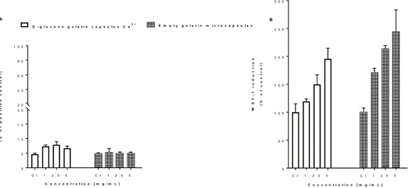
LDH and WST-1 reduction assays were performed for the actives species under study (results not shown) as well as for empty and calcium-loaded gelatin capsules. The obtained results did not show significant differences in LDH release, when compared to the control and no WST-1 decrease, which shows that Ca2+ is non/low toxic. In what concerns the anti-inflammatory agents tested, coumarin did not induce toxicity in L929 as assessed by both LDH and WST-1 assays. However, ibuprofen sodium increased LDH release and decreased WST-1 reduction in a concentration-dependent manner, being this effect more evident for the highest concentrations tested (950 and 1900 µM).
Figure 4. Effect of different concentrations of empty and Ca2+-loaded capsules on L929 cell viability as assessed by the LDH release (A) and WST-1 reduction (B) assays. Cells were exposed
to different concentrations (1-5 mg/mL) of the test agents for 24 h. Results are presented as mean ± SD of one experiment (n = 3 replicates per group).
As it can be seen, in Figure 4A, no changes in LDH release are observed for empty gelatin capsules but a concentration-dependent increase in WST-1 reduction (Figure 4B) was observed in the gelatin capsules-exposed cells, which is most likely due to the D-glucose content of the capsules and its consequent proliferative effect. The same effect can be observed for the calcium-loaded capsules, confirming the low toxicity of the gelatin capsules.
3.4 EIS Studies of PEI Coatings Containing Capsules – Coating Performance
The degradation of AZ31 in NaCl without coating (results not shown) can already be seen at early stages immersion by the low impedance magnitude values obtained (<0.5 kΩ cm2). One time constant at ~100 Hz is observed and attributed to electrochemical processes occurring at the metal interface. When a PEI coating is applied, the initial impedance magnitude goes up two orders of magnitude and a second time constant at high frequencies is detected (around 105 Hz), associated with the presence of this organic coating layer. However, with increase in immersion time, the porous nature of PEI leads to a fast decrease in the coating barrier properties. The addition of gelatin microcapsules loaded with different species causes an initial decrease in the impedance values across the whole frequency range (due to their hydrophilic nature or some sort of barrier weakening that facilitates conducting pathways for ingress of electrolyte). Nevertheless, over
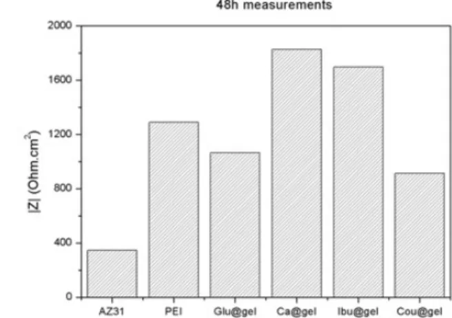
time, some systems seem to perform better than the bare PEI coating. Figure 5 depicts the EIS spectra for all the systems after 48h of immersion (left) and magnitude of impedance at 0.2 Hz (right).
Figure 5. (left) Bode plots of EIS (|Z| (Ohm.cm2) spectra and (right) impedance magnitude values at 0.2 Hz for all coatings systems studied.
It is possible to observe that coatings with Ca- and ibuprofen-loaded gelatin microcapsules are the best systems. While for Ca-containing gelatin the coating performs better than bare PEI across the whole frequency range, the ibuprofen-gelatin microcapsules leads to slightly lower barrier properties, though the second time-constant at intermediate frequencies shows higher impedance magnitude. This may be related to some sort of active effect of the capsules directly in the corrosion processes ongoing at the interface.
Visually (photos not shown) the different coating systems show some changes due to immersion and corrosion processes. The degradation of the coating layer leads, in some cases, to a detachment of the coating from the surface of the substrate.
4 CONCLUSIONS
Gelatin capsules were synthesized successfully by a fast, simple and environmentally-friendly approach. They were characterized physicochemically and although positive and promising results have been achieved, a more effective crosslinking of the gelatin backbone with D-glucose must be investigated, as well as a higher encapsulation/immobilization efficiency should be achieved in order to increase the amount of active species in the capsules.
Regarding the toxicity results, the selected species (calcium, ibuprofen and coumarin) as well as the respective gelatin capsules can be assumed as low toxic species. The toxicity associated with ibuprofen for higher concentrations is somehow expected taking into account its biological role. The dose-effect achieved within the final coatings will have to be determined in order to infer on possible toxic effects.
In what concerns PEI coatings, they were prepared, tested with gelatin microcapsules loaded with different species and applied on AZ31 Mg alloys. PEI coatings with Ca2+-loaded gelatin microcapsules showed higher impedance magnitude values at both high and low frequencies, when compared to PEI and PEI loaded with empty gelatin capsules. Although these are preliminary results it is possible to observe an improvement of barrier and active corrosion protection properties, respectively. Additionally, ibuprofen seems to play some role in terms of active corrosion protection as well, which is a promising double-feature for the ibuprofen gelatin capsules since they might act both as anti-inflammatory agents and corrosion inhibiting species for the underlying metallic substrate.
ACKNOWLEDGEMENTS
This work was financed by Portugal 2020 through European Regional Development Fund (ERDF) in the frame of Operational Competitiveness and Internationalization Programme (POCI), in the scope of the project MAGICOAT POCI-01-0145-FEDER-016597 / PTDC/CTM-BIO/2170/2014 and in the scope of the project CICECO- Aveiro Institute of Materials, POCI-01-0145-FEDER-007679 (FCT Ref. UID/CTM/50011/2013), and co-financed by national funds through the FCT/MEC.
[1] N. Li and Y. Zheng, Novel Magnesium Alloys Developed for Biomedical Application: A Review. Journal of Materials Science & Technology, Vol. 29 (6), pp. 489, 2013.
[2] M.P. Staiger, et al., Magnesium and its alloys as orthopedic biomaterials: A review. Biomaterials, Vol. 27 (9): 1728, 2006.
[3] H. Hornberger, et al, Biomedical coatings on magnesium alloys - A review, Acta Biomaterialia, Vol. 8, pp. 2442-2455, 2012.
[4] M.I. Sabir et al. A review on biodegradable polymeric materials for bone tissue engineering applications, Journal of Materials Science, Vol. 44, pp. 5713-5724, 2009.
[5] J.E. Gray and B. Luan, Protective coatings on magnesium and its alloys - a critical review, Journal of Alloys and Compounds, Vol. 336, pp.88-113, 2002.
[6] S. Shadanbaz and G.J. Dias, Calcium phosphate coatings on magnesium alloys for biomedical applications: A review. Acta Biomaterialia, Vol. 8 (1), pp. 20, 2012.
[7] G. Song, Control of biodegradation of biocompatible magnesium alloys. Corrosion Science, Vol. 49 (4): p. 1696, 2007.
[8] K.A. Yasakau et al., Handbook of Smart Coatings for Materials Protection, Woodhead Publishing, 2014. [9] N. Scharnagl and C. Blawert, Surface Coating and Modification of Metallic Biomaterials, Woodhead Publishing, 2015.
[10] P. Hiwale et al., In Vitro Release of Lysozyme from Gelatin Microspheres: Effect of Cross-linking Agents and Thermoreversible Gel as Suspending Medium, Biomacromolecules, Vol.12, pp. 3186–93, 2011. [11] U.C. Nwaogu et al., Effects of organic acid pickling on the corrosion resistance of magnesium alloy AZ31 sheet, Corrosion Science, Vol. 52, pp. 2143-2154, 2010.
[12] N. Scharnagl et al., Corrosion protection of magnesium alloy AZ31 by coating with poly(ether imides) (PEI)Surface & Coatings Technology, Vol. 203, pp. 1423–1428, 2009.
[13] R. Cortesi et al., Sugar cross-linked gelatin for controlled release: microspheres and disks, Biomaterials, Vol. 19, pp. 1641—1649, 1998.
![Figure 2. Schematic representation of the D-Glucose mediated gelatin crosslinking reaction mechanism (adapted from [8])](https://thumb-eu.123doks.com/thumbv2/123dok_br/15859657.1086695/3.892.63.851.187.376/figure-schematic-representation-glucose-mediated-crosslinking-reaction-mechanism.webp)