Angle Class I malocclusion treated with
extraction of first permanent molars*
Ivan Tadeu Pinheiro da Silva**
Angle Class I malocclusion is characterized by normal anteroposterior molar relationship, which may or may not be accompanied by skeletal changes—in the vertical or transverse planes—or dental changes. Bimaxillary dental protrusion, characterized by pronounced labial inclination of maxillary and mandibular incisors combined with excessive overjet, expose patients to dental trauma and compromise aesthetics. In deciding which teeth to extract for Class I correction the first or second premolars are usually selected due to their location in the dental arch. However, the extraction of a first permanent molar compro-mised by caries or extensive restoration may be an alternative that ensures the preservation of a healthy tooth instead of one that has already been manipulated. This case, treated in an unusual manner by the extraction of four first permanent molars, was presented to the Brazilian Board of Orthodontics and Dentofacial Orthopedics (BBO) as representative of category 2, as part of the requirements for obtaining the BBO diplomate title.
Abstract
Keywords: Angle Class I malocclusion. Tooth extraction. Corrective Orthodontics.
** Specialist in Pediatric Dentistry, EAP - Brazilian Dental Association, Ponta Grossa/PR. Specialist in Orthodontics and Facial Orthopedics, EAP - Brazilian Dental As-sociation, Curitiba/PR. Diplomate of the Brazilian Board of Orthodontics and Dentofacial Orthopedics.
* Case Report, category 2, approved by the Brazilian Board of Orthodontics and Dentofacial Orthopedics.
HISTORY AND ETIOLOGY
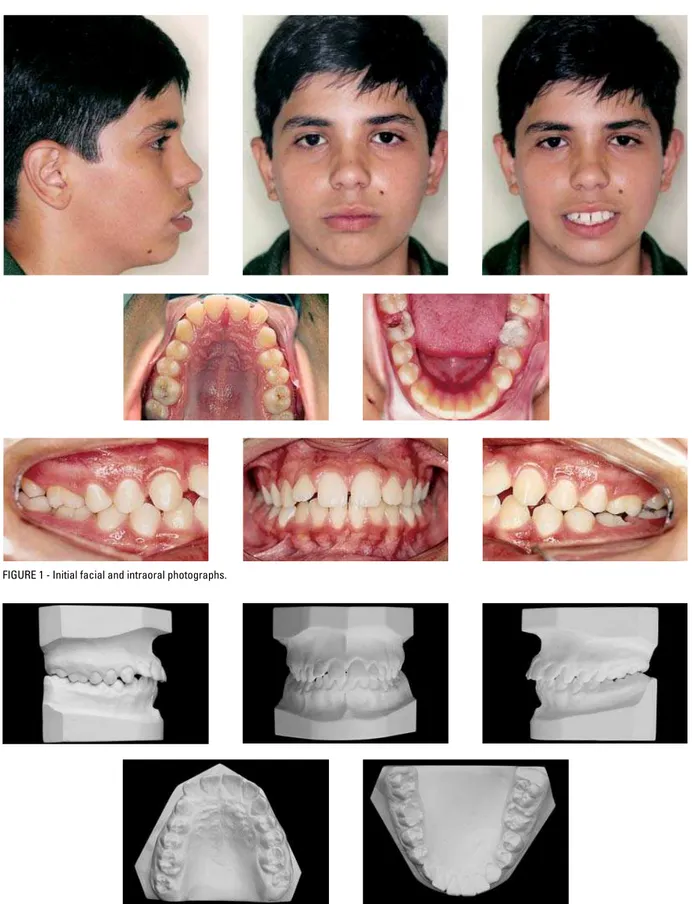
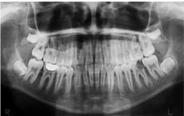
The patient, a Caucasian male, 13 years and four months old, presented for initial examination with the chief complaint of maxillary incisor pro-trusion. He was in good general health and report-ed a mreport-edical history of bronchitis and allergy. He had no sucking or postural habits and had normal swallowing and speech. Regarding oral health, his mandibular first molar crowns were significantly destroyed. The mandibular second molars and maxillary first molars showed carious lesions on the occlusal surface and the presence of dental calculi and gingivitis was observed.
DIAGNOSIS
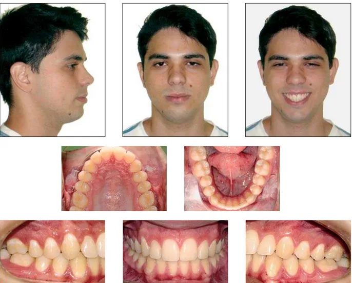
The patient’s facial aesthetics was compromised by a convex profile, lip protrusion, lack of passive lip seal and lower lip eversion. He presented a me-sofacial pattern, Class I molar relationship, slightly altered canine relationship with a Class II tendency, a 6 mm overjet, 4 mm overbite, severely projected maxillary incisors, a 1.4 mm Bolton discrepancy with excess in the mandibular anterior teeth and developing third molars (Figs 1, 2 and 3).
posi-FIGURE 1 - Initial facial and intraoral photographs.
A B FIGURE 3 - Initial panoramic radiograph.
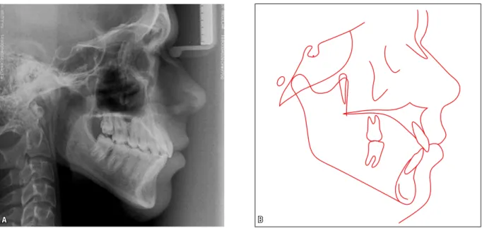
FIGURE 4 - Initial lateral cephalometric radiograph (A) and cephalometric tracing (B).
tioned mandible relatively to the cranial base (SNB=80°). He presented a divergent growth pattern (SN-GoGN = 38.5°) and a marked facial convexity (Convex. angle = 9.5º). His skeletal and cephalometric features can be evaluated in Figure 4 and Table 1.
TREATMENT GOALS
Treatment goals included improvement of fa-cial aesthetics, obtaining a balanced labial mus-culature and a stable occlusion from the func-tional point of view, maintaining the existing
relationship between molars, improvement in the relationship between canines, correction of maxil-lary incisor protrusion, reduction of overbite and overjet, maintaining healthy teeth and eliminating any teeth with destroyed crowns.
TREATMENT PLAN
The treatment plan provided for extraction of the mandibular first molars given their crown de-struction, and need of endodontic treatment and prosthetic rehabilitation, which would be conve-nient to avoid in such a young patient. In order to maintain mechanics symmetry while not depend-ing heavily on patient compliance, maxillary first molar extractions were also planned.
The planned retention consisted of a remov-able maxillary retainer and an canine to canine bonded lingual retainer in the mandibular arch.
TREATMENT PROGRESS
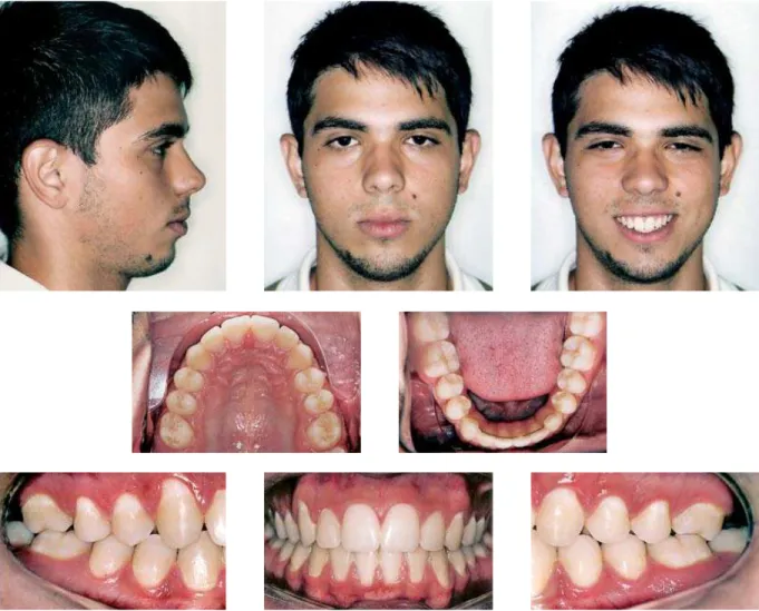
FIGURE 5 - Final facial and intraoral photographs.
electrical neuromuscular stimulation (TENS), confirming the coincidence of RC and MHI.10
After finishing the orthodontic treatment and ascertaining that all planned objectives had been achieved, the fixed appliances were removed. A removable maxillary retainer was used for reten-tion, to be used 24/7 in the first year, only nights in the second year, and three nights a week after that period, for an unlimited period of time. In the mandibular arch, a 0.036-in stainless steel lingual retainer was bonded to the canines.
Considering that orthodontic treatment was performed with extraction of the first perma-installed on both arches. Leveling and alignment
a pleasing smile line (Figs 5, 6, 7, 8, 9 and 10). Therefore, the profile improved substantially, in contrast to what Stalpers et al6 reported, describ-ing that orthodontic treatment involvdescrib-ing extrac-tion of maxillary first permanent molars exerts a minor effect on profile soft tissue.
The maxilla and mandible maintained their anteroposterior relationship, with the ANB angle remaining at 4º. Maxillary dentition improved with the correction of incisor protrusion and reduction of overjet. As can be seen in Table 1, the angular value of 1-NA decreased from 32º to 23º and its linear value fell from 9 mm to 4 mm.
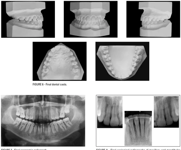
FIGURE 7 - Final panoramic radiograph. FIGURE 8 - Final periapical radiographs of maxillary and mandibular incisors.
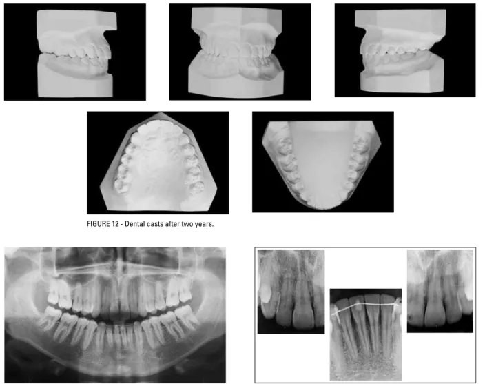
FIGURE 6 - Final dental casts.
nent molars, the space remaining for eruption of maxillary and mandibular third molars was increased, thereby reducing the likelihood of impaction.3 Thus, the patient was instructed to return periodically for monitoring third molars development and eruption.
RESULTS
A B
A B
FIGURE 9 - Final lateral cephalometric radiograph (A) and cephalometric tracing (B).
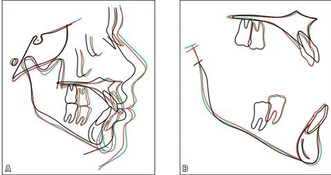
FIGURE 10 - Total (A) and partial (B) superimpositions of initial (black) and final (red) cephalometric tracings.
In the mandibular incisors a slight tipping decrease occurred, which caused a reduction in the IMPA and 1-NB angles, as well as in the 1-NB and 1-APo linear measurements. By reducing the interincisal angle and thereby decreasing the distance be-tween the S line and the upper and lower lips, the
to 36° (Table 1, Figs 11 and 12). The maxillary second molars, occupying the position of the first molars were not fully upright, but this inclination secured a greater settlement in the mesial mar-ginal ridge of the mandibular third molar, occu-pying the second molar position, as described by Andrews,1 and thus improving stability. The max-illary third molars, in the position of the second molars, were not placed in occlusion with their antagonists to avoid extending treatment time.
It should be stressed that the teeth used in the cephalometric tracings (initial, final and two years
after treatment) were the second permanent lars (Figs 4, 9, 10, 15 and 16). Thus, maxillary mo-lars were moved mesially by translation (bodily movement) and maxillary incisors retracted. Man-dibular second molars were moved mesially by translation to occupy the space of the first molars, as reported by Hom and Turley7 in their analysis of the effects of space closure on the area of man-dibular first molars in adults.
The relationship between the arches was maintained at normal molar occlusion with the second molars occupying the position of the
A B
FIGURE 13 - Panoramic radiograph two years after treatment completion. FIGURE 14 - Periapical radiographs two years after treatment completion.
FIGURE 15 - Lateral cephalometric radiograph (A) and cephalometric tracing (B) two years after treatment completion.
A B
FIGURE 16 - Total (A) and partial (B) superimpositions of initial (black), final (red) and two-year posttreat-ment (green) cephalometric tracings.
MEASUREMENTS Normal A B A-B
DIFFERENCE C
Skeletal Pattern
SNA (Steiner) 82° 84° 85° 0 85°
SNB (Steiner) 80° 80° 81° 1 82°
ANB (Steiner) 2° 4° 4° 0 3°
Convexity Angle (Downs) 0° 9.5° 8° 1.5 6°
Y axis (Downs) 59° 61° 60° 1 58.5°
Facial Angle (Downs) 87° 86.5° 89° 2.5 90°
SN-GoGn (Steiner) 32° 38.5° 36° 2.5 36°
FMA (Tweed) 25° 32° 30° 2 28.5°
IMPA (Tweed) 90° 97° 94° 3 93°
Dental Pattern
–
1 - NA (degrees) (Steiner) 22° 32° 23° 9 21.5°
–
1 - NA (mm) (Steiner) 4 mm 9 mm 4 mm 6 6 mm
–
1 - NB (degrees) (Steiner) 25° 37° 33° 4 31°
–
1 - NB (mm) (Steiner) 4 mm 9 mm 6.5 mm 2.5 7 mm
– 1
1 - Interincisal angle (Downs) 130° 108° 121° 13 123.5°
–
1 - APo (mm) (Ricketts) 1 mm 6 mm 3 mm 3 2.5 mm
Pr
oi
le Upper Lip - S Line (Steiner) 0 mm 3 mm 0.5 mm 2.5 1 mm
Lower Lip - S Line (Steiner) 0 mm 7 mm 2 mm 5 3 mm
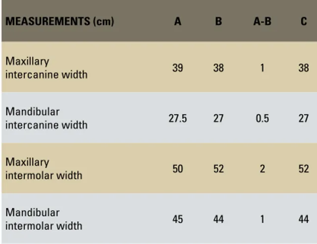
first molars and canines in normal occlusion. As a result, adequate stability would be ex-pected given the adequate intercuspation that was achieved. In Table 2 it can be observed that the intermolar and intercanine distances were maintained. Figures 11, 12, 13, 14, 15 and 16 depict that the final facial, skeletal and dental results obtained with treatment were stable two years after treatment.
FINAL CONSIDERATIONS
Angle Class I malocclusion2 is characterized by skeletal changes—in the vertical or transverse planes—, or dental changes. Bimaxillary dental protrusion, when coupled with excessive overjet, increases patient exposure to dental trauma while compromising aesthetics. When extractions are indicated the choice often falls on premolars due to their strategic position in the transition zone between the anterior and posterior segments. However, other approaches should be consid-ered, especially when the patient presents with caries, extensive restorations, periapical lesions or prostheses.4 Despite advances in prevention, first
molar loss rate is still high, affecting nearly 35% of children with mixed dentition.9 The character-istics of malocclusion in this patient, with his sig-nificant mandibular first molars coronal destruc-tion resemble those found by Normando,5 who reported an increased frequency of Class II ca-nine relationship in patients with this teeth miss-ing. Hom and Turley7 believe that space closure in the region of missing mandibular first permanent molars should be regarded as a therapeutic ap-proach. In 1899, Angle2 defined first permanent molars as “the key to normal occlusion,” consider-ing them essential for dentition stability, probably because these teeth are the first permanent teeth of the posterior segment and thus provide guid-ance for the eruption of the others.
In 1973, Jensen8 asserted that the extraction of the four first premolars followed by the ex-traction of the four third molars is equivalent to the loss of 25% of the total dental material. In his view, the latter was unnecessary since most of the space left by the third molar is not used to accommodate the remaining teeth. Moreover, the extraction of four first molars is equivalent to 12.5% of the dental material, and virtually the entire space is used.
It can therefore be concluded that this case was successful for both the patient and his le-gal guardians. Treatment goals were achieved, with the establishment of a normal occlusion in canines and second molars, in the position of the first molars. Maxillary incisors protrusion was eliminated while overjet and overbite were reduced, thereby improving facial aesthetics. Teeth with destroyed crowns were eliminated, which would otherwise require endodontics and prosthetics, and healthy teeth were pre-served. Muscle balance and functionally stable occlusion were accomplished.
MEASUREMENTS (cm) A B A-B C
Maxillary
intercanine width 39 38 1 38
Mandibular
intercanine width 27.5 27 0.5 27
Maxillary
intermolar width 50 52 2 52
Mandibular
intermolar width 45 44 1 44
1. Andrews LF. The six keys to normal occlusion. Am J Orthod. 1972 Sep;62(3):296-309.
2. Angle EH. Classiication of malocclusion. Dental Cosmos. 1899; 41(2):248-64.
3. Bayram M, Ozer M, Arici S. Effects of irst molar extraction on
third molar angulation and eruption space. Oral Surg Oral Med Oral Pathol Oral Radiol Endod. 2009 Feb;107(2):e14-20. 4. Diaz MCA, Pinzan A, Freitas MR. Extração de primeiros
molares permanentes – apresentação de um caso. Ortodontia. 1992;25(1):47-53.
5. Normando DCA. Alterações oclusais espontâneas decorrentes da perda dos primeiros molares permanentes inferiores. Rev
Dental Press Ortod Ortop Facial. 2003 maio-jun;8(3):15-23.
6. Stalpers MJ, Booij JW, Bronkhorst EM, Kuijpers-Jagtman AM,
Katsaros C. Extraction of maxillary irst permanent molars in
patients with Class II Division 1 malocclusion. Am J Orthod Dentofacial Orthop. 2007 Sep;132(3):316-23.
REFERENCES
7. Hom BM, Turley PK. The effects of space closure of the
mandibular irst molar area in adults. Am J Orthod. 1984 Jun;85(6):457-69.
8. Jensen ID. Extraction of irst molars in discrepancy cases. Am J
Orthod. 1973;64(2):115-36.
9. Silva OG Filho, Freitas SF, Cavassan AO. Oclusão: prevalência de oclusão normal e má oclusão na dentadura mista em escolares da cidade de Bauru (São Paulo). Parte I: relação sagital. Rev Odontol Univ São Paulo. 1990 abr-jun;4(2):130-7. 10. Silva ITP, Telles FS, Moro A. Diagnóstico ortodôntico em
relação cêntrica: comparação de medidas cefalométricas em relação cêntrica obtida pela “TENS” com medidas em máxima intercuspidação habitual. Rev Dental Press Ortod Ortop Facial. 2001 maio-jun;6(3):7-24.
Contact address
Ivan Tadeu Pinheiro da Silva
Rua Nove, nº 1519 – Q E 12 L10 / Setor Marista CEP: 74.150 - 130 – Goiânia / GO, Brazil E-mail: [email protected]