261
The Importance of the Medial Patellofemoral Ligament
in the Lateral Displacement and Inclination of the
Patella: A Radiographic Study in Cadavers
Edimar Fávaro1
Nilson Roberto Severino2
Tarciso Fávaro3
Arnaldo José Hernandez4
1. Medical Sciences College of the Santa Casa of São Paulo, Brazilian Society of Orthopedics and Traumatology (SBOT) and Brazilian Society of Sports Medicine. 2. Department of Orthopedics and Traumatology, Medical Sciences College of the Santa Casa of São Paulo.
3. Brazilian Society of Orthopedics and Traumatology (SBOT). 4. Department of Orthopedics and Traumatology and Medicine College of the University of São Paulo.
Mailing address:
Av. Santa Catarina, 187 – 04635-000 São Paulo, SP, Brasil
ABSTRACT
Objective: Acute patellar luxation is a complex disease that mainly affects young patients. Its physio-pathology is little known and the understanding of it and its therapeutic conduct are controversial. The medial patellofemoral ligament (MPFL) is the main static stabilizer for preventing lateral displacement of the patella. In order to assess the stability of the patellofemoral joint, the authors radiographically assessed the presence, or absence of lateral displacement and inclination of the patella before and after the MPFL section in the knees of cadavers. Methods: Thirty knees of cadavers were radiographed on the axial incidence of the patella by means of the technique described by Merchant before and after MPFL section. The Merchant congruence angle and Laurin lateral patellofemoral angle were measured. Results: The medial patellofemoral ligament presented mean length of 4.8cm, and width of 1.6 cm. In six anatomical pieces there was no change in the Laurin lateral patellofemoral angle (20%), in three anatomical pieces the change was one degree (10%), in 20 (67%) two degrees and in one anatomical piece it was four degrees (3%). Changes occurred between zero and two degrees in 97% of the knees of cadavers. In six anatomical pieces there was no change in the Merchant congruence angle (17%); in six anatomical pieces the change was one degree (20%), in 17 (57%) two degrees, in one anatomical piece it was three degrees (3%) and in one it was six degrees (3%). These changes occurred between zero and two degrees in 93% of the knees of cadavers. Conclusion: Analysis of the results obtained in this study allowed us to conclude that the medial patellofemoral ligament is important in the lateral inclination and displacement of the patella with knee flexed at 45º.
Keywords: medial patellofemoral ligament, radiography, displacement, study in cadaver, patella. LOCOMOTOR APPARATUS IN
EXERCISE AND SPORTS
ORIGINAL ARTICLE
INTRODUCTION
The study of the disorders between the patella and femur is of great relevance not only for their high prevalence but also for the expressive number of cases of difficult solution, especially in athletes, this context becomes more complex(1,2)
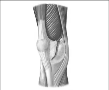
The current literature has emphasized the importance of the in-jury of the medial patellofemoral ligament (MPFL) in acute patellar luxation, since it is the main static stabilizer of the medial side of the patella and is the first structure injured in this condition(1-2,3,4) (figure 1).
Many authors use different imaging methods to refer to the MFPL injury as an essential factor in the patellar instability after an acute patellar luxation episode(5-7).
In order to contribute to the study of the patellofemoral joint, the authors radiographically assess through the Merchant incidence the presence or absence of lateral dislocation and inclination of the patella, before and after MPFL section in knees of cadavers. Thus, it is tried to verify the importance of this structure in the patellar positioning in relation to the femoral trochlea.
METHOD
This study, number 042/03, was approved by the Ethics and Research Committee of the Santa Casa Congregation of São Paulo and is in accordance with the criteria established by this institution. 30 fresh, adult, male knees of cadavers came from the Vital Health Record Service of the Capital of the University of São Paulo
(SVO) are used in this study. The time between the cadaver arrival and the research performance ranged from one to three days with mean of 1.5 days, mean age was of 50 years and three months, ranging from 27 to 74 years.
Inclusion criteria in this investigation are: anatomical piece with no macroscopic signs of decomposition, absence of previous surgi-cal approaches, absence of degeneration signs of the patellofemoral joint or knee deformities.
Exclusion criteria in this investigation are: accidental injury of MPFL during dissection, injury of the extensor apparatus during manipulation, cadaveric stiffness, even after manipulation, which makes free patellar excursion impossible.
Once selected, the knees of the cadavers are manipulated with the aim to decrease cadaveric stiffness, providing free patellar ex-cursion. Subsequently, radiograph of the patellar axial incidence for each studied knee is performed, before and after MPFL section, through the technique described by Merchant, which consists in promoting knee flexion at 45º, with the X-ray beams placed on the cranial to caudal position, at 30º angle with the horizontal, with the chassis being below the knee. A portable 50KV single-phase X-ray equipment brand name G.E.with 24 x 30cm film was used.
MPFL approach
262
Measurement of the Merchant and Laurin angles
Subsequently, the radiographs were submitted to measurement of the Merchant congruence angle, before and after the ligament section, to determine the patellar dislocation and the Laurin lateral patellofe-moral angle degrees in order to verify occasional patellar inclination.
STATISTICAL ANALYSIS
Previous descriptive statistics was performed and after its analy-sis the Wilcoxon test was used to compare the alteration before and after the MPFL section.
Significance level adopted was of 5%. The rounding was of two decimal digits. The significant values had an asterisk (*) added to facilitate their identification.
RESULTS
The medial patellofemoral ligament, located on the second me-dial layer of the knee, was identified in all anatomical pieces and presented mean of 4.8cm of length and 1.6cm of width.
The values obtained for the Laurin lateral patellofemoral and Merchant congruence angles, previous and after MPFL section are found in table 1.
The variation of these angles, termed “alteration”, was calculated and significant differences for these values were identified (table 2). Their maximum and minimum values are also found in table 2.
Tables 3 and 4 present the knee distribution by degree of alte-ration in the Laurin lateral patellofemoral angle as well as by degree of alteration of the patellar lateral dislocation (Merchant congruence angle), after MPFL section, respectively, and through its frequency, percentage and accumulated percentage.
In table 3 it can be observed that in six anatomical pieces alte-ration in the Laurin lateral patellofemoral angle did not occur and in three anatomical pieces the alteration was of one degree; in 20 anatomical pieces, two degrees and in one anatomical piece four degrees, with respective percentages of 20%, 10%, 67% and 3%; therefore, the greatest majority of alteration occurs between zero and two degrees, in 97% of the knees (table 3).
Besides that, it can be observed in table 4 that in five ana-tomical pieces no alteration in the Merchant congruence angle occurs; in six anatomical pieces the alteration was of one degree; in 17 anatomical pieces, two degrees; in one anatomical piece, three degrees; and, in one anatomical piece, six degrees, with res-pective percentages of 17%, 20%, 57%, 3% and 3%; therefore, the greatest majority of the alteration of this angle occurred between zero and two degrees (93% of the knees of the cadavers) (table 4).
DISCUSSION
Acute patellar luxation corresponds to about 3 % of all knee injuries(8,9), occurring mainly to the young population during
sports practice(9,10). Statistical studies demonstrate incidence
of 5.8 per 100,000 inhabitants and this number increases to 29 per 100,000 inhabitants between 10 years and 17 years of age(11,12). Hsiao et al(8) observed higher incidence of this injury in
military privates, who attributed the intrinsic physical fitness of this group and the character of their activities, which make the risks of musculo-skeletal injuries increase, condition which we consider comparable to professional athletes. Despite the low
Figure 1: Drawing representing the rupture of the patellar medial stabilizers. tubercle, about 10cm long, with the purpose to dissect the MPFL, which is then measured in length and width and sectioned close to the adductor tubercle (figures 2 and 3).
Figure 2. Anatomical piece (knee) with MPFL dissection. The points mark to the left the patellar medial border and to the right the adductor tubercle.
263
incidence of this condition, many patients still complain about pain and instability symptoms after one episode of acute luxa-tion(10,12-14). Atkin et al10, describe that after06 months from an
acute patellar luxation episode, more than half of the patients from his set presented limitation for extenuating physical acti-vities. This high index of unsatisfactory results in the treatment of acute patellar luxation, many of which practitioners of sports activities, stimulated us to investigate the patellar biomechanical functioning through its main medial static stabilizer, the medial patellofemoral ligament(1,2).
The MPFL was initially described by Kaplan, in 1957(15), but
he did not name it. Nevertheless, it was from the pioneer study by Warren and Marshal(16), in which the knee medial anatomy
and the MPFL were described, that a new phase in the com-prehension on the acute patellar luxation has begun. Later on, Feller et al.(17) confirmed that the MPFL is a distinct structure from
the second knee medial layer, with no variations between sides in the same individual. Such fact was also verified in our study, in which the MPFL was present in all knees and presented size mean range of 1.6cm of width and 4.8cm of length, being these values close to the ones found by other authors, who have also demonstrated that, despite its small size, this ligament resists considerable tensile pressure l(18).
In the last decade, important biomechanical investigations have been carried out confirming the importance of this ligament, demonstrating that it is the most important medial static structure for prevention of the patellar lateral dislocation, contributing with over 50% of this strength(19-20,21).
An interesting biomechanical essay was performed by Sandmeieret al.(22), in which they promoted the MPFL reconstruction
Knee of cadavers
Laurin lateral
patellofemoral angles Merchant congruence angles
Before After Before After
1 D 22 24 42 40
1 E 22 24 26 24
2 D 21 22 14 15
2 E 20 20 22 21
3 D 20 22 48 50
3 E 20 22 24 26
4 D 22 24 20 22
4 E 24 22 23 24
5 D 10 12 0 0
5 E 16 18 0 0
6 D 25 27 24 26
6 E 14 14 0 0
7 D 16 12 26 28
7 E 9 7 0 0
8 D 26 28 38 40
8 E 26 28 34 36
9 D 22 24 30 30
9 E 24 26 40 42
10 D 14 16 13 11
10 E 18 20 16 19
11 D 24 24 45 43
11 E 20 22 30 28
12 D 21 20 38 40
12 E 20 22 18 16
13 D 24 24 21 20
13 E 29 30 46 44
14 D 25 27 14 12
14 E 36 38 15 14
15 D 34 34 15 14
15 E 34 35 16 15
Table 1. Measurements of Laurin lateral patellofemoral and Merchant congruence angles in 30 knees of cadavers, before and after MPFL section.
Difference in the obtained angulation
Number of knees
of cadavers
Meandeviation Standard Median Minimum Maximum Wilcoxon
test p
Patellar lateral inclination
30 1.6 0.9 2 0 4 p < 0.001
Patellar lateral dislocation
30 1.6 1.2 2 0 6 p < 0.001
Table 2. Summed measurements of the alteration of the patellar lateral inclination angle (Laurin lateral patellofemoral angle) and of the patellar lateral dislocation (Mer-chant congruence angle) after MPFL section with descriptive level (p).
Alteration in the
inclination angle Frequency Percentage
Accumulated percentage
0 6 20 20
1 3 10 30
2 20 67 97
4 1 3 100
Total 30 100
Table 3. Frequency, percentage and accumulated percentage for the alteration in patellar lateral inclination (Laurin lateral patellofemoral angle), after MPFL section.
Alteration of the
dislocation angle Frequency Percentage
Accumulated percentage
0 5 17 17
1 6 20 37
2 17 57 93
3 1 3 97
6 1 3 100
Total 30 100
264
with the tendon of the gracilis muscle, observing reestablishment of normal patellofemoral exam after the ligament reconstruction.
An important and little studied aspect of the MPFL is concer-ned with the maintenance of its function in the different degrees of knee flexion. It is believed that this ligament plays a relevant role in the medial patellar stabilization, only in the initial degrees of knee flexion, where it would be clinically more vulnerable to suffer lateral dislocation. The radiographic incidence reported by Laurin, in which the patellar axial radiograph is performed with 20º of knee flexion, would be from this perspective, our first choice. However, according to the own author of the technique, the incidence with 20º of knee flexion is difficult to be obtained and requires careful instructions to the individual who will perform knee positioning for the radiography(23). Moreover, Vainionpää et al.(5), describe that
even in this position the patellar stability cannot be estimated due to countless false-negative findings.
The radiographic incidence by Merchant(24), which is used in
our study, is mentioned by Fulkerson(25) as widely accepted,
repro-ducible and clinically useful for the evaluation of the patellofemoral joint, is also used by other authors(26,27) for assessment of this joint.
In this study, the MPFL incision is done close to the adductor tubercle, since it is in this site that great part of the authors find the injured ligament after an acute patellar luxation episode(5,28,29).
Corroborating this fact, we observed that it is common greater sensitivity on the region of the adductor tubercle, after an acute patellar luxation episode, according to the classic sign described by Bassett(30).
The use of cadavers in this study created some limitations, such as: lack of muscular action, ideal positioning of the axial radiogra-phy, age and sex of the used cadavers. On the other hand, these same limitations are common in biomechanical assays involving the MPFL(18-21).
In order to evaluate the patellar dislocation, we used two cri-teria: visibilization of the patellar swerve and quantitative method through measurement of the Merchant congruence angle, which expresses the patellar dislocation and of the Laurin lateral patello-femoral angle, which measures patellar inclination. After the MPFL section, we have not visually observed important patellar disloca-tion or inclinadisloca-tion. Nonetheless, the evaluadisloca-tions of the radiographic measurements statistically present high significance level.
Our results suggest that the medial patellofemoral ligament, contrary to what the literature reports, can also play the role of stabilizing the patella medially, even with the knee at greater fle-xion degrees, at which it would presumably be relaxed and the patella centered on the femoral trochlea, submitted to a ‘butto-nhole’ effect. Our findings clash with the experiments performed by Nomura et al.(31) and Steensen et al.(32), who report that the
me-dial patellofemoral ligament is isometric up to 90º of knee flexion. When the angle values of dislocation and inclination presented in tables 3 and 4 are carefully evaluated in increasing order, it can be observed that they are low. As far as we are concerned, this can make their clinical application difficult since they are vulne-rable to measurement errors. Conversely, in this study no kind of traction to simulate the muscle action was performed, a current fact in the MPFL biomechanical assays, which could favor greater angle swerves.
Based on the data found in this work and in the literature suggesting the MPFL action both in knee flexion and extension, we are encouraged to think that it may play a role even more important in the cases which the trochlea is shallow, and there is no ‘butttonhole’ effect.
Although the aim of this investigation had not been to study surgical techniques, the MPFL is crucial to the understanding on different surgical positions which contemplate or not the approach of this ligament.
It is of vital importance to better comprehend the stability of the patellofemoral joint in order to propose a surgical technique closer to the knee normal biomechanics.
Due to massive controversy concerning this issue and the MPFL role, we believe further investigation on this topic should be carried out to better understand this entity.
CONCLUSION
The analysis of the results obtained in this study permits to conclude that the medial patellofemoral ligament presents im-portance in the patellar inclination and lateral dislocation with knee flexed at 45º.
All authors have declared there is not any potential conflict of interests concerning this article.
REFERENCES
1. Hernandez JH, Fávaro E, Laraya MHF. Luxação aguda da Patela. Rev Bras Ortop 2004;39:65-74.
2. Hernandez AJ, Fávaro E, Almeida A, Bonavides A, Demange MK, Camanho GL. Reconstruction of the Medial Patellofemoral Ligament in Skeletally Immature Patients: Description of Technique Techniques in Knee Surgery. 2009;8:42-6.
3. Camanho GL, Bitar AC, Hernandez AJ, Olivi R. Medial patellofemoral ligament reconstruction A novel technique using the patellar ligament. Arthroscopy 2007;23:108.e1-4. Epub 2006 Oct 16.
4. Camanho GL, Viegas Ade C, Bitar AC, Demange MK, Hernandez AJ. Conservative versus surgical treatment for repair of the medial Arthroscopy. 2009;25:620-5.
5. Vainionpaa S, Laasonen E, Patiala H, Rusanen M, Rokkannen P. Acute dislocation of the patella. Clinical, radiographic and operative findings in 64 consecutive cases. Acta Orthop Scand 1986;57:331-3.
6. Sanders TG, Morrison WB, Singleton BA, Miller MD, Cornum KG. Medial patello-femoral ligament
injury following acute transient dislocation of the patella: MR findings with surgical correlation in 14 patients. J Comput Assist Tomogr 2001;25:957-62.
7. Trikha SP, Acton D, O’Reilly M, Curtis MJ, Bell J. Acute lateral dislocation of the patella: correlation of ultrasound scanning with operative findings. Injury 2003;34:568-71.
8. Hsiao M, Owens BD, Burks R, Sturdivant RX, Cameron KL .Incidence of acute traumatic patellar dislocation among active-duty United States military service members .Am J Sports Med. 2010 Oct;38(10):1997-2004.
9. Stefancin JJ, Parker RD. First-time traumatic patellar dislocation: a systematic review. Clin Orthop Relat Res. 2007;455:93-101.
265
11. Fithian DC, Paxton EW, Stone ML, et al. Epidemiology and natural history of acute patellar dislocation. Am J Sports Med. 2004;32:1114-1121
12. Hawkins RJ, Bell RH, Anisette G. Acute patellar dislocations. The natural history. Am J Sports Med. 1986;14:117-120.
13. Cofield RH, Bryan RS. Acute dislocation of the patella: results of conservative treatment. J Trauma. 1977;17:526-531.
14. Camila Cohen; Ferretti, Mario; Cohen, Moises. Patella: Dislocation and Chronic Instability Kaleka, Techniques in Knee Surgery.2010; 9(3):139-144.1
15. Kaplan EB. Factors responsible for the stability of the knee joint. Bull Hosp Joint Dis 1957;18:51-9.
16. Warren LF, Marshall JL. The supporting structures and layers on the me- dial side of the knee. An anatomical analysis. J Bone Joint Surg Am 1979;61:56-62.
17. Feller JA, Feagin JA Jr, Garrett WE Jr. The medial patellofemoral ligament revisited: an anatomical study. Knee Surg Sports Traumatol Arthrosc 1993;1:184-6.
18. Mountney J, Senavongse W, Amis AA, Thomas NP. Tensile strength of the medial patellofemoral ligament before and after repair or reconstruction. J Bone Joint Surg Br 2005;87:36-40.
19. Conlan T, Garth WP Jr, Lemons JE. Evaluation of the medial soft-tissue restraints of the extensor mechanism of the knee. J Bone Joint Surg Am 1993;75:682-93.
20. Hautamaa PV, Fithian DC, Kaufman KR, Daniel DM, Pohlmeyer AM. Medial soft tissue restraints in lateral patellar instability and repair. Clin Orthop Relat Res 1998;(349):174-82.
21. Desio SM, Burks RT, Bachus KN. Soft tissue restraints to lateral patellar translation in the human knee. Am J Sports Med 1998;26:59-65.
22. Sandmeier RH, Burks RT, Bachus KN, Billings A. The effect of re- construction of the medial patello--femoral ligament on patellar tracking. Am Sports Med 2000;28:345-9.
23. Laurin CA, Dussault R, Levesque HP. The tangential X-ray investigation of the patellofemoral joint: X-ray technique, diagnostic criteria and their interpretation. Clin Orthop Relat Res 1979;144:16-26.
24. Merchant AC, Mercer RL, Jacobsen RH, Cool CR. Roentgenographic analysis of patellofemoral congru-ence. J Bone Joint Surg Am 1974;56:1391-6.
25. Fulkerson JP, Buuk DA, Post WR. Patologia da articulação patelofemoral. Cap. 4: Estudando a articulação patelofemoral através de imagens. 3º ed. Ed. Revinter Rio de Janeiro - RJ. Trad: Irma Fioravanti. Revisão: Jayme de Paulo Gonçalves. Ano 2000. P. 65-93.
26. O’Neil DB. Open lateral retinacular lengthening compared with arthroscopic release. A prospective, randomized outcome study. J Bone Joint Surg Am 1997;79:1759-69.
27. Nakagawa S, Kadoya Y, Kobayashi A, Tatsumi I, Nishida N, Yamano Y. Kinematics of the pa-Kinematics of the pa-tella in deep flexion. Analysis with magnetic resonance imaging. J Bone Joint Surg Am 2003;85:1238-42.
28. Avikainen VJ, Nikku RK, Seppanen-Lehmonen TK. Adductor magnus tenodesis for patellar dislocations: technique and preliminary results. Clin Orthop 1993;297:12-6.
29. Sallay PI, Poggi J, Speer KP, Garrett WE. Acute dislocation of the patella. A correlative pathoanatomic study. Am J Sports Med 1996;24:52-60.
30. Basset F.H.: “Acute dislocation of the patella, osteochondral fractures, and injuries to the extensor mechanism of the knee”. In: Burke E.: American Academy of Orthopedic Surgeons Instructional Course Lectures, St. Louis, CV Mosby, Inc., p. 40-49, 1976.
31. Nomura E, Horiuchi Y, Kihara M. A mid-term follow-up of medial patellofemoral ligament reconstruc-A mid-term follow-up of medial patellofemoral ligament reconstruc-tion using an artificial ligament for recurrent patellar dislocareconstruc-tion. Knee 2000;7:211-5.