180 Rev Bras Med Esporte – Vol. 23, No 3 – Mai/Jun, 2017
ABSTRACT
Introduction: Muscular fatigue during voluntary muscle contractions is a complex and multifactorial phenomenon associated with central changes and adaptations of the neuromuscular system. Objective: The aim of this study was to analyze the changes in both peak torque and hematological parameters in active and inactive healthy subjects following a muscular fatigue protocol. Methods: Twenty-one healthy adult males performed a muscle fatigue protocol (10 sets, 10 repetitions of knee extension at 75% of the maximal isometric torque (MIT), 120ºs-1 and 40 seconds of rest). Hematocrit (Ht), red blood cells (RBC) and leukocytes (L) analysis
were evaluated before (B) and after (A) a fatigue test. Results: The inactive group presented impairment only in concentric muscular action of -41±4% in relation to the active (-23±3%) group. No differences were found in hematological parameters in inactive group (Ht, B: 45.00±0.01 vs. A: 47.00±0.01; RBC, B: 442±40 vs. A: 487±41; L, B: 7,565±2,878 vs. A: 8,015±4,224) and the active group (Ht, B: 45.00±0.63 vs. A: 47.00±0.01; RBC, B: 477±30 vs. A: 559±37; L, B: 6,418±3,557 vs. A: 6,632±4,460). Conclusion: Differences were found only in concentric actions between groups. Additionally, there were no relationships found in hematological parameters between groups.
Keywords: muscle fatigue; muscle, skeletal; muscle strength; physiology.
RESUMO
Introdução: A fadiga muscular durante as contrações musculares voluntárias é um fenômeno complexo e mul-tifatorial associado a alterações centrais e adaptações do sistema neuromuscular. Objetivo: O objetivo deste estudo é analisar as alterações do pico de torque e dos parâmetros hematológicos em indivíduos saudáveis ativos e inativos depois de realizar um protocolo de fadiga muscular. Métodos: Vinte e um adultos saudáveis do sexo masculino real-izaram um protocolo de fadiga muscular (10 séries, 10 repetições de extensão do joelho a 75% do torque isométrico
máximo (TIM) a 120ºs-1 e 40 segundos de repouso). Hematócrito (Ht), glóbulos vermelhos (GV) e leucócitos (L) foram
analisados antes (A) e depois (D) do teste de fadiga. Resultados: O grupo inativo apresentou deterioração apenas na ação muscular concêntrica (-41 ± 4%) com relação ao grupo ativo (-23 ± 3%). Não foram encontradas diferenças nos parâmetros hematológicos no grupo inativo (Ht, A: 45,00 ± 0,01 vs. D: 47,00 ± 0,01; GV, A: 442 ± 40 vs. D: 487 ± 41; L, A: 7.565 ± 2.878 vs. D: 8.015 ± 4.224) e no grupo ativo (Ht, A: 45,00 ± 0,63 vs. D: 47,00 ± 0,01; GV, A: 477 ± 30 vs. D: 559 ± 37; L, A: 6.418 ± 3.557 vs. D: 6.632 ± 4.460). Conclusão: Foram encontradas diferenças apenas em ações concêntricas entre os grupos. Além disso, não foram encontradas relações quanto aos parâmetros hematológicos entre os grupos.
Descritores: fadiga muscular; músculo esquelético; força muscular; fisiologia.
RESUMEN
Introducción: La fatiga muscular durante las contracciones musculares voluntarias es un fenómeno complejo
y multifactorial asociado con cambios centrales y adaptaciones del sistema neuromuscular.Objetivo: El objetivo de
este estudio es analizar los cambios del par máximo y los parámetros hematológicos en sujetos sanos activos e inac-tivos después de un protocolo de fatiga muscular. Métodos: Veintiún adultos sanos del sexo masculino realizaron un protocolo de fatiga muscular (10 series, 10 repeticiones de extensión de la rodilla en el 75% del par isométrico máximo
(PIM) a 120ºs-1 y 40 segundos de descanso). Hematocrito (Ht), glóbulos rojos (GR) y leucocitos (L) fueron evaluados antes
(A) y después (D) de la prueba de fatiga. Resultados: El grupo inactivo presentó deterioro sólo en la acción muscular concéntrica (-41 ± 4%) con respecto al grupo activo (-23 ± 3%). No se encontraron diferencias en los parámetros hematológicos en el grupo inactivo (Ht, A: 45,00 ± 0.01 vs. D: 47,00 ± 0,01; GR, A: 442 ± 40 vs. D: 487 ± 41; L, A: 7.565 ± 2.878 vs. D: 8.015 ± 4.224) y en el grupo activo (Ht, A: 45,00 ± 0,63 vs. A: 47,00 ± 0,01; GR, A: 477 ± 30 vs. D: 559 ± 37; L, A: 6.418 ± 3.557 vs. D: 6.632 ± 4.460). Conclusión: Se encontraron diferencias sólo en las acciones concéntricas entre los grupos. Además, no hubo relaciones con respecto a los parámetros hematológicos entre los grupos.
Descriptores: fatiga muscular; músculo esquelético; fuerza muscular; fisiología.
ALTERATIONS IN PEAK TORQUE OCCUR WITHOUT
HEMATOLOGICAL CHANGES AFTER MUSCLE FATIGUE
AS ALTERAÇÕES NO PICO DE TORQUE OCORREM SEM MODIFICAÇÕES EM PARÂMETROS
HEMATOLÓGICOS APÓS FADIGA MUSCULAR
LOS CAMBIOS EN EL PICO DE PAR OCURREN SIN MODIFICACIONES EN PARÁMETROS HEMATOLÓGICOS
DESPUÉS DE FATIGA MUSCULAR
Frank Shiguemitsu Suzuki1
(Physical Education Professional) Marcelo Martins Kalytczak2
(Physical Education Professional) Rodrigo Augusto Ferreira Palomares1
(Physical Education Professional) Marcos Rodolfo Paunksnis1
(Physical Education Profissional) Sergio Matias Silva1
(Physical Education Professional) Fabiano Politi2
(Physiotherapist) Adriana Jaime Sbampato3
(Biologist) Andrey Jorge Serra2
(Physical Education Professional) Alexandre Lopes Evangelista3
(Physical Education Professional) Angélica Castilho Alonso1
(Physiotherapist) Aylton Figueira Junior1
(Physical Education Professional) Duncan Buchan4
(Physical Education Professional) Julien Steven Baker4
(Physical Education Professional) Danilo Sales Bocalini1
(Physical Education Professional) 1. Universidade São Judas Tadeu (USJT), Programa de Pos-Graduação em Educação Física e Ciências do Envelhecimento. Laboratório de Fisiologia Translacional, São Paulo, Brazil.
2. Universidade Nove de Julho (UNINOVE), Programa de Pos-Graduação em Ciências da Reabilitação e Biofotônica Aplicada a Ciências da Saúde São Paulo, Brazil. 3. Universidade Nove de Julho (UNINOVE), Departamento de Educação Física, São Paulo, Brazil. 4. Institute of Clinical Exercise and Health Science. University of the West of Scotland, Hamilton, Lanarkshire, Scotland.
Correspondence: Danilo Sales Bocalini Rua Ary Barroso, 68, apto 105, Torre 1, Ferrazópolis, São Bernardo do Campo, São Paulo, Brazil. 09790-240. [email protected]
ORIGINAL ARTICLE
ARTIGO ORIGINAL
ARTÍCULO ORIGINAL
181
Rev Bras Med Esporte – Vol. 23, No 3 – Mai/Jun, 2017
INTRODUCTION
Conceptually, muscle fatigue corresponds to an inability to maintain skeletal muscle strength levels for a certain period of time1. Studies2
investi-gating muscle fatigue have been published that consider the multiple facets of the associated fatigue mechanisms, with a view to understanding the fatigue process. Some of the mechanisms involved in the fatigue process2,
include changes in electromyography3, mechanical alterations4, changes
in blood biochemistry5 have received much attention. However, research
related to hematological change is still inconclusive6. Some studies7 have
outlined this research limitationduring studies investigating biochemical associations and fatigue profiles following exercise.
In addition, fatigue profiles of individuals with different levels of fitness and levels of physical activity have received little attention in the scientific community. Knowledge about the relationship between physical activity as a risk factor for chronic degenerative disease is already well established in the literature8. The level and intensity of physical activity has been
recognized as a component that influences muscle performance9,10 by
promoting force development and endurance. However, information relating to the mechanisms influencing performance when this variable is considered remains inconclusive.
Therefore, considering the lack of information in this area, especially the relationship between the level of physical activity and changes in hematological parameters, the objective of this study, was to investi-gate any changes in force development following a fatigue protocol. A secondary objective was to consider muscle action and changes in hematological parameters of active and inactive individuals undergoing a specific muscle fatigue protocol.
MATERIALS AND METHODS
Following approval by the Research Ethics Committee of the São Judas Tadeu University (number 786.06), twenty male adults aged ≥ 18 years and physically independent signed an Informed Consent form to participate voluntarily in this study. The International Physical Activity Questionnaire (IPAQ) - short version was used to estimate the level of physical activity conform previously publication of our group10.
Ques-tions were based on the level of physical activity performed weekly. The questions explored frequency and duration of exercise, including moderate walking, vigorous exercise and time spent sitting down. Sub-jects were considered active if they reached or exceeded 150 minutes of physical activity per week. Subjects who did not reach 150 minutes per week were classified as inactive. Classifications were based on the guidelines outlined by the World Health Organization. Subjects with diabetes mellitus, current smokers, individuals with musculoskeletal injuries and diagnosed clinical case pathologies and/or cardiovascular changes confirmed by medical evaluation, were excluded from the study.
Anthropometric measurements
Anthropometric assessment was performed following the model previously outlined by our group11,12. Briefly, height was measured with
a Cardiomed stadiometer (WCS model), with a 115/120 cm range. The measurement was performed with the cursor placed at a 90o angle with
the scale in the upright position, and feet placed together in contact with the stadiometer. Subjects were instructed to hold their breath and keep the head parallel to the ground. Weight was measured with a calibrated electronic scale (Filizola - Personal Line Model 150), with a 100 grams range, and maximum capacity of 150 kg. Body mass index (BMI) was calculated and recorded from the equation weight/height1. Fatigue induction protocol
The completion of the dynamic fatigue protocol followed the model used of our group10. Briefly, all subjects attended the Laboratory on four
occasions to perform an Isokinetic evaluation protocol using a Biodex System 3 (Biodex, Inc.,Shirley, NY). On the first day, the subjects signed consent forms and completed the IPAQ. On the second day, anthropo-metric data (height, and weight) was evaluated. On the third day, the subjects were placed on the Isokinetic Dynamometer in a comfortable, upright, seated position for familiarization procedures. Straps were used to fix the thigh, pelvis, and trunk to prevent extraneous body movement. The axis of the dynamometer was aligned with the axis of right knee joint rotation. Arms were placed across the chest with hands grasping the straps. All subjects performed eight repetitions of sub maximum concentric/eccentric knee extension/flexion at 120ºs1. The knee joint
range of motion was 90 to 10º of knee flexion. The calibration of the Isokinetic Dynamometer was performed according to the specifications of the manufacturer before each test, and finally, on the fourth day the fatigue protocol was performed. All testing and experimental procedures were performed at the same time of day to exclude diurnal variation. The fatigue protocols were performed at an intensity of 70% of maximum isometric contraction, which was determined on the third day.
Evaluation of hematological indicators
To establish hematological changes, blood was collected with a 5 ml syringe using methodologies outlined previously10. Subsequently, the blood
was transferred into tubes containing EDTA and kept in motion to avoid coagulation. The hematocrits were analyzed with the microhematocrit technique, which consists of collecting blood in a microcapillary tube of up to ¾ of its capacity, sealing one end with plasticine, then centrifuging for five minutes at 10,000 rpm in a microcentrifuge. The reading was re-corded from a table for hematocrit, considering between 40 and 50% of red blood cells (RBC) as normal values. For the RBC count, 20 ml of blood was diluted in 5 ml of citrate formaldehyde, with subsequent counting in a Neubauer chamber. For the establishment of leukocyte count, the same amount of blood was diluted in 400Ol Turk, with subsequent counting in a Neubauer chamber. Cells were counted in a manual counter (model SYSMEX-SF- 3000) in the specific areas for each cell type with appropriate calculations of dilutions performed. A slide containing a blood smear was used to verify the morphology and the usefulness of cells. Smears were made by dragging a drop of blood by contact with another slide, and the material was placed to drynaturally. After complete drying, the slides were subjected to the Fast Panoptic method, with a methanol bath for five seconds for fixation, eosin for five seconds, and ten seconds in Methylene Blue for coloring. Then, slides were washed with deionized water and observed under an optical microscope.
Statistical analysis
The analyses were performed using the SPSS for Windows (version 12.0; SPSS, Chicago, IL, USA). The D’Agostino-Pearson test was used for data normalization. For comparison group analysis was used the Student’s t test or two-way ANOVA for repeated measures, followed by a post- hoc Bonferroni test as needed. Data are expressed as mean and standard error of the mean, and the significance level was set at p <0.05.
RESULTS
After analyzing the IPAQ questionnaire, seminal time spent on phy-sical activity of the inactive group corresponded to 121 ± 36 minutes, which was significantly lower (p = 0.001) than the active group (657 ± 267 min). In relation to anthropometric parameters no differences were found between groups, as shown in Table 1.
182 Rev Bras Med Esporte – Vol. 23, No 3 – Mai/Jun, 2017
curve slope were found only in the eccentric peak torque between groups (active: -6.30 ± 0.61, inactive: -9.55 ± 12:29; Nm/series). No difference was found in the eccentric peak torque between active (-13.24 ± 0.62 Nm/ series) and inactive groups (-14.28 ± 0.77 Nm/series).
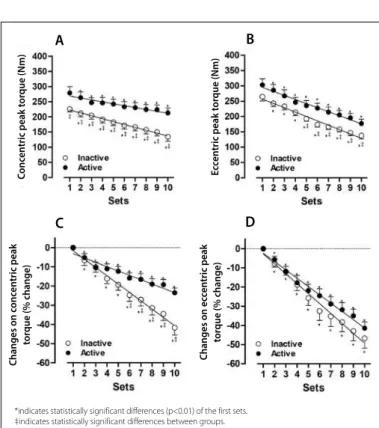
Additionally, a significant percentage reduction (p <0.001) was found after ten sets at torque maintenance of both groups in both the concentric (active: -23 ± 2, inactive: -41 ± 4; %) and the eccentric (active: -41 ± 3, inactive: -46 ± 3; %) muscular action as shown in Figures 1C and 1D. Differences between the percentage of maintenance of muscle strength in concentric peak torque are observed from set 5 between inactive groups (set 5: -19 ± 3, set 6: -25 ± 3; set 7: -27 ± 3, set 8 : -31 ± 4, set 9: -34 ± 4, set 10: -41 ± 4; %) and active groups (set 5: -12 ± 2, set 6: -15 ± 3, set 7: -17 ± 3, set 8: -19 ± 3, set 9: -20 ± 3, set 10: -23 ± 3; %).
Table 2 presents the hematological parameters. In spite of increases in all analyzed parameters, no significant difference was established between groups, before and following the fatigue protocol.
Table 2. Hematological parameters.
Inactive Active Before After % Before After % Hematocrit (%) 45.00 ± 0.01 47.00 ± 0.01 4 ± 1 45.00 ± 0.63 47.00 ± 0.01 5 ± 2 Red blood cells
(count) 442 ± 40 487 ± 41 9 ± 4 477 ± 30 559 ± 37 10 ± 8 Leukocyte
(cells/µL) 7565 ± 2878 8015 ± 4224 9 ± 58 6418 ± 3557 6632 ± 4460 1 ± 18
Values expressed as mean ± standard deviation.
DISCUSSION
The reduction in muscle performance has been the focus of nume-rous studies13,14 especially when considering the mechanisms involved in
its development. When we refer to tension development, our data agree with the study previously published10 by our group, demonstrating that
active individuals have higher fatigue resistance than inactive individuals. Taking into account the responses of different muscle actions, our data indicate a reduction of torque capacity in both groups in eccentric and concentric action. However, only significant differences were found between groups when comparing concentric actions.
The reduction in muscular strength (peripheral) is related to several mechanisms. These include: increase in concentrations of ADP, Pi, AMP, Ca2+ and H+, resulting in changes in sarcoplasmic reticulum function
to release and recapture Ca2+, with decreased concentrations of
phos-phocreatine and glycogen. Also, reduced velocity of conduction and amplitude of action potentials due to the potassium flow, changes in threshold of excitability of muscle fibers for action potentials, and a delayed relaxation phase after a contractile movement are contributory factors13,15. To our knowledge only a few studies7,9 investigated the
differences between specific groups in response to fatigue.
Differences between muscle actions have already been investigated in the literature16–21 demonstrating greater impairment in concentric
muscle activation3,19,20, increased metabolic burden evaluated by lactate
concentration3 in concentric muscle actions and of elastic components
specifically in the titin protein16,21 have been recognized as indicators
of this difference.
Considering hematological changes and physical exercise, many of the available studies have used horses22. To our knowledge, there are
few studies23 that investigated the effects of fatigue on hematological
parameters in humans. In 1977 Novosadová24 evaluated changes
bet-ween rest and exercise and found no difference betbet-ween the conditions. However, studies found significant changes in parameters such as blood viscosity7 and leukocyte profile23.
The proportion of blood represented by red blood cells is called hematocrit. For adult men and women, the mean hematocrit concen-tration corresponds to 42 and 38 respectively, and these values can vary considering the presence of anemia, degree of bodily activity, and the altitude at which the person resides. In our study, the hematocrit-in-dependent concentration of the groups has not changes significantly (Table 2) following the fatigue protocol. Additionally, the values found in our study were similar to others7. Thus, it is possible to consider the
relationship between the variation in hematocrit concentration and the osmotic balance and cell hydration, which are characteristics present in predominantly anaerobic exercises7. Anaerobic performance
demons-trates little change in water content and concomitant changes in blood viscosity when compared to aerobic exercises25.
Such changes in viscosity with concomitant changes in red blood cells may contribute to alterations in the oxygen transport system but these alterations are found mainly in aerobic exercise25. The primary function
of red blood cells (erythrocytes) is the transportation of hemoglobin that carries oxygen from the lungs to the tissues. When free in the plasma of human beings, about 3% of total hemoglobin overflows through the capillary membrane into the interstitial space or through the kidney glomerular membrane to the glomerular filtrate. This happens each time that blood passes through the capillaries. Thus, hemoglobin should remain within the red blood cells to perform its functions effectively.
Red blood cells have other functions besides hemoglobin transport. For example, they contain a large amount of carbonic anhydrase, an enzyme that catalyzes the reversible reaction between carbon dioxide and water to form carbonic acid, increasing by thousands of times the
Table 1. Anthropometric parameters.
Inactive Active Significance Age (years) 26 ± 6 28 ± 7 p = 0.636 Height (cm) 1.74 ± 1.0 1.81 ± 1.0 p = 0.057 Weight (kg) 80 ± 13 87 ± 18 p = 0.316 BMI (kg/cm²) 26 ± 3 26 ± 4 p = 0.991
Values expressed in mean ± standard deviation.
Figure 1. Values expressed as mean ± standard deviation of concentric peak torque (Panel A), Eccentric peak torque (Panel B), Changes (%) of concentric (Panel C) and eccentric (Panel D) peak torque during the muscle fatigue induction protocol of inactive (○) and active subjects (●).
*indicates statistically significant differences (p<0.01) of the first sets. ‡indicates statistically significant differences between groups.
A
C
B
D
C
onc
en
tric peak t
or
que (N
m)
C
hanges on c
onc
en
tric peak
tor
que (% change)
C
hanges on ec
cen
tric peak
tor
que (% change)
E
cc
en
tric peak t
or
que (N
183
Rev Bras Med Esporte – Vol. 23, No 3 – Mai/Jun, 2017
velocity of this reaction26. The velocity of this reaction helps transport
large quantities of CO2 in the form of bicarbonate ions from the tissues to lungs, where it is converted to CO2 and eliminated to the atmosphe-re as a product of body metabolism26. The hemoglobin in the cells is
considered an excellent basic acid buffer, and responsible for most of the blood acid-base buffering capacity.
The figures for red blood cells in the present study did not differ between active and inactive individuals (Table 2), and were similar to other studies in humans27 and animals28. However, values remain
indif-ferent when referring to anaerobic exercises, especially when performed close to exhaustion.
When referring to leukocyte concentration, no differences were noted after completion of the fatigue protocol in both groups (Table 2). Leukocy-tes are formed partly in the bone marrow (granulocyLeukocy-tes, monocyLeukocy-tes and some lymphocytes), and partly in the lymphatic tissue (lymphocytes and plasma cells). The leukocyte action is directed at areas of severe infection and inflammation, promoting fast and powerful defense mechanisms against infectious agents.
The increased concentration of leukocytes is closely related to in-creased inflammation and serious infections29. When referring to physical
exercise, especially depending on intensity and duration several Dias et al.30 showed increase, decrease or maintenance of leukocyte values.
Although not statistically different, the leukocyte values of inactive individuals were higher than those of active individuals, possibly as a result of the immune protection promoted by regular practice of physical
activity30. Studies23,27,29 have showed that in acute situations, the practice
of strenuous exercise can increase the inflammatory process allowing increased leukocyte content. Considering data from our study, we did not demonstrate significant increases in leukocyte content, which may indicate absence of inflammation following the fatigue protocol. Ho-wever, the values in this study were similar to other30. Additionally, the
increase of lymphocytes is controlled by catecholamine concentration, particularly in activities of short duration31.
CONCLUSION
In conclusion, the neuromuscular response differs when analyzing eccentric and concentric actions. However, the concentric action had more impact on the development of muscle strength during the fatigue protocol, which was different between active and inactive individuals. Additionally, no hematological changes were found following the fatigue protocol in both groups.
ACKNOWLEDGEMENT
The authors thank CAPES (Coordenação de Aperfeiçoamento de Pessoal de Nível Superior) fellowships addressed to Suzuki FS. The fund providers had no role in decision to publish and preparation of the paper.
All authors declare no potential conflict of interest related to this article.
REFERENCES
1. Enoka RM. Neuromechanics of human movement. 4th ed. Champaign, IL: Human Kinetics; 2008. 2. Abbiss CR, Laursen PB. Models to explain fatigue during prolonged endurance cycling. Sport Med.
2005;35(10):865–98.
3. Gonzalez-Izal M, Cadore EL, Izquierdo M. Muscle conduction velocity, surface electromyography variables, and echo intensity during concentric and eccentric fatigue. Muscle Nerve. 2014;49(3):389–97. 4. Katsiaras A, Newman AB, Kriska A, Brach J, Krishnaswami S, Feingold E, et al. Skeletal muscle fatigue ,
strength , and quality in the elderly: the Health ABC Study. J Appl Physiol. 2005;99(1):210–6. 5. Bertuzzi RC de M, Silva AEL, Abad CCC, Pires F de O. Lactate metabolism: a review on bioenergetics and
muscle fatigue. Rev Bras Cineantropom Desempenho Hum. 2009;11(2):226–34.
6. Olkoski MM, Fuke K, Matheus SC, Soares FAA, Portella R, Da Rosa EJF, et al. Physical and biochemical responses to training performed in and out of the water in indoor soccer players. Motriz. 2013;19(2):432–40. 7. Petrício AIM, Porto M, Burini RC. Changing in hemodynamics, basic acid equilibrium and enzymatics in
the exhaustive exercise with weights. Ativ Fís Saúde; 2001;6(3):17–26.
8. González-Gross M, Meléndez A. Sedentarism, active lifestyle and sport: Impact on health and obesity prevention. Nutr Hosp. 2013;28(Suppl 5):89-98.
9. Bogdanis GC. Effects of physical activity and inactivity on muscle fatigue. Front Physiol. 2012;3(3):1–15. 10. Suzuki FS, Kalytczak MM, Casarin CAS, Serra AJ, Evangelista AL, Marchetti PH, et al. Physical activity level
does not influence the neuromuscular fatigue in adults. Rev Bras Med Esporte. 2016;22(2):97–101. 11. Serra AJ, Amaral AM, Rica RL, Barbieri NP, Reis Junior D, Silva Junior JA, et al. Determination of body density
by generalized equations: easiness and simplification in the method. ConScientiae Saúde. 2009;8(1):19–24. 12. Martins A, Ceschini F, Battazza R, Rodriguez D, Allegretti G, Bocalini D, et al. A Low-volume weight training protocol reduces abdominal fat and increases muscle strength in 12 weeks. J Exerc Physiol. 2016;19(1):96–106.
13. Hunter SK, Critchlow A, Enoka RM. Muscle endurance is greater for old men compared with strength--matched young men. J Appl Physiol. 2005;99(3):890–7.
14. Corvino RB, Caputo F, de Oliveira AC, Greco CC, Denadai BS. Rate of force development in different muscle contraction velocities. Rev Bras Med Esporte. 2009;15(6):428–31.
15. Hunter SK. Sex differences and mechanisms of task-specific muscle fatigue. Exerc Sport Sci Rev. 2009;37(3):113–22.
16. Ide BN, Dechechi CJ, Lopes CR, Brenzikofer R, Macedo DV. Eccentric muscle movements - Why Generate more power? Why Generate more trauma? Rev Bras Prescr Fisiol Exerc. 2011;5(25):61–8.
17. Kawakami Y, Kanehisa H, Ikegawa S, Fukunaga T. Concentric and eccentric muscle strength before, during and after fatigue in 13 year-old boys. Eur J Appl Physiol Occup Physiol. 1993;67(2):121–4. 18. Mullaney MJ, McHugh MP. Concentric and eccentric muscle fatigue of the shoulder rotators. Int J
Sports Med. 2006;27(9):725–9.
19. Pasquet B, Carpentier A, Duchateau J, Hainaut K. Muscle fatigue during concentric and eccentric contractions. Muscle Nerve. 2000;23(11):1727–35.
20. Tesch PA, Dudley GA, Duvoisin MR, Hather BM, Harris RT. Force and EMG signal patterns during repeated bouts of concentric or eccentric muscle actions. Acta Physiol Scand. 1990;138(3):263-71. 21. Trappe TA, Carrithers JA, White F, Lambert CP, Evans WJ, Dennis RA. Titin and nebulin content in human
skeletal muscle following eccentric resistance exercise. Muscle Nerve. 2002;25(2):289–92. 22. Veiga APM, Lopes STDA, Franciscato C, Oliveira LSS De, Merini LP. Hematological values, plasma protein and
fibrinogen of criollo horse – their variation with sex, age and management. Acta Sci Vet. 2006;34(10):275–9. 23. Prestes J, Frollini AB, Guereschi M, Dias R, Donatto FF. Effects of two acute exercise sessions performed in the same day in different intensities on total leukocytes counting, circulating and tissue lymphocytes. Rev Bras Ciênc Saúde. 2007;3(11):27–33.
24. Novosadová J. The changes in hematocrit, hemoglobin, plasma volume and proteins during and after different types of exercise. Eur J Appl Physiol Occup Physiol. 1977;36(3):223–30.
25. Ferreira FG, Alves K, Costa NMB, Santana ÂMC, Marins JCB. Effect of physical conditioning level and oral hydration on hydric homeostasis in aerobic exercise. Rev Bras Med Esporte. 2010;16(3):166–70. 26. Ribeiro F, Campbell CSG, Mendes G, Arsa G, Moreira SR, da Silva FM, et al. Exercise lowers blood pressure
in university professors during subsequent teaching and sleeping hours. Int J Gen Med. 2011;4:711–6. 27. Ellwanger RB, Brentano MA, Kruel LFM. The effect of different velocities of strength training in indirect
markers of muscle damage. Rev Bras Educ Fís Esp. 2007;21(4):259–70.
28. Leandro C, Nascimento E, Manhães-de-Castro R, Célia MMB. Physical exercise and immune system: mechanisms and integra- tion processes. Rev Port Cien Desp. 2002;2(5):80–90.
29. Foschini D, Prestes J, Charro MA. Relationship between physical exercise , muscle damage and delayed--onset muscle soreness. Rev Bras Cineantropom Desempenho Hum. 2007;9(1):101–6.
30. Dias R, Frollini AB, Prestes J, Teixeira LFM, Cereja DMP, Baganha RJ, et al. Strength exercise and immunological parameters: leukocytes counting, inflammation and regeneration. Rev Bras Ciênc Mov. 2008;16(3):100–7. 31. Minetto M, Rainoldi A, Gazzoni M, Terzolo M, Borrione P, Termine A, et al. Differential responses of serum and salivary interleukin-6 to acute strenuous exercise. Eur J Appl Physiol. 2005;93(5–6):679–86.