Abstract
The purpose of this study was to investigate the effects of shade and material type and shape in dental polymer composites on the hardness and shrinkage stress of bulk and two-layered restoration systems. For this purpose, some bulk and layered specimens from three different shades of dental materials were prepared and light-cured. The experiments were carried out on three types of materials: conventional restorative composite, nanohybrid compo-site and nanocompocompo-site. Micro-indentation experiment was per-formed on the bulk and also on each layer of layered restoration specimens using a Vicker’s indenter. The interface between the two layers was studied by scanning electron microscopy (SEM). The results revealed significant differences between the values of hardness for different shades in the conventional composite and also in the nanohybrid composite. However, no statistically signif-icant difference was observed between the hardness values for different shades in the nanocomposite samples. The layered resto-ration specimens of different restorative materials exhibited lower hardness values with respect to their bulk specimens. The reduc-tion in the hardness value of the layered convenreduc-tional composite samples was higher than those of the nanocomposite and nanohy-brid composite specimens indicating more shrinkage stresses gen-erated in the conventional composite restorations. According to the SEM images, a gap was observed between the two layers in the layered restorations.
Keywords
Shade effect, polymer dental materials, Layered restorative sys-tems, Micro-indentation.
Effect of Dental Restorative Material Type and Shade
on Characteristics of Two-Layer Dental Composite Systems
1 INTRODUCTION
The demand for aesthetic, strength, life lasting and easy-to-use dental restorative systems has led to the development of polymer-based dental composite materials. At the present time, dental
compo-Atefeh Karimzadeh a Majid R. Ayatollahi b A.R. Bushroa c,d
a Fatigue and Fracture Laboratory, Center
of Excellence in Experimental Solid Mechanics and Dynamics, School of Mechanical Engineering, Iran University of Science and Technology, Narmak, Teh-ran 16846, ITeh-ran,
b Fatigue and Fracture Laboratory,
Cen-ter of Excellence in Experimental Solid Mechanics and Dynamics, School of Me-chanical Engineering, Iran University of Science and Technology, Narmak, Tehran 16846, Iran, [email protected]
c Department of Mechanical Engineering,
Faculty of Engineering, University of Malaya, Kuala Lumpur 50603, Malaysia, [email protected].
d Centre of Advance Manufacturing and
Mechanical Engineering, Faculty of Engineering, University of Malaya, Kuala Lumpur 50603, Malaysia.
http://dx.doi.org/10.1590/1679-78252562
sites are widely used for filling the tooth cavities, veneering to mask discoloration, correcting con-tour, making dental implants and bonding orthodontic brackets . Dental composite materials consist of a polymeric matrix and inorganic ceramic filler particles which are generally produced by light curing. The polymeric matrix is flowable before curing, which makes the composite to fully pene-trate into the tooth cavity. During the curing procedure, the polymerization is activated, which allows the resin matrix to solidify and to change its mechanical properties rapidly and significantly. Volumetric shrinkage also occurs in polymerization due to the decrease of intermolecular separations in the monomers of polymeric matrix (Li et al., 2011). In the restorative systems where the restora-tions are constrained along the interfaces, the polymerization shrinkage causes some shrinkage stresses in the composite and in the tooth. In these cases, shrinkage stress may result in pulling the material away from the cavity walls (Ferracane and Mitchem, 2003, Braga and Ferracane, 2004) which is the main reason for marginal debonding and then micro-leakage within the composite res-torations (Davidson et al., 1984). Moreover, shrinkage forces on cusps produce cuspal deformation, enamel cracks and crazes (Marzouk and Ross, 1989) which cause reduction in the fracture resistance of the cusp (Wieczkowski Jr et al., 1988). Prediction or measurement of the shrinkage stresses is a difficult procedure because the each tooth cavity has small and complicated geometry and also there are many factors that affect the restorative composite properties. Up to now, some experiments have been developed to estimate the shrinkage stress of dental restorative composites (Ferracane, 2005, Gonçalves et al., 2008, Li et al., 2011, Simon et al., 2008).
Indentation experiments have received much attention from numerous researchers for determin-ing the mechanical properties of different biomaterials, see for example (Şakar-Deliormanli and Güden, 2006, Kruzic et al., 2009, Ayatollahi and Karimzadeh, 2012, Karimzadeh and Ayatollahi, 2012, Towler et al., 2001, Karimzadeh et al., 2014, Oréfice et al., 2003). According to these studies, indentation tests are independent of the specimen geometry and size and give reliable results. Therefore, such tests can be considered as suitable alternatives for estimation of shrinkage stress and determination of the mechanical properties of restorations.
Recently, layering restoration techniques have been applied by various clinicians. The base of these techniques is restoring the lost tooth structure layer by layer using composite materials with different or similar shades. Two major advantages of layering restoration techniques are the creation of a more natural-looking restoration and the reduction in the magnitude of polymerization shrink-age stresses (Park et al., 2008, Li et al., 2011, Lee et al., 2007). Previous studies (Kwon et al., 2012, Park et al., 2008, Van Ende et al., 2013, Bicalho et al., 2013, Arakawa, 2010) have suggested that the layered or incremental filling method produces lower shrinkage stresses compared to the bulk filling method.
In addition to the conventional dental restorative composites, dental nanocomposites and nano-hybrid composites have been recently introduced by changing the size and volume fraction of fillers in the dental composites. Dental nanocomposites contain nano-fillers with dimensions of 4 to 20 nm added to the composite resins. In the nanohybrid adhesives, in addition to nanometer particles, 0.2 to 1 micrometer particles are also added to the composite resins.
com-posite, nanocomposite and nanohybrid restorative materials are available to achieve desired proper-ties of natural tooth structure. According to some research studies performed on different shades of dental restorative materials, the polymerization process of the dental restorations and their mechan-ical and physmechan-ical properties are affected by the type of shades utilized (Aguiar et al., 2005, Cesar et al., 2001, Della Bona et al., 2007, Guiraldo et al., 2009). However, very few researches have been conducted on the mechanical properties of various shades of nanocomposite and nanohybrid compo-site.
In this study, the effects of material shade on hardness of three dental restorative polymers, i.e. conventional composite, nanocomposite and nanohybrid composite were investigated. In addition to hardness, the shrinkage stresses in two-layer restorative systems of these materials were studied. Besides, the interface between the two layers was inspected by scanning electron microscopy (SEM). The hypothesis is that (1) different shades of these restorative materials have various hardness val-ues, and darker shades have higher hardness and (2) using different combinations of material shades affects the shrinkage stress in layered restoration systems.
2 MATERIALS AND METHODS
2.1 Materials
Three different types of dental polymer composites (i.e. conventional composite, nano-composite and nano-hybrid composite) with three different shades including enamel (A1), dentine (A4) and body (C2) shades were utilized. The characteristics of the used materials are indicated in Table 1.
Dental Material Type of resins Filler particles Filler
content Shade
Composite BIS-GMA
1, UDMA2,
BIS-EMA3 Zirconia/silica 60%V A1,A4,C2
Nanohybrid composite
BIS-GMA,
UD-MA,PEGDMA4, TEGDMA5 Silica, Zirconia/silica 67.8%V A1,A4,C2
Nanocomposite BIS-GMA, UDMA,
TEGD-MA, PEGDTEGD-MA, BIS-EMA Zirconia, silica 63.3%V A1,A4,C2
Table 1: Characteristics of dental restorative composites used in the experiments.
2.2 Sample Preparation
Two categories of specimens, bulk and layered, were prepared for the tests. For the bulk category, nine cylindrical specimens of 5 mm diameter and 4 mm thickness were prepared from each type of material and shade. Each sample was light-cured using a LED light with minimum intensity of 400 mW/cm2 according to its manufacturer's instruction.
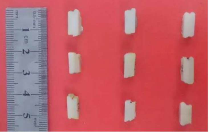
and thickness was applied on the first strip and light-cured. The light-curing procedure was per-formed on the materials in accordance with the manufacturer’s instruction. Figure 1 shows some layered specimens prepared from different shades of each restorative material.
Figure 1: Layered specimens prepared from different shades of each restorative material.
In order to obtain a smooth surface, all samples were ground with 800 to 3000 grit sandpapers two days after their polymerization, and then polished by diamond suspension with meshes of 3 microns to 1 micron to make sure that the surface was sufficiently smooth.
2.3 Experiment
The micro-indentation experiment is a method for determining material hardness or resistance of material to penetration. It is also called micro hardness test. The micro hardness experiment can be utilized when test specimen is very small and when small regions in a composite sample or plating should be measured. This test uses an established method in which an indenter tip is pressed into specific sites of the material by applying an increasing normal load. When the penetration depth of the indenter tip or the indentation load reaches its pset maximum value, the normal load is re-duced till partial or complete relaxation occurs.
In this study, micro-indentation experiment was performed on the bulk and layered samples by using a micro indentation instrument (SHIMADZU, Japan) and Vickers indenter. In the layered samples, the indentation was done at each layer. In the micro-indentation experiment, a normal load should be selected from the predefined load values of the instrument to make a perfect indenta-tion hole without any crack or damage around it. The value of normal load depends generally on the properties of the sample material. In this study, a normal load of 490.3 mN was applied for all specimens to make a perfect indentation hole. The indentation hole was observed by a high resolu-tion optical microscope with magnificaresolu-tion of 40x.
2.4 Imaging
some images of the interface. Each specimen was placed on a carbon double-side tape and an accel-erated voltage of 15 kv was used for SEM.
3 RESULTS AND DISCUSSION
3.1 Micro Hardness
Hardness (H) could be calculated by dividing the normal load (F) by the area of the surface where load is imposed on (A).
F H
A
= (1)
When Vicker’s indenter is applied, A is calculated from: 2
2 sin( / 2)
d A
a
= (2)
where d is the average diagonal length remained by the indenter and α is the angle between oppo-site faces of the indenter which is equal to 136° for the Vicker’s indenter.
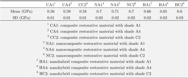
Table 2 shows the mean value and standard deviation (SD) obtained directly from the micro-indentation test for the hardness of each bulk sample.
CA11 CA42 CC23 NA14 NA45 NC26 HA17 HA48 HC29
Mean (GPa) 0.56 0.59 0.58 0.7 0.71 0.7 0.66 0.65 0.6 SD (GPa) 0.01 0.01 0.01 0.03 0.02 0.02 0.02 0.02 0.03
1 CA1: composite restorative material with shade A1
2 CA4: composite restorative material with shade A4
3 CC2: composite restorative material with shade C2
4 NA1: nanocomposite restorative material with shade A1
5 NA4: nanocomposite restorative material with shade A4
6 NC2: nanocomposite restorative material with shade C2
7 HA1: nanohabrid composite restorative material with shade A1
8 HA4: nanohybrid composite restorative material with shade A4
9 HC2: nanohybrid composite restorative material with shade C2
Table 2: Mean value and standard deviation of micro hardness obtained for the bulk samples.
Moreover, in the nanohybrid restoration group significant differences are seen between A1 and C2 shades (p-value<<0.05) and between A4 and C2 (p-value=0.004) ones.
Based on Table 2, the highest value of hardness was obtained for the nanocomposite. This ob-servation can be interpreted by the filler size and its volume fraction. According to previous re-search studies, hardness could be increased by applying smaller particles and higher filler contents (Oberholzer et al., 2003, Lodhi, 2006). The particle size of the nanocomposite is smaller than the other two materials and its volume fraction is higher than the conventional composite. Therefore, its hardness is also expected to be higher.
In the conventional composite group, the darker shade A4 was significantly harder than its lighter shade A1. While for the nanohybrid composite, higher hardness was obtained for the lighter shade A1 and for the nanocomposite group no significant difference was observed between the hard-ness values of different shades. Since the darker shade can absorb light and lighter shade spreads it (Sakaguchi et al., 1992, Aguiar et al., 2005), more hardness value was expected for the darker shade. It has been previously shown that shade may have different effects on the hardness of light-cured restorative composites. For example, Aguiar et.al. (Aguiar et al., 2005) found that darker shade of dental composite has lowest hardness value compared to the lighter shade. On the contra-ry, a higher hardness value was reported for the darker shade of dental composite in the research studies performed by Lodhi (Lodhi, 2006) and Pierce et.al. (Price et al., 2005). Previous studies have indicated that at each depth of light-cured restorative materials different factors have domi-nant effects on material hardness (Rueggeberg et al., 1993, Lodhi, 2006). The target layer in the present study is the top surface which is the nearest layer to the light-curing device. According to Rueggeberg et.al. evaluation (Rueggeberg et al., 1993), the influential factors at the top surface of the light-cured restorative materials, with respect to their importance are filler type, exposure dura-tion and resin shade. Therefore, at the top surface, the shade is not of very important influence on hardness.
The size, load and distribution of fillers affect the light scattering and its absorption, hence they have influence on the polymerization shrinkage of restorations (Leonard et al., 2001, Ruyter and Øysæd, 1982, Aguiar et al., 2005). These factors vary greatly in different types of composites and their effects should be investigated separately with highly controlled conditions. As a result, the mechanical behavior of different material shades should be explored for the same types of restora-tive materials.
In terms of clinical applications, similar hardness values for different shades of the nano-composite could be a good achievement for dental restorative behavior. However, the variations of hardness in the conventional composite and nanohybrid composite were also quite small for clinical applications.
3.2 Shrinkage Stress
stress generated due to the polymerization shrinkage in the restoration systems is a kind of residual stress which influences the hardness value.
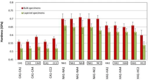
In this part of research, hardness was measured in each layer of two-layer restorative systems made of different shades. By comparing the material hardness obtained for the bulk restorative ma-terials with the corresponding value for the individual layer in the layered restoration specimen, the effect of shrinkage stress can be studied. The diagram of the hardness values of bulk samples and those obtained from each layer in the layered specimens has been exhibited in Figure 2.
Figure 2: The hardness values of bulk samples and those obtained from each layer in layered specimens.
In Figure 2, the hardness of each individual layer in layered specimen has been shown by the right (green) bars and the hardness of bulk specimens has been indicated by the left (red) bars, as mentioned in the chart legend at the top corner of the figure. For each layered specimen, the data related to each of the two layers are written next to each other in a box beneath the horizontal axis, while the vertical axis shows the hardness values by the green bars. For those layered specimens in which the two layers are made of the same materials with the same shades, such as (CA1-CA1 or NA1-NA1 or HA1-HA1), the hardness of only one layer is shown in Figure 2, because both layers have equal hardness values.
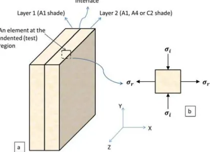
It is seen from Figure 2 that the hardness of each individual layer in the layered specimens is less than the corresponding value of its bulk sample, which can be attributed to the polymerization shrinkage in the constrained samples causing shrinkage or contraction stresses. In the layered spec-imens, the shrinkage stress is created perpendicular to the interface of the two layers. The indenta-tion test is also performed on the sample secindenta-tion which is not constrained. The indentaindenta-tion region and stress configuration are schematically depicted in Figure 3.
hardness of the layers in the layered specimens compared to the corresponding values of bulk sam-ples.
Figure 3: Schematic picture of stress configuration in a layered specimen at the indented or test region a) layered specimen with a square element at the indented (test) region and b) magnified square
element with applied stresses caused by the indenter and shrinkage of the materials.
Since all conditions related to the sample preparation, the storage condition and the experi-mental procedure were carefully controlled, the material hardness of the layers in each layered spec-imen is predominately affected by the shrinkage stress. Although, the difference between the hard-ness values of layers and their bulk samples is not equal to the value of shrinkage stress, it can be used for comparing the effect of shrinkage stress in specimens of different materials and shades.
Comparing the hardness difference in the layered specimens of different restorative materials relative to their bulk samples indicates that more reduction has occurred in the hardness values of conventional composite specimens (around 7.1% to 10.3%) in comparison with the nanocomposite (around 5.7% to 10%) and the nanohybrid composite specimens (around 4.5% to 6.6%) all depend-ing on the shades of layers. This implies that more shrinkage stress was generated in the conven-tional composite restorations.
Several factors can affect the polymerization contraction and the shrinkage stress of dental re-storative materials. The composition of resin matrix of a rere-storative material is one of the factors which influence the polymerization shrinkage. The resin matrix of nanocomposite and nanohybrid composite samples contains TEGDM which has higher shrinkage than other resins. The reported shrinkage for TEGDMA is about 12.5%, while its value for the BisGMA is equal to 5.2% and for typical resins is ranged between 2% and 3% (Labella et al., 1999, Gonçalves et al., 2011, Peutzfeldt, 1997, Stansbury, 1992). Therefore, more shrinkage would cause more stress in the constrained nano-composite and nanohybrid nano-composite samples compared to the conventional nano-composite one.
fraction among the examined materials belongs to the nanohybrid composite (67.8 %). Therefore, nanohybrid composite is supposed to have the lowest shrinkage stress between the three materials.
In restricted dental restorative materials, two factors must be considered. First, the level of re-striction imposed on the material, which could be estimated by the percentage of composite surface that is bonded to the substrate in relation to its total surface area. Second factor is the compliance of bonding substrate (Braga et al., 2005). Since all restoration systems used in the present study are in the same shape, the percentage of constrained area has no effect on the variation of hardness. Hence the compliance of restorative layers which could be considered as the substrate in the layered restorative systems should be evaluated. Our previous study shows that the elasticity modulus of dental restorative nanohybrid composite is higher than the corresponding values for the nanosite and the conventional componanosite samples (Ayatollahi et al., 2015). Thus, the nanohybrid compo-site layer has less compliance compared to the other materials which increases the induced shrink-age stress in its layered specimens. Although the shrinkshrink-age stress of conventional composite is more than the other two materials, its compliance is more than the corresponding value for the nanocom-posite and nanohybrid comnanocom-posite. It means that other factors have dominant effects on the shrink-age stress of nanocomposite and nanohybrid composite specimens.
The three tested restorative materials consist of different filler sizes which might affect their shrinkage stress. Satterthwaite et.al (Satterthwaite et al., 2012) showed that the shrinkage stress changes in a complex manner when the sizes of filler particles vary. However, more comprehensive studies on the effect of filler sizes with different combinations in their sizes and types are needed. Since in our study different factors such as resin matrix, substrate compliance and filler type change in the specimens, no judgment can be done about the effect of filler size on the shrinkage stress in different types of restorative materials.
As a result, in the restorative systems the interaction between their compliance, resin matrix component and filler volume fraction affects the shrinkage stress variation. The investigation of dominant factor depends greatly on the material type and the restoration system properties which should be found out from experiment and cannot be interpreted by knowing the general behavior of restorative systems. For example, in the nanohybrid two-layer restoration the volume fraction of fillers can be considered as a dominant factor while in the nanocomposite restoration the resin ma-trix component is the main factor.
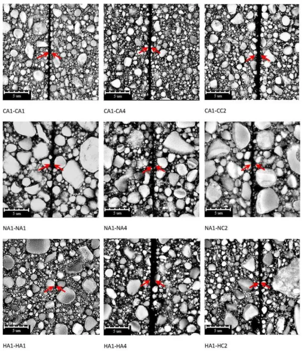
Figure 4: SEM images of the layered restoration systems.
two-layer specimens compared to the two-layered specimens of nanohybrid composite. Meanwhile, the nano-hybrid composite has shown greater hardness than the conventional dental composite and its hard-ness reduction in the layered specimens is also lower than that of the conventional dental composite. Consequently, considering its higher hardness and lower shrinkage effect, nanohybrid composite can be recommended for the layered restoration treatments.
3.3 SEM Analysis
Each layered sample was inspected by a SEM instrument to study the interface between the two layers of the layered restoration systems and to investigate the quality of their interfacial seal. Fig-ure 4 shows the SEM images of the interface zone in all the layered specimens wherein the gap be-tween the two layers is pointed by red arrows.
According to Figure 4, an interfacial gap exists between the two layers in all specimens, and the thickness of gap varies for different samples. The interfacial gap formation can be attributed to the poor bond strength between the two restoration layers which was not strong enough to resist against the polymerization shrinkage of the resin-based dental restorative materials and could not provide an adequate interfacial seal for these restoration systems (Erickson, 1992, Samet et al., 2006). Table 3 presents the mean values of the gap thickness measured from the SEM images.
CA 1-CA 1 CA 1-CA 4 CA 1-CC2 NA1-NA1 NA1-NA4 NA1-N C 2 HA1-HA1 HA1-HA4 HA1-H C 2
Mean (μm) 0.53 0.55 0.78 0.54 0.62 0.58 0.38 1.16 0.61 SD (μm) 0.12 0.15 0.17 0.16 0.18 0.11 0.1 0.4 0.17
Table 3: The mean value and standard deviation of the gap thickness for each layered restoration specimen.
Based on Table 3, the gap thickness of the sample which consists of two nanohybrid composite with A1 shade (HA1-HA1), is lower than the other specimens. Since the same preparation proce-dures were applied to all samples, this observation could be due to the stronger bonding between the two layers of the HA1-HA1 specimen. Thus, better seal is obtained by using the A1 shade of nanohybrid composite for the layered restoration systems. This is in agreement with the results of previous studies which have shown the hybrid restorative composites generated fewer voids than the conventional dental restorative composites (Kugel and Perry, 2002, Samet et al., 2006). Indeed, the possibility of interfacial gap occurrence in layered restorations is reduced when fewer voids are cre-ated. However, it is noteworthy that from the esthetic aspect, restoring only by one shade of dental restorative material can not satisfy the natural optical properties of teeth.
presence of a gap in restoration systems may increase the potential for mechanical failures by the bacterial growth (Moorthy et al., 2012). To improve the bond strength between the composite re-storative systems, using a bonding agent is recommended in order to make a strong chemical bond-ing between the two restorative layers.
4 CONCLUSION
Micro-indentation technique was used to measure the hardness values of three polymer restoration materials of different shades. The experiments were performed both on bulk materials and on two-layer restorative systems. The results indicated that the effect of material shades on hardness was dependent on the type of restorative material used. Since there are several factors involved in the degree of polymerization and the mechanical properties of the dental restorations, separate compari-sons should be performed on each material. In the two-layer restorative systems, the shade and the material type affected the shrinkage stress of different restorative layers, but the level of influence was again dependent on the materials used. More reductions occurred in the hardness values of the conventional dental composite specimens (around 7.1% to 10.3%) in comparison to those of the nanocomposite (around 5.7% to 10%) and the nanohybrid composite (around 4.5% to 6.6%) all depending on the shades of layers. As a result of this study, the nanohybrid composite can be rec-ommended for layered restorations in clinical applications, because of its higher hardness (equal to 0.6 GPa to 0.66 GPa depending on the shade) and lower shrinkage effect in comparison to the con-ventional composite and nanocomposite dental materials. While the SEM images showed a gap be-tween the two layers in the layered restorations, the thinnest gap with a thickness about 0.38 μm, was observed in the nanohybrid layered specimens.
Acknowledgement:
The authors would like to acknowledge the Ministry of Higher Education, Malaysia for providing a high impact research (HIR) grant with number UM.C/625/1/HIR/MOHE/ENG/27. This research is also partly funded under the University of Malaya under UMRG programme grant number of UM.TNC2/RC/AET/261/1/1/RP017-2012A.
References
Aguiar, F. H. B., Lazzari, C. R., Lima, D. A. N. L., Ambrosano, G. M. B., Lovadino, J. R. (2005). Effect of light curing tip distance and resin shade on microhardness of a hybrid resin composite. Brazilian Oral Research 19: 302-306.
Arakawa, K. (2010). Shrinkage forces due to polymerization of light-cured dental composite resin in cavities. Polymer Testing 29: 1052-1056.
Ayatollahi, M. R., Karimzadeh, A. (2012). Determination of Fracture Toughness of Bone Cement by Nano-Indentation Test. International Journal of Fracture 175: 193-198.
Ayatollahi, M. R., Karimzadeh, A., Nikkhooyifar, M., Yahya, M. Y. (2015). Effects of temperature change and bev-erage on mechanical and tribological properties of dental restorative composites. Materials Science and Engineering C 54: 69-75.
Braga, R. R., Ballester, R. Y., Ferracane, J. L. (2005). Factors involved in the development of polymerization shrinkage stress in resin-composites: a systematic review. Dent Mater 21: 962-70.
Braga, R. R., Ferracane, J. L. (2004). Alternatives in Polymerization Contraction Stress Management. Critical Re-views in Oral Biology & Medicine 15: 176-184.
Calheiros, F. C., Sadek, F. T., Braga, R. R., Cardoso, P. E. C. (2004). Polymerization contraction stress of low-shrinkage composites and its correlation with microleakage in class V restorations. Journal of Dentistry 32: 407-412. Cesar, P. F., Miranda, W. G., Braga, R. R. (2001). Influence of shade and storage time on the flexural strength, flexural modulus, and hardness of composites used for indirect restorations. The Journal of prosthetic dentistry 86: 289-296.
Chen, H. Y., Manhart, J., Hickel, R., Kunzelmann, K. H. (2001). Polymerization contraction stress in light-cured packable composite resins. Dental Materials 17: 253-259.
Condon, J. R., Ferracane, J. L. (2000). Assessing the effect of composite formulation on polymerization stress. Jour-nal of the American Dental Association (1939) 131: 497-503.
Da Silva Telles, P. D., Aparecida, M., Machado, M., Nor, J. E. (2001). SEM study of a self-etching primer adhesive system used for dentin bonding in primary and permanent teeth. Pediatr Dent 23: 315-20.
Davidson, C. L., De Gee, A. J., Feilzer, A. (1984). The Competition between the Composite-Dentin Bond Strength and the Polymerization Contraction Stress. Journal of Dental Research 63: 1396-1399.
Della Bona, Á., Rosa, V., Cecchetti, D. (2007). Influence of shade and irradiation time on the hardness of composite resins. Brazilian Dental Journal 18: 231-234.
Deyhle, H., Schmidli, F., Krastl, G., Müller, B. Evaluating tooth restorations: micro-computed tomography in practi-cal training for students in dentistry. 2010. 780417-780417-9.
Dieter Jr. George E. (1961). Mechanical Metallurgy, Mcgrawhill.
Erickson, R. L. (1992). Surface interactions of dentin adhesive materials. Oper Dent Suppl 5: 81-94.
Ferracane, J. L. (2005). Developing a more complete understanding of stresses produced in dental composites during polymerization. Dental Materials 21: 36-42.
Ferracane, J. L., Mitchem, J. C. (2003). Relationship between composite contraction stress and leakage in Class V cavities. American journal of dentistry 16: 239-243.
Gonçalves, F., Azevedo, C. L. N., Ferracane, J. L., Braga, R. R. (2011). BisGMA/TEGDMA ratio and filler content effects on shrinkage stress. Dental Materials 27: 520-526.
Gonçalves, F., Pfeifer, C. S., Ferracane, J. L., Braga, R. R. (2008). Contraction Stress Determinants in Dimethacry-late Composites. Journal of Dental Research 87: 367-371.
Guiraldo, R. D., Consani, S., Consani, R. L., Berger, S. B., Mendes, W. B., Sinhoreti, M. A. (2009). Light energy transmission through composite influenced by material shades. The Bulletin of Tokyo Dental College 50: 183-190. Jeong, T. S., Kang, H. S., Kim, S. K., Kim, S., Kim, H. I., Kwon, Y. H. (2009). The effect of resin shades on micro-hardness, polymerization shrinkage, and color change of dental composite resins. Dent Mater J 28: 438-45.
Karimzadeh, A., Ayatollahi, M. R. (2012). Investigation of mechanical and tribological properties of bone cement by nano-indentation and nano-scratch experiments. Polymer Testing 31: 828-833.
Karimzadeh, A., Ayatollahi, M. R., Bushroa, A. R., Herliansyah, M. K. (2014). Effect of sintering temperature on mechanical and tribological properties of hydroxyapatite measured by nanoindentation and nanoscratch experiments. Ceramics International 40 9159–9164.
Kwon, Y., Ferracane, J., Lee, I.-B. (2012). Effect of layering methods, composite type, and flowable liner on the polymerization shrinkage stress of light cured composites. Dental Materials 28: 801-809.
Labella, R., Lambrechts, P., Van Meerbeek, B., Vanherle, G. (1999). Polymerization shrinkage and elasticity of flow-able composites and filled adhesives. Dental Materials 15: 128-137.
Lee, M.-R., Cho, B.-H., Son, H.-H., Um, C.-M., Lee, I.-B. (2007). Influence of cavity dimension and restoration methods on the cusp deflection of premolars in composite restoration. Dental Materials 23: 288-295.
Leonard, D., Charlton, D., Roberts, H., Hilton, T., Zionic, A. (2001). Determination of the minimum irradiance required for adequate polymerization of a hybrid and a microfill composite. Oper Dent 26: 176-180.
Li, H., Li, J., Yun, X., Liu, X., Fok, A. S.-L. (2011). Non-destructive examination of interfacial debonding using acoustic emission. Dental Materials 27: 964-971.
Li, M.-Y. (ed.) (2012). Contemporary Approach to Dental Caries, InTech.
Lodhi, T. A. 2006. Surface hardness of different shades and types of resin composite cured with a high power led light curing unit Master of Science, University of the Western Cape
Marzouk, M. A., Ross, J. A. (1989). Cervical enamel crazings associated with occluso-proximal composite restorations in posterior teeth. American journal of dentistry 2: 333-337.
Moorthy, A., Hogg, C. H., Dowling, A. H., Grufferty, B. F., Benetti, A. R., Fleming, G. J. P. (2012). Cuspal deflec-tion and microleakage in premolar teeth restored with bulk-fill flowable resin-based composite base materials. Journal of Dentistry 40: 500-505.
Oberholzer, T. G., Grobler, S. R., Pameijer, C. H., Hudson, A. P. G. (2003). The effects of light intensity and meth-od of exposure on the hardness of four light-cured dental restorative materials. International Dental Journal 53: 211-215.
Oréfice, R. L., Discacciati, J. A. C., Neves, A. D., Mansur, H. S., Jansen, W. C. (2003). In situ evaluation of the polymerization kinetics and corresponding evolution of the mechanical properties of dental composites. Polymer Testing 22: 77-81.
Oztas, N., Olmez, A. (2005). effect of one versus two-layer applications of a self-etching adhesive to dentine of prima-ry teeth: a SEM study. The Journal of Contemporaprima-ry Dental Practice 6: 1-7.
Park, J., Chang, J., Ferracane, J., Lee, I. B. (2008). How should composite be layered to reduce shrinkage stress: Incremental or bulk filling? Dental Materials 24: 1501-1505.
Peutzfeldt, A. (1997). Resin composites in dentistry: the monomer systems. European Journal of Oral Sciences 105: 97-116.
Price, R. B. T., Felix, C. A., Andreou, P. (2005). Knoop hardness of ten resin composites irradiated with high-power LED and quartz-tungsten-halogen lights. Biomaterials 26: 2631-2641.
Rueggeberg, F. A., Caughman, W. F., Curtis, J. W., Davis, H. C. (1993). Factors affecting cure at depths within light-activated resin composites. American journal of dentistry 6: 91-95.
Ruyter, I. E., Øysæd, H. (1982). Conversion in different depths of ultraviolet and visible light activated composite materials. Acta Odontologica Scandinavica 40: 179-192.
Sakaguchi, R. L., Douglas, W. H., Peters, M. C. R. B. (1992). Curing light performance and polymerization of com-posite restorative materials. Journal of Dentistry 20: 183-188.
Şakar-Deliormanli, A., Güden, M. (2006). Microhardness and fracture toughness of dental materials by indentation method. Journal of Biomedical Materials Research Part B: Applied Biomaterials 76B: 257-264.
Samet, N., Kwon, K. R., Good, P., Weber, H. P. (2006). Voids and interlayer gaps in Class 1 posterior composite restorations: a comparison between a microlayer and a 2-layer technique. Quintessence Int 37: 803-9.
Shortall, A. C. (2005). How light source and product shade influence cure depth for a contemporary composite. Journal of Oral Rehabilitation 32: 906-911.
Simon, Y., Mortier, E., Dahoun, A., Gerdolle, D. (2008). Video-controlled characterization of polymerization shrink-age in light-cured dental composites. Polymer Testing 27: 717-721.
Stansbury, J. W. (1992). Synthesis and Evaluation of Novel Multifunctional Oligomers for Dentistry. Journal of Dental Research 71: 434-437.
Tosha, K. (2002). Influence of Residual Stresses on the Hardness Number in the Affected Layer Produced by Shot Peening. 2nd Asia-Pacific Forum on Precision Surface Finishing and Deburring Technology. Seoul, Korea.
Towler, M. R., Bushby, A. J., Billington, R. W., Hill, R. G. (2001). A preliminary comparison of the mechanical properties of chemically cured and ultrasonically cured glass ionomer cements, using nano-indentation techniques. Biomaterials 22: 1401-1406.
Van Ende, A., De Munck, J., Van Landuyt, K. L., Poitevin, A., Peumans, M., Van Meerbeek, B. (2013). Bulk-filling of high C-factor posterior cavities: Effect on adhesion to cavity-bottom dentin. Dental Materials 29: 269-277.
Wieczkowski Jr, G., Joynt, R. B., Klockowski, R., Davis, E. L. (1988). Effects of incremental versus bulk fill tech-nique on resistance to cuspal fracture of teeth restored with posterior composites. The Journal of Prosthetic Dentis-try 60: 283-287.