Stem Cells in Response to Retinoic Acid Induced
Differentiation
Jayantha B. Tennakoon
1☯, Hongran Wang
2☯, Cristian Coarfa
2☯, Austin J. Cooney
2*, Preethi H.
Gunaratne
1,3,4*1 Department of Biology and Biochemistry, University of Houston, Houston, Texas, United States of America, 2 Department of Molecular and Cellular Biology, Baylor College of Medicine, Houston, Texas, United States of America, 3 Department of Pathology, Baylor College of Medicine, Houston, Texas, United States of America, 4 Human Genome Sequencing Center, Baylor College of Medicine, Houston, Texas, United States of America
Abstract
Loss of Dicer, an enzyme critical for microRNA biogenesis, results in lethality due to a block in mouse embryonic
stem cell (mES) differentiation. Using ChIP-Seq we found increased H3K9me2 at over 900 CpG islands in the Dicer
-/-ES epigenome. Gene ontology analysis revealed that promoters of chromatin regulators to be among the most
impacted by increased CpG island H3K9me2 in ES (Dicer-/-). We therefore, extended the study to include H3K4me3
and H3K27me3 marks for selected genes. We found that the ES (Dicer-/-) mutant epigenome was characterized by a
shift in the overall balance between transcriptionally favorable (H3K4me3) and unfavorable (H3K27me3) marks at
key genes regulating ES cell differentiation. Pluripotency genes Oct4, Sox2 and Nanog were not impacted in relation
to patterns of H3K27me3 and H3K4me3 and showed no changes in the rates of transcript down-regulation in response to RA. The most striking changes were observed in regards to genes regulating differentiation and the
transition from self-renewal to differentiation. An increase in H3K4me3 at the promoter of Lin28b was associated with
the down-regulation of this gene at a lower rate in Dicer-/-ES as compared to wild type ES. An increase in H3K27me3
in the promoters of differentiation genes Hoxa1 and Cdx2 in Dicer-/-ES cells was coincident with an inability to
up-regulate these genes at the same rate as ES upon retinoic acid (RA)-induced differentiation. We found that siRNAs
Ezh2 and post-transcriptional silencing of Ezh2 by let-7g rescued this effect suggesting that Ezh2 up-regulation is in
part responsible for increased H3K27me3 and decreased rates of up-regulation of differentiation genes in Dicer-/-ES.
Citation: Tennakoon JB, Wang H, Coarfa C, Cooney AJ, Gunaratne PH (2013) Chromatin Changes in Dicer-Deficient Mouse Embryonic Stem Cells in Response to Retinoic Acid Induced Differentiation. PLoS ONE 8(9): e74556. doi:10.1371/journal.pone.0074556
Editor: Atsushi Asakura, University of Minnesota Medical School, United States of America Received August 24, 2012; Accepted August 5, 2013; Published September 9, 2013
Copyright: © 2013 Tennakoon et al. This is an open-access article distributed under the terms of the Creative Commons Attribution License, which permits unrestricted use, distribution, and reproduction in any medium, provided the original author and source are credited.
Funding: This work was supported by National Institute of General Medical Sciences (NIGMS)- P01 GM081627 pilot grant to PHG and NIGMS- P01 GM081627 grant to AJC. The funders had no role in study design, data collection and analysis, decision to publish, or preparation of the manuscript. Competing interests: AJC is a PLOS ONE editorial board member. However the authors state that this does not alter the authors' adherence to all the PLOS ONE policies on sharing data and materials.
* E-mail: [email protected] (PHG); [email protected] (AJC)
☯ These authors contributed equally to this work.
Introduction
Dicer is an RNAse III type endoribonuclease with specific enzymatic activity that cleaves double stranded RNA molecules [1]. Dicer-dependent microRNAs (miRNAs) are initiated through RNA polymerase II activity to generate a primary miRNA that forms a stem and loop structure which is cleaved by the combined activities of two enzymes of the microprocessor complex, Drosha and DGCR8/Pasha [2]. The resultant stem loop precursors are then transported to the cytoplasm through Exportin-5 action where Dicer catalyzes the second cleavage event to produce 18 to 25 nucleotide mature miRNA duplex structures [2]. Mature miRNAs are known to
regulate gene expression by either cleaving mRNA molecules by binding to respective 3’ UTR elements or by translational repression [2–4]. The conventional role of miRNAs in posttranscriptional gene silencing is well established, however the full extent of the role of Dicer-dependent pathways beyond their established role in regulatory mechanisms such as epigenetic silencing critical for biological processes has yet to be determined. In spite of several studies having shown small RNA molecules regulating epigenetic modifications in plants, flies and fission yeast [5–7] it remains unclear whether similar regulation occurs in higher order mammals.
development [8–10]. Critical networks that Dicer-dependent miRNAs regulate in maintaining mammalian stem cell pluripotency and cellular transition by functioning as repressors of transcription factors, chromatin modifiers and cell signaling molecules was reported by Marson et al. in 2008 [11]. Moreover it is known that miRNAs are needed for proper maintenance of DNA methylation in mES cells [12]. The currently proposed mechanistic principle is that DNA methylation is invoked in ES cells by down regulation of repressors of DNA methyltransferases 3A and 3B (Dnmt3) such as Rbl in part by miRNAs in the miR-290-295 cluster ensuring availability of DNA methyltransferases for de novo
DNA methylation [12]. The critical roles which histone modifications play in determining cellular differentiation and stem cell plasticity is all the more emphasized through observations made by Boyer and co-workers [13] that polycomb group proteins repress key developmental regulators in mES cells through repressive histone H3K27me3 modifications in the pluripotent state. Bernstein et al. 2006 reported that the co-occupation of key developmental regulators by transcriptionally unfavorable H3K27me3 as well as transcriptionally favorable H3K4me3 modifications render these subsets of genes to be repressed during the pluripotent state poised for activation upon differentiation [14].
We hypothesized that Dicer-dependent pathways, either directly or indirectly through miRNA mediated regulation, guide epigenetic changes in mES cells and affect transcript levels of genes that are critical for stem cell function. To address our hypothesis we mapped genome-wide changes in H3K9me2 using ChIP-Seq analysis of WT ES cells and Dicer-/-ES cells.
We found a significant increase in H3K9me2 at over 900 CpG islands in Dicer-/-ES. Gene ontology analysis indicated that
CpG islands of chromatin modifiers were significantly impacted. Therefore, we extended this work to study additional histone marks including H3K27me3 and H3K4me3 in the promoter regions of genes critical for the maintenance and differentiation of ES cells. H3K9me2 and H3K27me3 are considered context dependent repressive marks while H3K4me3, which is favorably linked with polymerase recruitment is typically associated with transcriptional activation. Focusing on key genes involved mES self-renewal and pluripotency (Oct4,
Nanog, Sox2 and Ronin), differentiation (Cdx2 and Hoxa1) and genes regulating the transition between these states (Lin28b and Gcnf) we systematically measured levels of transcripts and associated changes in the above mentioned histone modifications in Dicer-/- ES as compared to WT ES [15–17]. .
Taken together our data showed that loss of Dicer impacts the balance between transcriptionally favorable and unfavorable histone modifications affecting the rates of changes and expression levels of mRNA of key developmental genes. miRNA mediated rescue experiments, which we subsequently carried out showed that changes in chromatin state, and mRNA expression associated with loss of Dicer is reversible by targeted silencing of Ezh2. In summary, our data show that Dicer directly or indirectly regulates gene expression in mES cells at the epigenetic as well as post-transcriptional levels and point to a critical role Dicer plays in mammalian embryogenesis.
Results
a: Loss of Dicer has a strong impact on H3K9me2 in the epigenomic landscape in mES cells
It has been shown that small RNAs regulate epigenetic changes in plants, flies and fission yeast [5–7]. However a potential role for Dicer-dependent pathways and miRNAs in mammals in particular remain hitherto unexplored. It has also been shown that miRNAs regulate DNA methylation via the miR-290-295 candidates functioning as repressors of Rbl2, which in turn is a repressor of Dnmt3 [12]. H3K9me2 modifications are widely considered epigenetic marks that demarcate genetic regions bound to undergo DNA methylation [18,19]. In order to assess Dicer’s effect on potential upstream events regulated via H3K9me2 modifications we carried out genome-wide H3K9me2 ChIP-Seq analysis using the Illumina platform. We then generated genome-wide H3K9me2 maps on the mES cell genome using the PASH algorithm [20]. Of the regions showing differential H3K9me2 modification we found increased H3K9me2 in Dicer-/- ES cells in 71% of promoter
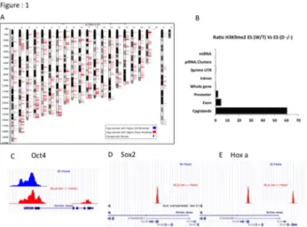
regions and 98% of CpG islands, (Figure 1A). Comparison of ratios of H3K9me2 occupation by the number of tags mapping to different genomic features revealed that on a global scale Dicer-/- ES cells showed fold changes of 60 at CpG islands, 5 at
exons and 2 at promoters and <1 in whole genes, introns, 3’ UTR whole gene, 3’ UTR piRNA and miRNA clusters respectively (Figure 1B). These observations strongly suggested either a direct or indirect role for Dicer-dependent pathways in regulating the mES cell epigenome via H3K9me2 modifications. In order to see if the biological processes that are impacted by the large number of genes associated with increased CpG island H3K9me2 and the small number of genes associated with decreased CpG island H3K9me2 in Dicer-/- ES are distinct we carried out GO analyses on the gene
sets. From data shown in table 1 we see that genes with equivalent levels of CpG island H3K9me2 in WT and Dicer-/- ES
and genes exhibiting higher levels of CpG island H3K9me2 in WT were associated with housekeeping functions such as nucleosome and cellular component assembly, amino acid biosynthesis, protein acetylation and cation transport. In sharp contrast genes associated with higher levels of CpG island H3K9me2 in Dicer-/- ES cells included chromatin regulators,
genes regulating DNA damage and repair and cell cycle. The same was true when we examined key genes involved in ES cell self-renewal and differentiation. Pluripotency genes such as Oct4 and Nanog showed equivalent levels of H3K9me2 in WT and Dicer-/- ES (Figure 1C). In contrast, genes in the Hox
cluster that are induced during ES cell differentiation showed increased CpG island H3K9me2 in Dicer-/- ES (Figure 1E). To
more closely assess the effects of loss of Dicer on promoter H3K9me2 enrichment levels of key developmental genes we carried out ChIP-qPCR assays and found statistically insignificant subtle differences in H3K9me2 levels between the two cell lines subject to RA induced differentiation Figure S1 (A to G).
modifying enzyme gene expression levels are comparable in both cell lines.
To ascertain how loss of Dicer affects the transcript levels of histone modifying and DNA modifying enzymes, which contribute downstream to elevated or reduced levels of corresponding epigenetic modifications we carried out qRT-PCR analysis on a panel of epigenetic modifier genes in WT and Dicer-/- ES cells. While we found no significant differences
in H3K4me3, and H3K36me3 modifying enzymes Setd2 and
Ash1l gene transcript levels (Figure 2A and 2C) we found slightly elevated statistically insignificant levels of the H3K9me2 modifying enzyme G9a transcripts in Dicer-/- ES cells (Figure
2B). Most notably the transcript levels of H3K27me3 modifying enzyme Ezh2 [21] was consistently overexpressed in Dicer
-/-ES cells compared to WT cells from day 0 through day 6 of RA induced differentiation (Figure 2D).
c: Pluripotency genes Oct4, Sox2 and Nanog are not impacted by loss of Dicer but Ronin levels are significantly lower in Dicer-/- ES cells
Dicer-deficient embryos tend to show morphological abnormalities as early as day 6.5 of development [8,9]. It has also been shown that Dicer-deficient ES cells are defective in their ability to properly differentiate [9]. In order to assess the degree to which Dicer-deficient ES cells differentiate and to uncover mechanisms through which Dicer potentially regulates
Figure 1. Dicer’s effect on H3K9me2 distribution patterns
in mES cells. A) Differential distribution of H3K9me2 in mouse
chromosomes. Blue bars represent loci where higher enrichment levels were observed in mES cells while red bars represent loci where enrichment levels were higher in Dicer
-/-ES cells. Specific genomic loci can be found in table S3. B) Bar graph showing ratio of H3K9me2 occupation in Dicer-/- ES cells
compared to WT ES cells in different genomic elements. C, D and E) Sequence tags from H3K9me2 ChIP-Seq experiment mapped to the UCSC genome browser to show enrichment at
Oct4 and Sox2 and Hoxa gene cluster promoter regions of WT ES and Dicer-/- ES cells. Blue peaks represent WT ES cells and
red peaks represent Dicer-/- ES cells. doi: 10.1371/journal.pone.0074556.g001
Table 1. Gene ontology terms of differentially methylated CpG island H3K9me2 sites.
Dicer Specific ES Specific Common ES-Dicer
Chromatin regulator BP00014:Amino acid
biosynthesis GO:0000786~nucleosome
DNA damage BP00066:Protein acetylation
GO:0006334~nucleosome assembly
DNA repair BP00143:Cation transport
GO:0022607~cellular component assembly GO:0000166~nucleotide
binding
BP00201:Skeletal development
GO:0022829~wide pore channel activity
GO:0000785~chromatin BP00204:Cytokinesis GO:
0065003~macromolecular complex assembly
GO:0003676~nucleic acid binding
BP00289:Other metabolism
MF00101:Guanyl-nucleotide exchange factor phosphoprotein
GO:0003677~DNA binding GO:0048471~perinuclear region of cytoplasm GO:0004672~protein kinase
activity
MF00060:Damaged DNA-binding protein
GO:0005488~binding MF00178:Extracellular matrix Nucleosome core GO:0005515~protein binding
GO:0005524~ATP binding GO:0005622~intracellular GO:0005634~nucleus GO:0005694~chromosome GO:0005737~cytoplasm GO:0005921~gap junction GO:0006139~nucleobase, nucleoside, nucleotide and nucleic acid metabolic process GO:0006259~DNA metabolic process
GO:0006323~DNA packaging GO:0006325~establishment and/or maintenance of chromatin architecture GO:0006333~chromatin assembly or disassembly GO:0006464~protein modification process GO:0006468~protein amino acid phosphorylation GO:0006793~phosphorus metabolic process GO:0006796~phosphate metabolic process GO:0007049~cell cycle GO:0007242~intracellular signaling cascade GO:0008152~metabolic process
cellular transition we carried out ChIP-qPCR assays in the promoter regions of key genes regulating self-renewal and differentiation for transcriptionally favorable H3K4me3 versus transcriptionally unfavorable H3K27me3 marks side by side with qRT-PCR assays to determine gene expression. We particularly chose the H3K27me3 mark because Ezh2 mRNA levels were high in Dicer-/- ES cells and H3K4me3 mark as it is
associated with active polymerase recruitment. Upon RA
Table 1 (continued).
Dicer Specific ES Specific Common ES-Dicer
GO:0016310~phosphorylation GO:
0016773~phosphotransferase activity, alcohol group as acceptor
GO:0017076~purine nucleotide binding
GO:0022403~cell cycle phase doi: 10.1371/journal.pone.0074556.t001
Figure 2. Ezh2 transcripts in Dicer-/- ES cells are
overexpressed compared to WT ES cells, while Setd2, G9a
and Ash1l levels remain relatively similar in both cell
lines. A, B C and D bar graphs showing expression levels of
the histone methyl transferases Setd2, G9a, Ash1l and Ezh2 in WT and Dicer-/- ES cells upon RA treatment through days 0
(D0) to day 6 (D6). Note that the transcripts of enzymes Setd2
and Ash1l, which promote H3K36me3 and H3K4me3 modifications respectively show comparable results while H3K9me2 promoting G9a is slightly elevated at a statistically insignificant level in Dicer-/- ES cells and Ezh2 is significantly
high in Dicer-/- ES cells. The symbol* indicates that the results
of a given time point were significantly different between the two cell lines at a confidence level of 0.05 when a students t-test was performed.
doi: 10.1371/journal.pone.0074556.g002
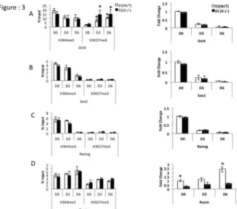
induction transcripts of pluripotency factors Oct4 Sox2 and
Nanog gradually went down in both WT and Dicer-/- ES cells at
comparable rates (Figure 3A–C right panels). However transcripts of Ronin (Figure 3D right panel) was found to be significantly lower in Dicer-/- ES cells. ChIP-qPCR on promoter
regions of the pluripotency genes Oct4, Sox2, Nanog and Ronin showed no consistent change in H3K4me3 and H3K27me3 marks in Dicer-/- ES cells compared to WT ES cells
(Figure 3A to 3D left panels). However, an increase of H3K27me3 marks in response to RA treatment was noticeable in both cell lines at the Oct4 promoter (Figure 3A Left panel).
Figure 3. Enrichment levels of transcriptionally favorable
versus transcriptionally unfavorable histone modifications in pluripotency factor gene promoter regions and their
mRNA expression in mES cells. A, B, C and D left
ChIP-qPCR and right panels qRT-PCR results of relative mRNA levels in Oct4, Sox 2, Nanog and Ronin in WT and Dicer-/-ES
cells upon RA induced differentiation through days 0 (D0) to 6 (D6). A and B right panels transcripts of Oct4 and Sox2 go down both in mES and Dicer-/-ES cells upon RA treatment (A
and B left panels). Transcriptionally favorable (H3K4me3) and unfavorable (H3K27me3) presence at Oct4 and Sox2
promoters in WT ES and Dicer-/- ES (C right panel). Expression
levels of Nanog go down upon RA induction while transcriptionally favorable H3K4me3 marks gradually go down in both cell lines (C left panel) (D left panel). H3K4me3 and H3K27me3 occupation at the Ronin promoter (D right panel). Transcripts of Ronin go up at a higher rate in Dicer-/- ES cells
compared to WT ES cells. Figure 3A Left panel * indicates that the difference for a given cell line at a given time point was significant when compared to day zero of the same cell line at 0.05 confidence levels when a students t-test was performed. Figure 3D right panel * indicates that the difference at a given time point between the two cell lines were significant at 0.05 confidence levels when a students t-test was performed.
d: Transcriptionally favorable histone modifications in promoter regions impede the down-regulation of
Lin28b in Dicer-/- ES
Lin28b and Gcnf are genes known to facilitate the transition from ES self-renewal to differentiation. Lin28b decreases and
Gcnf increases upon RA-induced differentiation [22–24]. We examined both; epigenetic changes in promoter regions and transcript levels of these genes in Dicer-/- ES cells treated with
RA and compared this with WT. In Dicer-/- ES cells the
promoter region of Lin28b exhibited high enrichment of H3K4me3 compared to ES cells while the Gcnf promoter region showed slightly elevated yet statistically insignificant levels of enrichment (Figure 4A and B left panels). At the transcript level Lin28b failed to go down at the same rate in Dicer-/- ES cells upon RA induction as compared to WT (Figure
4A right panel). It is possible that the presence of higher levels of H3K4me3 marks at their promoter regions favoring transcription in Dicer-/- ES cells is in part responsible for this. Gcnf as well was observed to go up at a faster rate in Dicer
-/-ES cells upon RA induction as compared to WT (Figure 4B right panel).
e: Transcriptionally unfavorable histone modifications impede the up-regulation of Hoxa1 and Cdx2 during RA-induced differentiation in Dicer-/- ES cells
In order to examine the impact of Dicer-dependent histone modifications on genes promoting ES cell differentiation we carried out ChIP-qPCR and qRT-PCR analysis on Cdx2 and
Hoxa1. Here we noted an entirely different outcome to the other genes analyzed so far. Dicer-/- ES cells retained the ability
to induce transcripts upon RA-treatment but failed to do so at the same rate as WT cells (Figure 4C and 4D right panels). In addition the promoter regions of Cdx2 and Hoxa1 had significantly higher levels of H3K27me3 levels in promoter regions in Dicer-/- ES cells compared to WT cells, which likely
explains the attenuated up-regulation of these genes upon RA-induction of Dicer-/- ES cells. Comparable and high H3K4me3
marks were found in both cell lines (Figure 4C and 4D left panels).
f: Reduced transcript levels of Hoxa1 and Cdx2 in Dicer-/- ES cells can be rescued by overexpressing
let-7g miRNA or siRNAs targeting Ezh2
Based on 1) the failure of Hoxa1 and Cdx2 to be fully transcriptionally induced upon RA treatment in Dicer-/- ES cells,
2) increased enrichment of H3K27me3 in the promoter regions of Hoxa1 and Cdx2 in Dicer-/- ES cells, and 3) significant
increase in transcript levels of Ezh2 that has been established to regulate H3K27me3 in WT ES cells we hypothesized that a key developmentally regulated miRNA lacking in Dicer-/- ES
cells due to miRNA biogenesis defects may play a mechanistic role in facilitating higher levels of Ezh2 transcripts and hence higher enrichment of H3K27me3 marks in Hoxa1 and Cdx2 in promoter regions. Moreover Lin28b, which is known to play a critical role in facilitating cell fate through a double negative feed back loop between let-7g and itself [23], was found to be substantially increased in Dicer-/- ES cells and let-7g is
predicted to target Ezh2. From these observations we
hypothesized that the loss of let-7g in Dicer-/- ES cells led to the
increase in its predicted target Ezh2, which in turn led to increased H3K27me3 at the Hoxa1 and Cdx2 promoter regions. It is possible that failure to fully induce genes that are critical for embryonic differentiation such as Hoxa1 and Cdx2
are a result of the failure to down-regulate epigenetic modifiers such as Ezh2 which potentially play an important role in the embryonic lethality of the Dicer-/- embryos.
To functionally validate our hypothesis we overexpressed mature let-7g in Dicer-/- ES cells and assayed the expression
levels of developmentally significant genes, which are critical in determining cell fate. Upon over-expression of mature let-7g, both Lin28b and Ezh2 transcript levels were down-regulated in Dicer-/- ES cells (Figure 5A and 5B), while Cdx2 and Hoxa1
transcript levels were increased (Figure 5A). In sharp contrast, overexpression of let-7g has very little effect on pluripotency genes (Figure S2: A). In order to assess the effect of Ezh2 by itself on the expression levels of genes in the absence of concomitant down-regulation of Lin28 with Ezh2 we used
Figure 4. Transcriptionally favorable H3K4me3
modifications in promoter regions increase expression
levels of Lin28b in Dicer-/- ES cells relative to WT ES cells,
while H3K27me3 histone modifications disfavor Hoxa1 and
Cdx2 expression in Dicer-/- ES cells relative to WT. ES. AB
C and D left panels, ChIP- qPCR results on promoter regions and qRT-PCR results (Right panles) of Lin28b, Gcnf, Hoxa1
and Cdx2 respectively upon RA induction from day 0 (D0) through day 6 (D6). Note higher enrichment levels of H3K4me3 in the promoter regions of Lin28b (A left panel) which affect increased expression levels in Dicer-/- ES cells (A right panel).
As shown in C and D left panels higher occupation of H3K27me3 in Hox a1 and Cdx2 attenuates upregulation of transcripts in Dicer-/- ES cells compared to WT ES cells (C and
D right panels). The symbol * indicates that the difference at a given time point between the two cell lines were significant at 0.05 confidence levels when a students t-test was performed.
siRNAs targeting Ezh2. We found that siRNAs to Ezh2 were sufficient to increase levels of Cdx2 and Hoxa1 transcripts in Dicer-/- ES cells (Figure 6A). However, siRNAs targeting Ezh2
had no significant effect on Lin28b, Gcnf and the pluripotency genes (Figure 6B and Figure S3: A) To test the impact of let-7g and siRNAs to Ezh2 on patterns of H3K27me3 at Hoxa1 and
Cdx2 loci in Dicer-/- ES cells we carried out treatments with
let-7g and siEzh2 along with respective controls and chromatin immunoprecipitated genomic DNA from the cell lines using an H3K27me3 specific antibody. A significant reductions of H3K27me3 was found at Hoxa1 and Cdx2 in Dicer-/- ES cells
compared to mock and control treated samples (Figure 7A).
Discussion
The role of Dicer, in posttranscriptional regulation of embryonic stem cell genes is well established. Sinkkonen and co-workers have shown that regulating DNA methylation in ES cells require the miR-290-295 cluster which facilitates appropriate DNA methylation at developmentally critical genes by downregulating Rbl a known repressor of the enzymes DNA methyltransferases 3A and 3B in ES cells [12]. Here we report that Dicer-dependent histone methyl modifications can affect transcription of key developmental genes by functioning as an additional layer of epigenetic regulation in conjunction with DNA methylation. To examine the role of Dicer in regulating histone modifications that are critical to self-renewal and differentiation of ES cells we first examined genome-wide
Figure 5. Transfecting Dicer-/- ES cells with let-7g miRNA
rescues Hoxa1 and Cdx2 transcript levels during retinoic
acid induced differentiation. A) Hoxa1 and Cdx2 transcript
levels increase upon transfecting Dicer-/- ES cells with miRNA
let-7g while Ezh2 transcript levels go down. B) Lin28b transcripts are significantly reduced in Dicer-/-ES cells upon
transfecting with miRNA let-7g. * indicates that the difference at a given time point between Dicer-/- ES cells and Dicer-/- ES cells
with let-7g were significant at 0.05 confidence levels when a students t-test was performed.
doi: 10.1371/journal.pone.0074556.g005
changes in H3K9me2 in the presence and absence of Dicer
and subsequently measured local changes in H3K27me3 and H3K4me3 in the promoters of key regulators of self-renewal and pluripotency (Oct4, Nanog, Sox2 and Ronin), differentiation (Cdx2 and Hoxa1) and genes promoting the transition between these states (Lin28b and Gcnf). The critical role, which Dicer plays in mammalian embryogenesis, is highlighted by the fact that mouse embryos lacking Dicer do not successfully progress beyond day 6.5 in development [8,9]. Using the ability to maintain Dicer-deficient ES cells in long-term culture and RA-mediated differentiation cues to our advantage we specifically show that loss of Dicer can lead to changes in the landscape of the embryonic stem cell epigenome by impacting histone modifications in genes critical for embryonic stem cell differentiation.
Loss of Dicer in particular led to a global increase in H3K9me2 modifications at over 900 CpG islands. In order to examine how H3K9me2 modifications work with other histone modifications at the promoters of genes critical for stem cell function we also integrated data for H3K4me3 and H3K27me3 modifications. Upon carefully evaluating genes regulating different aspects of ES cell functions (pluripotency, differentiation and transition between the two states) we uncovered a higher level of complexity where the presence and comparative abundance of transcriptionally favorable (H3K4me3) marks versus transcriptionally unfavorable (H3K27me3) marks affect mRNA expression levels. We further
Figure 6. Transfecting Dicer-/- ES cells with siRNAs
specifically targeting Ezh2 rescues Hoxa1 and Cdx2
transcript levels during retinoic acid induced
differentiation. A) Ezh2 transcript levels reduce while Hoxa1
and Cdx2 transcript levels increase upon transfecting Dicer
-/-ES cells with siRNA specific to Ezh2. B) Lin28b transcripts remain significantly unchanged upon transfecting Dicer-/-ES
cells with siRNA specifically targeting Ezh2. * Indicates that the differences at a given time point between Dicer-/- ES cells and
Dicer-/- ES cells with siEzh2 were significant at 0.05 confidence
levels when a students t-test was performed.
found that maintaining this intricate balance is Dicer -dependent. An increase in H3K27me3 in the promoters of differentiation genes Hoxa1 and Cdx2 in Dicer-/-ES cells was
coincident with an inability to up-regulate these genes at the same rate as WT ES cells upon RA-induced differentiation. siRNAs and microRNA let-7g rescued this effect by down-regulating Ezh2 suggesting that Ezh2 up-regulation is in part responsible for increased H3K27me3 and decreased rates of up-regulation of differentiation genes in Dicer-/-ES cells. The
presence or absence of Dicer had no impact on H3K4me3/ H3K27me3, transcript levels or rate of down-regulation of pluripotency genes Oct4, Nanog and Sox2. By contrast, the promoter of Lin28b regulating the transition from self-renewal to differentiation was associated with a significant increase H3K4me3 in Dicer-/-ES coincident with
down-regulation of Lin28b at a lower rate. Lin28b mediated repression of let-7g, has been shown to block ES cells from differentiating [23]. Gcnf, established to be critical for the repression of Oct4 and Nanog [22,25,26] was found to be up-regulated at a higher rate in Dicer-/-ES cells.
A putative model to explain our findings is shown in Figure 8. It is already known that the transition of ES cells from the stem cell state to the differentiated state in response to RA requires the down-regulation of Lin28b and the subsequent differentiation of ES cells require the upregulation of differentiation genes such as Hoxa1 and Cdx2. Our data suggest that Dicer plays a critical role in balancing the transcriptionally favorable and unfavorable histone modifications at these genes that regulate RA-induced
Figure 7. Transfecting Dicer-/- ES cells with let-7g miRNA
and siRNA targeting Ezh2 reduces H3K27me3 at Hoxa1
and Cdx2 during retinoic acid induced
differentiation. Cells were treated with let-7g miRNA and
siRNA-targeting Ezh2 along with a scrambled negative control as described in the methods section and chromatin immunoprecipitated using an H3K27me3 specific antibody. Thereafter enrichment of H3K27me3 at Hoxa1 and Cdx2 loci were assayed by quantitative real time PCR. * Indicates that the differences at a given time point between Dicer-/- ES cells
and Dicer-/- ES cells treated with siEzh2, or let-7g mimic were
significant at 0.05 confidence levels when a students t-test was performed.
doi: 10.1371/journal.pone.0074556.g007 differentiation of ES cells. We propose that one of the majorimpacts of loss of Dicer is the disruption of let-7g mediated
post-transcriptional repression of Ezh2 during RA-induced differentiation. This in turn results in a cascade of events whereby increased H3K27me3, leads to the attenuated up-regulation of genes critical for differentiation such as Hoxa1
and Cdx2 in Dicer-/-ES. In addition genes regulating the
transition from stem cell state to the differentiated state are also perturbed in their response to RA-induction. Lin28b, which is typically down-regulated upon RA-induced ES cell differentiation, is down-regulated at a lower rate in Dicer-/-ES
cells. The 3’ UTR based repression of Ezh2 by let-7g mimics introduced into Dicer-/-ES cells is sufficient to rescue the
increase in H3K27me3 and the resulting attenuation in up-regulation of Hoxa1 and Cdx2. In addition, the rapid increase in levels of let-7g levels upon RA induction leads to a decrease in levels of Lin28b via 3’ UTR based repression in WT ES cells. Since Lin28b is a translational enhancer of pluripotency factors the ES cells are now free to differentiate. Collectively, the attenuated up-regulation of genes critical for differentiation such as Hoxa1 and Cdx2 and the attenuated down-regulation of Lin28b render Dicer deficient ES cells unable to differentiate upon retinoic acid induction.
Understanding the interactions between transcription factors, miRNAs and the epigenetic landscape is a fundamental requirement to develop strategies aimed at cellular reprogramming. Our observations suggest that a single miRNA (let-7g) alone can significantly alter epigenetic histone modifications at genes critical for maintenance of the stem cell state and their ability to differentiate when exposed to the appropriate cues. Our findings suggest that Dicer mediated pathways involving miRNAs, PcGs and epigenetic marks, could
Figure 8. Putative model explaining Dicer mediated
regulation of mES cell differentiation. When ES cells are
induced to differentiate by RA treatment let-7g levels increase. Hence let-7g suppresses Ezh2 resulting in reduced H3K27me3 favoring transcription of Hoxa1 and Cdx2 differentiation genes. As let-7g increases during differentiation Lin28b levels are reduced. As a result translational enhancement of pluripotency factors by Lin28b is reduced, this event in turn favors differentiation.
be potentially manipulated to direct cellular fate in engineering cells for therapeutic applications in future.
Materials and Methods
Cell Culture
mES cells were cultured on gelatin coated plates using DMEM (Gibco, Invitrogen, Carlsbad California catalog number-31053) containing 15% FBS (Invitrogen, Carlsbad California catalog number-10439-024), 1X Penstrep (Gibco, Grand Island New York catalog number-15140-122), 1X Glutamine (Gibco, Grand Island New York catalog number-25030-024), 1X non-essential amino acids (Gibco, Grand Island New York catalog number-11140-035), 1X Pyruvate Solution (Gibco, Grand Island New York catalog number- cat#11360), 0.1mM 2-mercaptoethanol (Sigma, St. Louis Missouri catalog number-M7522) and 1000U/ml LIF (LIF 2010, Millipore, Billerica, Massachusetts) during day 0 by seeding at a density of 250,000 cells per well. LIF containing media was replaced with RA containing media to induce differentiation after day 0. Thereafter the cells were maintained in RA containing medium by feeding on a daily basis. Mouse WT ES and Dicer-/- ES cells were a generous gift from Gregory
Hannon, Cold Spring Harbor Laboratories [10].
miRNA and siRNA transfections
Mature let-7g miRNAs and Ezh2 siRNAs were purchased from Invitrogen Inc, Carlsbad, USA. Cells were transfected with let-7g and siEzh2 RNAs on day 0 on six well format plates using standard protocols for Lipofectamine 2000 transfection reagent (Catalog number11668-019, Invitrogen, Carlsbad, California). Each well was transfected with 5 µl of Lipofectamine 2000 reagent and let-7g miRNA at a final concentration of 10 nM and a final concentration of 50 nM siEzh2. In order to sustain overexpression the cells were re-transfected using the same protocols at day 3 after RA induction.
RNA Extraction
RNA was extracted from the cell lines using a Qiagen miRNAeasy mini extraction kit (Catalog number 217004), Qiagen, Maryland, USA) according to manufactures instructions. In order to make sure that the RNA samples were free of DNA contamination an on column DNAse treatment was carried out according to manufactures’ protocols using a Qiagen DNAse reagent kit.
Quantitative real time PCR
Quantitative real-time reactions were performed on Chromatin immunoprecipitated DNA and reverse transcribed RNA samples on a 96 well format Applied Biosystems 7500 real time PCR machine using SYBR green dye according to manufacturers protocols (catalog number 4385112, Applied Biosystems, New Jersey, USA). Mouse 18S ribosomal subunit gene mRNA was used as an endogenous control. mRNA fold changes of candidate genes were calculated by normalizing against the ES day zero samples in respective experiments of
a given panel. Total mRNA was extracted from cell samples using a Qiagen RNA easy kit (catalog number 74104, Qiagen, California, USA). In order to ensure the RNA samples were free of genomic contamination an on column DNAse digestion treatment was carried out using a Qiagen RNase free DNase kit (catalog number 79254, Qiagen, California, USA). CDNA synthesis of isolated RNA was carried out using a Taqman Reverse transcription kit according to manufacturers protocol (catalog number N8080234, Applied Biosystems, New Jersey, USA).
Chromatin Immunoprecipitation
Chromatin immunoprecipitation was performed on genomic DNA samples from cell lines using antibodies specific to H3K4me3, HK9me2 and H3K27me3 as previously described [27]. 5x10⁶ cells were used for each ChIP assay. The cells
were Formaldehyde (1%) cross-linked for 10 minutes. The cell lysates were sonicated to shear DNA to lengths between 200 and 1000 bp. The following antibodies were used for the chromatin immunoprecipitation assays: Rabbit anti-Dimethyl-H3K9 (Cat# 39765, Active Motif 914 Palomar Oaks Way # 150, Carlsbad, CA) 92008-6509Rabbit anti-Trimethyl- H3K4 (Cat# 07-473, Upstate Biotechnology ,inc., Lake Placid, NY), Rabbit
anti-Trimethyl-H3K27 (Cat# 07-449, Upstate
Biotechnology ,inc., Lake Placid, NY ). The DNA/antibody complexes were pulled down by the protein A/G plus-agarose (cat# sc- 2003, Santa Cruz Biotechnology, Inc., Santa Cruz, CA) Enrichment of a given histone modification in the promoter regions of respective genes were calculated as a percentage of the input samples used for each experiment.
Solexa Library preparation
Libraries for ChIP-Seq analysis were prepared using standard protocols of the Solexa Illumina paltform provided by the manufacturers’ as well as our previous descriptions [27].
Genomic Mapping
Genomic mapping of chromatin immunoprecipitated sequence libraries for ES and Dicer-/- were performed using the
PASH algorithm as previously described [20]. Uniquely mapping reads were selected, then read coverage was computed over 100bp windows tiling across the entire genome. The read density maps were normalized employing a quantile normalization step. For each genomic window the difference between ES and Dicer-/- and the associated p-values were
computed; windows with a false discovery rate (FDR) below 0.001 were selected, and then a segmentation algorithm was applied. Finally, genomic feature sets such as gene promoters, gene exons, microRNAs, piRNA clusters, CpG islands and others were annotated for individual elements with enriched bindings in either ES or Dicer-/-. The virtual genome map in
Figure 1A was generated using the VGP feature of the Genboree tool (http://www.genboree.org/java-bin/login.jsp).
Primers
(http://frodo.wi.mit.edu/primer3/). Primers for qRT-PCRs as listed in table S1 were designed to span intronic regions whenever possible to avoid background resulting from genomic contamination of RNA samples. Primers for ChIP-qPCR were designed to span promoter regions of the assayed genes derived from the Cold Spring Harbor mammalian promoter database (http://rulai.cshl.edu/CSHLmpd2/). The complete lists of primers used for ChIP-qPCR assays are listed in table S2.
Statistical analysis
Statistical analyses were carried out using Graph pad Prism and Excel software. Students’ t-tests were carried out to assess significance in differences in quantitative real time PCR results while false differential ratios were calculated to assure quality of chromatin immunoprecipitated sequence mapping. Unless otherwise stated all statistical analyses were carried out and tested at confidence levels of 0.05 or less.
Supporting Information
Figure S1. (A, B, C, D, E, F and G) ChIP-qPCR results
showing H3K9me2 enrichment in Oct4, Sox2, Nanog, Lin28b, Gcnf, Cdx2 and Hoxa1 promoters respectively.
(TIFF)
Figure S2. (A) Pluripotency factors Oct4, Sox2 and Nanog
remain relatively unchanged upon transfecting Dicer-/- ES cells
with let-7g.
(TIFF)
Figure S3. (A) Transfecting Dicer-/- ES cells with siRNA
targeting Ezh2 has no significant effect on pluripotency gene mRNA.
(TIFF)
Table S1. List of primers used for qRT-PCR assays.
(DOC)
Table S2. List of primers used for ChIP-qPCR assays.
(DOC)
Table S3. Genomic Loci of H3K9me2 in ES and Dicer-/- ES
cells. (XLS)
Author Contributions
Conceived and designed the experiments: JBT AJC PHG. Performed the experiments: JBT HW. Analyzed the data: JBT CC PHG. Contributed reagents/materials/analysis tools: AJC PHG. Wrote the manuscript: JBT.
References
1. Bernstein E, Caudy AA, Hammond SM, Hannon GJ (2001) Role for a bidentate ribonuclease in the initiation step of RNA interference. Nature 409: 363-366. doi:10.1038/35053110. PubMed: 11201747.
2. Bartel DP (2004) MicroRNAs: genomics, biogenesis, mechanism, and function. Cell 116: 281-297. doi:10.1016/S0092-8674(04)00045-5. PubMed: 14744438.
3. Gan J, Tropea JE, Austin BP, Court DL, Waugh DS et al. (2006) Structural insight into the mechanism of double-stranded RNA processing by ribonuclease III. Cell 124: 355-366. doi:10.1016/j.cell. 2005.11.034. PubMed: 16439209.
4. Macrae IJ, Zhou K, Li F, Repic A, Brooks AN et al. (2006) Structural basis for double-stranded RNA processing by Dicer. Science 311: 195-198. doi:10.1126/science.1121638. PubMed: 16410517.
5. Bao N, Lye KW, Barton MK (2004) MicroRNA binding sites in Arabidopsis class III HD-ZIP mRNAs are required for methylation of the template chromosome. Dev Cell 7: 653-662. doi:10.1016/j.devcel. 2004.10.003. PubMed: 15525527.
6. Lippman Z, Martienssen R (2004) The role of RNA interference in heterochromatic silencing. Nature 431: 364-370. doi:10.1038/ nature02875. PubMed: 15372044.
7. Reinhart BJ, Bartel DP (2002) Small RNAs correspond to centromere heterochromatic repeats. Science 297: 1831. doi:10.1126/science. 1077183. PubMed: 12193644.
8. Bernstein E, Kim SY, Carmell MA, Murchison EP, Alcorn H et al. (2003) Dicer is essential for mouse development. Nat Genet 35: 215-217. doi: 10.1038/ng1253. PubMed: 14528307.
9. Kanellopoulou C, Muljo SA, Kung AL, Ganesan S, Drapkin R et al. (2005) Dicer-deficient mouse embryonic stem cells are defective in differentiation and centromeric silencing. Genes Dev 19: 489-501. doi: 10.1101/gad.1248505. PubMed: 15713842.
10. Murchison EP, Stein P, Xuan Z, Pan H, Zhang MQ et al. (2007) Critical roles for Dicer in the female germline. Genes Dev 21: 682-693. doi: 10.1101/gad.1521307. PubMed: 17369401.
11. Marson A, Levine SS, Cole MF, Frampton GM, Brambrink T et al. (2008) Connecting microRNA genes to the core transcriptional
regulatory circuitry of embryonic stem cells. Cell 134: 521-533. doi: 10.1016/j.cell.2008.07.020. PubMed: 18692474.
12. Sinkkonen L, Hugenschmidt T, Berninger P, Gaidatzis D, Mohn F et al. (2008) MicroRNAs control de novo DNA methylation through regulation of transcriptional repressors in mouse embryonic stem cells. Nat Struct Mol Biol 15: 259-267. doi:10.1038/nsmb.1391. PubMed: 18311153. 13. Boyer LA, Plath K, Zeitlinger J, Brambrink T, Medeiros LA et al. (2006)
Polycomb complexes repress developmental regulators in murine embryonic stem cells. Nature 441: 349-353. doi:10.1038/nature04733. PubMed: 16625203.
14. Bernstein BE, Mikkelsen TS, Xie X, Kamal M, Huebert DJ et al. (2006) A bivalent chromatin structure marks key developmental genes in embryonic stem cells. Cell 125: 315-326. doi:10.1016/j.cell. 2006.02.041. PubMed: 16630819.
15. Huff JT, Plocik AM, Guthrie C, Yamamoto KR (2010) Reciprocal intronic and exonic histone modification regions in humans. Nat Struct Mol Biol 17: 1495-1499. doi:10.1038/nsmb.1924. PubMed: 21057525. 16. Andersson R, Enroth S, Rada-Iglesias A, Wadelius C, Komorowski J
(2009) Nucleosomes are well positioned in exons and carry characteristic histone modifications. Genome Res 19: 1732-1741. doi: 10.1101/gr.092353.109. PubMed: 19687145.
17. Tilgner H, Nikolaou C, Althammer S, Sammeth M, Beato M et al. (2009) Nucleosome positioning as a determinant of exon recognition. Nat Struct Mol Biol 16: 996-1001. doi:10.1038/nsmb.1658. PubMed: 19684599.
18. Jenuwein T (2002) Molecular biology. An RNA-guided pathway for the epigenome. Science 297: 2215-2218. doi:10.1126/science.1077903. PubMed: 12351775.
19. Jenuwein T (2006) The epigenetic magic of histone lysine methylation. FEBS J 273: 3121-3135. doi:10.1111/j.1742-4658.2006.05343.x. PubMed: 16857008.
21. Bannister AJ, Kouzarides T (2011) Regulation of chromatin by histone modifications. Cell Res 21: 381-395. doi:10.1038/cr.2011.22. PubMed: 21321607.
22. Gu P, Xu X, Le Menuet D, Chung AC, Cooney AJ (2011) Differential recruitment of methyl CpG-binding domain factors and DNA methyltransferases by the orphan receptor germ cell nuclear factor initiates the repression and silencing of Oct4. Stem Cells 29: 1041-1051. doi:10.1002/stem.652. PubMed: 21608077.
23. Newman MA, Thomson JM, Hammond SM (2008) Lin-28 interaction with the Let-7 precursor loop mediates regulated microRNA processing. RNA 14: 1539-1549. doi:10.1261/rna.1155108. PubMed: 18566191. 24. Viswanathan SR, Daley GQ, Gregory RI (2008) Selective blockade of
microRNA processing by Lin28. Science 320: 97-100. doi:10.1126/ science.1154040. PubMed: 18292307.
25. Fuhrmann G, Chung AC, Jackson KJ, Hummelke G, Baniahmad A et al. (2001) Mouse germline restriction of Oct4 expression by germ cell nuclear factor. Dev Cell 1: 377-387. doi:10.1016/ S1534-5807(01)00038-7. PubMed: 11702949.
26. Hummelke GC, Cooney AJ (2001) Germ cell nuclear factor is a transcriptional repressor essential for embryonic development. Front Biosci J Virtual Libr 6: D1186-D1191. doi:10.2741/Hummelke. PubMed: 11578963.