Arq Neuropsiquiatr 2010;68(2):306-308
306
Letter
Cholesteatoma as a late complication
of paraganglioma surgery
The role of difusion-weighted MR imaging
Cecília C.B. Brito1, Rosane S. Machado2,
Felippe Felix3, Emerson L. Gasparetto4
Correspondence
Cecília Castelo Branco Brito Hospital Universitário Clementino Fraga Filho / UFRJ
Rua Prof. Rodolpho Paulo Rocco, 255, Departamento de Radiologia / Subsolo 21941-617 Rio de Janeiro RJ - Brasil E-mail: cgmelo09@yahoo.com.br
Received 17 February 2009 Received in final form 3 August 2009 Accepted 8 September 2009
COLESTEATOMA COMO COMPLICAÇÃO TARDIA DE CIRURGIA DE PARAGANGLIOMA: O PAPEL DA RESSONÂNCIA MAGNÉTICA COM DIFUSÃO
Departaments of Radiology and Otorhinolaryngology, Federal University of Rio de Janeiro School of Medicine, Rio de Janeiro RJ, Brazil: 1MD, Resident of the Department of Radiology; 2MD, Resident of Department of Otorhinolaringology; 3MD, Staff
of the Department of Otorhinolaringology; 4MD, PhD, Associated Professor of the Department of Radiology.
Paraganglioma jugulotympanic (PJT) are originated by extension of paranglio-ma jugulare along the course of Jacobson`s nerve and are composed by cells of the ex-tra-adrenal neuroendocrine system1,2.
Ac-quired cholesteatomas are non-neoplastic keratinising lesions illed by desquamation debris seen more commonly in the tem-poral bone that may develop from
vari-ous mechanisms3,4. The association
be-tween these entities is extremely rare and to the best of our knowledge, only one pre-vious series mentioned the development of cholesteatoma in the postoperative of paraganglioma5.
We report a case of cholesteatoma pre-senting as a late complication of PJT resec-tion, emphasizing the importance of difu-sion-weighted magnetic resonance (MR) imaging (DWI) for the diagnosis of this rare association.
CASE
A 45-year-old woman presented with a four-week history of purulent discharge with rays of blood in the left retroauricu-lar region. Four years earlier she had a left radical mastoidectomy for total resection of a PJT, with closure by Rambo technique. Also, during the last year she had two epi-sodes of left mastoiditis that required sur-gical drainage, performed out of our insti-tution. he physical examination revealed a retroauricular istula and peripheral fa-cial palsy in the left side, both related to the previous procedure.
he high-resolution computed
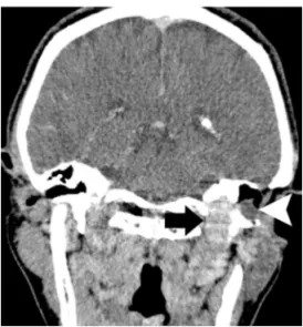
tomog-raphy (HRCT) scan showed a well-deined soft tissue mass with contrast-enhance-ment in the left jugular foramen with ex-tension to the temporal bone and below to the skull base, producing erosion and expansion of the jugular foramen. In ad-dition, a non-enhancing soft tissue lesion was seen in the surgical cavity, in close contact with the enhancing mass (Fig 1).
he MRI corroborated the HRCT scan indings, demonstrating a heterogeneous left jugular foramen lesion, extending to the middle ear, with low signal intensity on T1-weighted images, iso/high signal in-tensity on T2-weighted images,
Arq Neuropsiquiatr 2010;68(2)
307
Cholesteatoma: following paraganglioma surgery. MRI Brito et al.
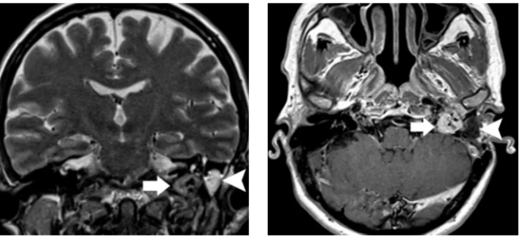
ing low-voids within the mass and intense contrast-en-hancement, measuring 1.9 × 1.5 cm. he non-enhancing lesion seen in HRCT scan demonstrated at MRI hypoin-tense signal intensity on T1-weighted sequences, hyper-intense signal intensity on T2-weighted images, with no enhancement after intravenous gadolinium administra-tion, measuring 1.5 × 1.1 cm (Figs 2 and 3). In the coro-nal singshot turbo spin- echo (SS TSE) DWI, this le-sion clearly exhibited a hyperintense signal (Fig 4). he diagnosis of residual PJT associated with cholesteatoma was suggested.
he patient underwent a revision mastoidectomy, for the resection of the cholesteatoma, and the histological study conirmed this diagnosis. hen, she was referred to the Oncology Department for treatment of the residual PJT with radiotherapy. At that time she signed informed consent allowing the publication of the case report.
DISCUSSION
Paragangliomas are extremely rare, accounting for 6% of all neoplasm in the head and neck region. he imaging aspects allow the diagnosis with conidence. he HRCT scan is important at the diagnosis to evaluate the exten-sion of the leexten-sion, mainly related to the skull base. PJT appears as locally aggressive and destructive neoplastic lesion of foramen jugular and middle ear, with intense contrast enhancement. MRI provides better deinition of extension and characterization of PJT and also is su-perior to demonstrate involvement of neural and vascu-lar structures. At MRI, they have a low intensity signal on T1-weighted images, a high intensity signal on T2-weighted images and enhance strongly after contrast. In addition, demonstrate focal intratumoral hemorrhages or slow- low appearing as high signal intensity and low
voids produced by high-low blood vessels, mainly in le-sion bigger than 1 cm, producing a characteristic “salt and pepper” appearance in both T1 and T2 weighted images (1,2). Corroborating these indings, in our case, the MRI showed a heterogeneous mass with low signal intensi-ty on T1-weighted images, iso/high signal intensiintensi-ty on T2-weighted images, presenting low-voids and contrast- enhancement.
In most cases of PJT, surgical resection of the tumor is the therapy of choice. However, even recent surgical series demonstrate a high incidence of incomplete resec-tion, because of their highly vascular structure and their extremely critical location. Unfortunately, the spectrum of associated morbidity remains considerable, including deicits of the IXth to XIIth cranial nerves, paralyzed
vo-cal cords, facial nerve palsy and hearing deterioration or loss, cerebrospinal luid leaks and so on5,6.
he development of cholesteatoma in the postoper-ative of paraganglioma cavity is a rare complication, and only one case was previously mentioned in the literature5.
he origin of acquired cholesteatomas in a post-surgi-cal context is uncertain, and is supported by iatrogenic, post-traumatic or implantation theories, that considers the possibility of iatrogenic implantation of epithelial cells into the middle ear, as after non-cholesteatomatous con-ditions or injury to the temporal bone3,4. We believed that
the use of Rambo procedure, in witch abdominal wall fat is placed into the surgical cavity, or unsatisfactory closure of the new cavity, might incidentally have coursed with implantation of skin cells into the surgical cavity and con-tributed to the development of cholesteatoma.
In patients with cholesteatoma, the HRCT remains the imaging technique of choice for the diagnosis and sur-gical planning. hey appear as a soft- tissue mass usually
Fig 2. Coronal T2- weighted MR image dem-onstrates an isointensive lesion with low-voids in the left jugular fossa and middle ear (arrow), compatible with residual PJT. Anoth-er lesion with hypAnoth-erintensive signal could be seen into the surgical cavity (arrowhead). Note the close relation between them.
Fig 3. Axial post-gadolinium T1-weighted MR image shows the intense enhancement of the lesion at left jugular fossa/middle ear, presenting the characteristics low voids (ar-row). Notice also the non-enhancing cho-lesteatoma (arrowhead) in close contact with the PJT.
Arq Neuropsiquiatr 2010;68(2)
308
Cholesteatoma: following paraganglioma surgery. MRI Brito et al.
associated with erosion of bone components of the mid-dle ear. In the MRI, they present with iso / hipointense signal intensity on T1-weighted images and moderate-ly hyperintense signal intensity on T2-weighted images, with or without peripheral enhancement after gadolinium administration. Recently, the DWI has become the use of MRI fundamental in diagnosis of cholesteatoma and pre-second look recidivate cholesteatoma7-10.
he cholesteatoma appears as a marked hyperintense lesion at DWI with no false-positives reported in the lit-erature. he sensitivity of SS TSE DWI in detection of cholesteatoma is higher compared with other DWI se-quences, such as echo-planar imaging (EPI), being able to detect lesions as small as 2 mm9-10. he exact cause of the
hyperintensity signal in cholesteatomas is not complete-ly understood, but most of the studies attributed this ab-normality to T2 shine-through efect7-10. In our case, the
SS TSE DWI was essential for the diagnosis of cholestea-toma in a surgical cavity of a previous resected PJT.
In summary the development of cholesteatoma in the surgical cavity of a post-operative paraganglioma is ex-tremely rare. In the present report, the use of SS TSE DWI was essential for the diagnosis of this unusual com-plication of PJT surgery, explaining the reason of the
pa-tient symptoms and helping the planning of the best ther-apeutic option.
REFERENCES
1. Rao AB, Koeller KK, Adair CF. Paragangliomas of head and neck: radiologic-pathologic correlation. Radiographics 1999;19:1605-1632.
2. Mattos LP, Ramina R, Borges W, et al. Intradural jugular foramen tumors. Arq Neuropsiquiatr 2004,62:997-1003.
3. Persaud R, Hajiof D, Trinidade A, et al. Evidence-based review of aetiopatho-genic theories of congenital and acquired cholestetoma. J Laryngol Otol 2007;121:1013-1019.
4. Youngs R. Epithelial migration in open mastoidectomy cavities. J Laryngol Otol 1995;109:286-290.
5. Forest JA, Jackson CG, McGrew BM. Long-term control of surgically treated glomus tympanicum tumors. Otol Neurol 2001; 22:232-236.
6. Gerosa M,Visca A, Rizzo P, Foroni R, Nicolato A, Bricolo A. Glomus jugulare tu-mors: the option of gamma knife radiosurgery. Neurosurgery 2006;59:561-569. 7. Vercruysse JP, De Foer B, Pouillon M, Somers T, Casselman JW, Ofeciers E. The value of difusion-weighted MR imaging in the diagnosis of primary ac-quired and residual cholesteatoma: a surgical veriied study of 100 patients. Eur Radiol 2006;16:1461-1467.
8. Aikele P, Kittner T, Ofergeld C, Kaftan H, Huttenbrink KB, Laniado M. Difusion-weight MR imaging of cholesteatoma in pediatric and adult patients who have undergone middle ear surgery. Am J Roentgenol 2003;181:261-265. 9. De Foer B, Vercruysse JP, Bernaerts A, et al. The value of single-shot turbo
spin-echo difusion-weight MR imaging in the detection of middle ear cho-lesteatoma. Neuroradiology 2007;49:841-848.

