Orthodontic traction:
Possible consequences for maxillary canines
and adjacent teeth
Part 1: Root resorption in lateral
incisors and premolars
Alberto Consolaro*
Some professionals are reluctant to indicate orthodontic traction, especially for upper ca-nines. Among the most common reasons for restricting the indication of orthodontic trac-tion are:
1) Root resorption in lateral incisors and premolars.
2) External cervical resorption of the canines under traction.
3) Alveolodental ankylosis of the canine(s) involved in the process.
4) Calcific metamorphosis of the pulp and aseptic pulp necrosis.
These conditions do not result primarily and specifically from orthodontic traction, and can be avoided if certain technical precautions are fol-lowed. For a better understanding of what these technical precautions are and how they work preventively against the possible consequences of orthodontic traction, we need a biological foun-dation. This is the goal of this series of studies on orthodontic traction, especially of upper canines, and its possible consequences.
Development, structure and functions of the dental follicle
The dental follicle occupies the radiolucent space around the crowns of unerupted teeth (Figs 1 and 2). It is firmly attached to the sur-face of the crown by the reduced epithelium of the enamel organ (Fig 3). This thin and delicate epithelial component is sustained and nourished by a thick layer of connective tissue with a variable density of collagen, sometimes loosely, sometimes even hyalinized. The outer portion of dental follicles binds to the surround-ing bone (Figs 2 and 3). In measurements of the pericoronal space in periapical radiographs and orthopantomographs, or panoramic radiographs, the thickness of the dental follicle can reach up to 5.6 mm and still maintain normal structure and organization2,4 (Fig 3).
By removing the follicle and detaching it from the surrounding bone a tissue fragment is obtained which is organized like a thin film and is therefore also known as pericoronal mem-brane. The isolated tissue fragment represented
FIGURE 1 - Typical image of the pericoronal space and normal follicle: homogeneous radiolucency with no overlapping radiopaque or radiolu-cent points; clear bone limit with solid, uniform line (arrows); uniform thickness, regular contour with maximum thickness ranging between 1 and 5.6 mm.2,4
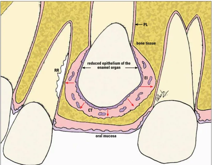
FIGURE 2 - Epithelial structures of the dental follicle—such as the reduced epithelium of the enamel organ and the epithelial islands/cords remnants of the dental lamina (EI)—constantly release epidermal growth factor (EGF, red arrows) in the connective tissue (CT). This mediator, along with other EGF-activated mediators, induces pericoronal bone resorption, an essential phenomenon in the occurrence of tooth eruption. When the path of an unerupted tooth compresses the vessels of the periodontal ligament (PL) of adjacent teeth—with or without orthodontic traction—cementoblasts die on the spot and the root is resorbed (RR) to give rise to the follicle and its moving crown.
RR
reduced epithelium of the enamel organ
oral mucosa
bone tissue
CT
EI PL
In the middle of the collagen fibers and other components of the extracellular matrix of fol-licular connective tissue there are islands and cords of epithelial cells, remnants of the dental lamina (Fig 3), whose number varies according to patient age.2
Gubernacular cord development
A B
future mandible or maxilla. Soon thereafter, it is fragmented by apoptosis, but some of these cells persist on a scheduled basis. The remnants of dental lamina cells are organized in the form of islands and epithelial cords forming a veritable single row that rises from the reduced epitheli-um of the enamel organ toward the oral mucosa. This epithelial cord is called the gubernaculum dentis, or gubernacular cord.
Once the tooth germs have become estab-lished and the dental lamina has undergone frag-mentation, the neighboring mesenchyme gives rise to bone tissue. The tooth germs and the cord of epithelial islands remain unscathed as bone forms around them into the alveolar crypt. Around the gubernacular cords, a delicate bony canal develops, called the gubernacular canal.
The function of the gubernacular canal and cord lies in directing the tooth—once the crown is fully developed—toward the occlusal-most re-gion of the alveolar process. As the tooth erupts towards the mucosa, the dental follicle will in-corporate the islands and cords of the epithelial
cells of the gubernacular cord into its connective tissue, while increasing the presence of its epi-thelial component in this region (Fig 3).
Development of the alveolar crypts and gubernacular canal
The epithelial cells need to be in constant pro-liferation and synthesis given their constant des-quamation in skin and mucosal linings and also because of its intense production of secretions such as milk, saliva and tears. This constant prolifera-tion stimulus is provided by individual epithelial cells, which release to their neighbors—via specific receptors—what is called the Epidermal Growth Factor (EGF) mediator. Although bone cells have EGF receptors, in these cells EGF stimulates bone resorption. Other mediators have their action trig-gered by EGF (Fig 1), such as TGF-beta, which stimulates the formation of clasts, and CSF-1 and IL-1, which recruit their precursors.
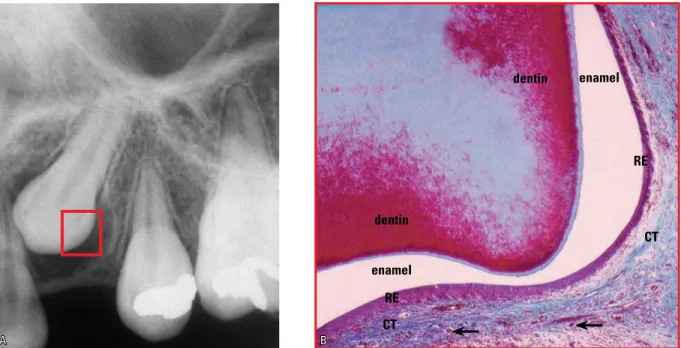
The bone tissue is maintained at a distance from the epithelial tissues because the released EGF stimulates bone resorption, as occurs in the FIGURE 3 - The pericoronal space and dental follicle of upper canines are more laterally bulging due to the coronary anatomy, as shown in A. The reduced epithelium of the enamel organ (RE) is firmly adhered to the enamel of unerupted teeth, while the epithelial islands remnants of the dental lamina and gubernaculum cord (arrows) are distributed across the connective tissue (CT) of the dental follicle.
dentin
dentin
RE
CT
RE
CT
enamel
When bone is formed by the mesenchyme, the tooth germs are circumscribed. The alveolar crypts and the gubernacular canal are simulta-neously established, since the tooth germs and gubernacular islands and cords are epithelial tis-sues that release EGF, which constantly stimu-lates bone resorption in the neighboring tissues. The foregoing explanation allows us to as-sert that:
1. The follicle is an epithelial component comprised of (a) the reduced epithelium of the enamel organ, firmly adhered to the crown, and (b) the cords and islands of odontogenic cells derived from the dental lamina (Figs 2 and 3).
2. The connective tissue comprises the largest volume of follicles and, outside the pericoro-nal space, it takes on the form of a membrane and/or pouch.
3. The epithelial component continuously releases EGF and thus preserves the peri-coronal space by stimulating bone resorp-tion and thus keeping the bone away from the enamel (Fig 2).
4. The cascading release of EGF and other me-diators is essential for the mechanism of tooth eruption. The forces derived from the devel-opment of teeth and growth vectors stimulate increased secretion of EGF and promote bone resorption, directing tooth eruption in the oc-clusal direction (Fig 2).
When a tooth root is experimentally re-moved1 but the crown and dental follicle are
preserved, the tooth will erupt normally. Like-wise, the tooth will erupt when the crown is removed and the dental follicle and tooth root are left in its place. When metal or silicone rep-licas replace unerupted teeth but the follicle is preserved, the artificial teeth or replicas will still erupt. The dental follicle is an essential and
Criteria for evaluating pericoronal space images: image, thickness, contour and boundaries
The image of the pericoronal space (Figs 1, 3, 4 and 7) should:
(a) Be homogeneously radiolucent, devoid of radiopaque points or radiolucent micro lodge type areas, as these may denote a source of odontogenic tumors.
(b) Have its boundaries with the adjacent bone defined by a uniform and continuous radiopaque line. If this line is discontinued and/or riddled with images that resemble the gnawing of a mouse, it may represent a source of odontogenic cysts and tumors. (c) Have its contour characterized by
uni-form pericoronal space thickness, posi-tioned symmetrically to the dental crown. When some areas grow thicker than oth-ers, in the form of embroidery and wavy contours, this may characterize a source of odontogenic cysts and tumors.
(d) Have a thickness ranging from 1 mm to less than 5.6 mm.2,4 Beyond these limits, one
should suspect the presence of a dentiger-ous cyst or some other follicular disease. In assessing the image of the pericoronal space, one should note that:
1) Diseases derived from the dental follicle can go unnoticed and may be present even when the pericoronal space displays normal apparent thickness.
2) Changes derived from the dental follicle take place only occasionally, and are percentage-wise very rare, considering the frequency of un-erupted teeth in patients.
The concept of pericoronal folliculopathies
A B
mediators will be increased, thereby stimulating the organization and function of bone modeling units (BMUs) (Fig 2).
From the standpoint of imaging, if an un-erupted tooth is located very close to the root of another tooth and if the former's trajectory is active due to the eruption and the presence of growth vectors, resorption is usually induced (Figs 2, 4, 5 and 6). This scenario is very often found in the relationship between the region of the canines and the upper lateral incisors (Figs 4, 5 and 6), as well as between third molars and the distal surface of second molars.
Extraction of the unerupted tooth triggers process regression and re-covering of the resorbed area by new cementoblasts, with deposition of a new layer of cementoblasts and reattachment of periodontal fibers. This behavior often occurs with the lower third and second molars. Such oc-currence will only take place if the environment is not contaminated by bacteria.
In cases of upper canines, orthodontic and or orthopedic appliances redirect the eruptive path and/or also the growth vectors involved. Root resorption will cease in neighboring teeth, whereas the surface will be repaired by new cementoblasts and renewed cementum follicle can be termed pericoronal
folliculopa-thy, namely:
• Acute and chronic pericoronaritis. • Paradental cyst.
• Inflammatory follicular cyst. • Dentigerous cyst (Fig 7). • Eruption cyst.
• Hyperplastic dental follicle.
However, many other odontogenic cysts and tumors also originate from the dental follicle but are not exclusive to that structure or loca-tion. Odontogenic keratocysts, ameloblastomas, odontogenic fibroma, odontoma, etc. also origi-nate from the dental follicle.
Pericoronal space of unerupted teeth and root resorption of adjacent teeth
The dental follicle is rich in mediators that stimulate bone resorption locally, especially EGF (Fig 2). When maxillary growth vectors and eruptive forces bring the crown of an un-erupted tooth close to the root of an un-erupted tooth, there occur the compression of periodon-tal vessels and the death of cementoblasts that cover the surface, protecting it from resorption (Figs 5 and 6). Thus, the root surface will be exposed and the amount of local resorption
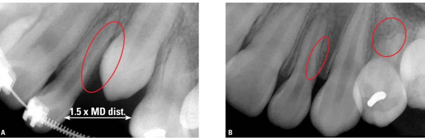
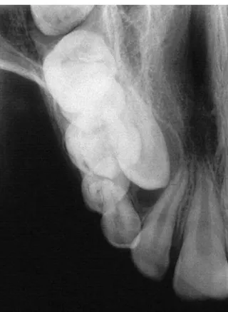
FIGURE 4 - Example of unerupted maxillary canine that did not reach the occlusal plane (A). Once the space in the dental arch reached 1.5 times the mesiodistal distance of the crown—to accommodate the bulging dental follicle typical of the maxillary canine—the tooth moved naturally to its place in the dental arch (B). But the existing proximity of the upper canine and its dental follicle caused lateral resorption (circles) in the roots of the lateral incisor and first premolar.
A B
formation (Fig 4). This situation is often found in the relationship of canines with the upper lateral incisors.
A conduct that must necessarily be adopt-ed to avert the resorption of teeth adjacent to the unerupted tooth—when such unerupted tooth is not being extracted but rather retracted orthodontically—lies in increasing dental arch space so that the unerupted tooth lodges in the area along with its crown and especially its follicle. The opening of space eliminates com-pression of the periodontal ligament of adja-cent teeth while cementoblasts and cementum re-cover the roots of these teeth (Figs 4 and 6). Thus, the dental follicle of the erupted tooth re-mains farther away from the root surface so that its mediators no longer act as stimulators of re-sorption. Instead, they only stimulate pericoro-nal bone resorption to allow eruption to occur in the desired path.
Size, thickness and shape of follicles in maxillary canines compared with other teeth
The thickness and shape of follicles allow their pericoronal spaces to have a more or less uniform contour of the incisal and occlusal surfaces with their cusps (Fig 1). However, the unique shape of upper canines—with their rather convex lateral surfaces forming a cusp, as it were, at their incisal edge, which ends in an acute angle—provides a very specific pericoronal space shape (Fig 3).
The dental follicle of maxillary canines ap-pears to bulge and widen laterally, more so than the other teeth (Figs 3 and 6). Radiographic im-ages and Computed Tomography (CT) scans clearly show that the lateral thickness of the pericoronal spaces of upper canines is greater than in other teeth, especially if compared with incisors, and even with premolars.
The dental follicle of the upper canines and their resulting pericoronal spaces are so bulging FIGURE 5 - In some cases, detection
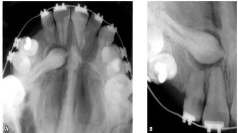
of the resorption caused by unerupted teeth—including maxillary canines—in adjacent teeth only occurs when it is al-ready too late, as was the case of this up-per lateral incisor. But sometimes, it can also involve the central incisors.
in some cases that added to all the probable im-age distortion, deciding between a diagnosis of normality or incipient dentigerous cyst poses a challenge (Figs 3, 6 and 7).
In assessing the need to whether or not open the space between upper lateral incisors and premolars to allow upper canines to naturally lodge in the upper arch, this lateral bulging of their pericoronal space should be considered.
This consideration should be emphasized be-cause the dental follicle does not represent only a soft tissue that covers the crown and could be easily compressed under traction, but rather
because it is the tissue or organ responsible for tooth eruption. Thanks to its large number of me-diators, the dental follicle stimulates pericoronal bone resorption, actively producing tooth move-ment in the occlusal direction (Figs 2, 3 and 4).
The follicle is composed of soft tissues and although it can be physically compressed be-tween the canine crown and the roots of the lat-eral incisors and premolars, this maneuver dur-ing traction may impose a biological cost. Re-sorption of these lateral roots cause, to a lesser or greater degree, structural impairment (Fig 4). Compression of the dental follicle of maxillary canines occurs in conjunction with compression of vessels of the periodontal ligament of adja-cent teeth and eventual death of cementoblasts that protect those roots from clasts and other BMU components.
In following the clinical guidelines to de-termine how much space must be provided to enable unerupted upper canine traction, profes-sionals are encouraged to calculate the mesio-distal distance of the crown and multiply that measurement by 1.5. This action will ensure greater integrity of the lateral roots of adjacent teeth (Fig 4).
One should be aware, however, that creating this space is not clinically possible in all cases. Using any measurement lower than the one aforementioned may result in highly successful traction, with no damage to lateral incisors and premolars, but the risks are greater. The exact-ness of mathematics cannot always be systemati-cally applied in making biological decisions. The recommended criterion and measurement serve as a starting point for decision making relevant to each case. In cases where it can be applied fully, assurance regarding the preservation of neighboring roots will certainly increase.
In assessing the damage caused by root re-sorption in maxillary lateral incisors due to the proximity of unerupted canines, it seems ap-propriate to cite the literature.5,6 The presence FIGURE 7 - The image of the pericoronal space of the maxillary canine
incisors were located close to canines that had remained unerupted for longer than normal. The same cases were evaluated using tomographic sections and reconstructions, and disclosed 25% impairment. CT is the best method to accurately assess the damage caused by canine traction to the roots of upper lateral incisors.
Dental follicle development and functions
In its early stages, the enamel organ resem-bles a bell and is lined by what are known as the inner and outer epithelia. Between these lia there are two other thicker layers of epithe-lial cells, which are known as stellate reticulum and intermediate stratum. As the enamel organ forms this mineralized tissue on the inside of the bell, it becomes narrower or thinner and the four epithelial layers will flatten to form a single epithelium that is firmly adhered to the enamel surface and receives the name of reduced epi-thelium of the enamel organ (Figs 2 and 3).
The reduced epithelium of the enamel organ and, as a result, the dental follicle, have the fol-lowing main functions:
a) "Hide" or protect enamel resorption by clastic cells (Fig 3).
b) Prevent the bone from developing directly on the enamel surface.
c) Support tooth eruption by releasing me-diators that are typical of epithelia, such as EGF. The reduced epithelium of the enamel organ and odontogenic epithelial islands and cords are actively involved in pericoronal bone resorption, essential if tooth eruption is to follow a path that leads to the alveolar mucosal surface, thanks to the release of EGF (Fig 2).
d) Constitute the primary junctional epithe-lium by merging with the oral mucosa, and allow teeth to erupt in the oral environment without exposing the internal environment of the body,
Final considerations
Root resorption of upper lateral incisors and premolars (Figs 4, 5 and 6) is among the pos-sible consequences of unerupted upper canine traction. In planning treatment of unerupted ca-nines, one is advised to assess the thickness of the dental follicle, bearing it in mind when creating space to accommodate it in the dental arch. The aim here is to seek either normal canine eruption or orthodontic traction of said teeth. The lateral compression of the dental follicle during erup-tion—with or without canine traction—against the roots of the lateral incisors and/or premolars may cause these teeth to resorb, as a result of the compression of periodontal vessels and the death of cementoblasts.
In planning the space to be obtained in the dental arch to ensure that the unerupted tooth fits properly, it must be assumed that the dental follicle of maxillary canines—given their unique anatomy—tend to bulge and broaden laterally, more than any other teeth.
The amount of space in the dental arch that would offer the least risk of root resorption for adjacent teeth during orthodontic traction is equivalent to 1.5 times the mesiodistal distance of upper canines, although this measure is not al-ways amenable to application in all clinical cases. In forthcoming studies, we will discuss the other possible consequences of orthodontic traction of unerupted teeth, especially canines, among which the following are noteworthy: (1) External cervical resorption in canines un-der traction, (2) Alveolodental ankylosis of ca-nines, (3) Calcific metamorphosis of the dental pulp and aseptic pulp necrosis.
1. Cahill DR, Marks SC Jr. Tooth eruption: evidence for the central role of the dental follicle. J Oral Pathol. 1980 Jul;9(4):189-200.
2. Consolaro A. Caracterização microscópica de folículos pericoronários de dentes não irrompidos e parcialmente irrompidos. Sua relação com a idade. [tese]. Bauru (SP): Universidade de São Paulo; 1987.
3. Consolaro A, Consolaro MFMO, Santamaria M Jr. A anquilose não é induzida pelo movimento ortodôntico. Os restos
epiteliais de Malassez na isiologia periodontal. Rev Clín
Ortod Dental Press. 2010 abr-maio;9(2):101-10.
REFERENCES
4. Damante JH. Estudo dos folículos pericoronários de dentes não irrompidos e parcialmente irrompidos. Inter-relação
clínica, radiográica e microscópica. [tese]. Bauru (SP):
Universidade de São Paulo; 1987.
5. Ericson S, Kurol J. Radiographic examination of ectopically erupting maxillary canines. Am J Orthod Dentofacial Orthop. 1987 Jun;91(6):483-92.
6. Otto RL. Early and unusual incisor resorption due to impacted maxillary canines. Am J Orthod Dentofacial Orthop. 2003 Oct;124(4):446-9.
Contact address Alberto Consolaro