Solitary median maxillary central incisor
syndrome: Case report
Eduardo Machado*, Patricia Machado**, Betina Grehs***, Renésio Armindo Grehs****
Introduction: The presence of a single median maxillary central incisor is an uncommon
event in the population. The prevalence of the Solitary Median Maxillary Central Inci-sor (SMMCI) syndrome is about 1:50,000 live births, occurring more in women. This alteration in the development of the dental occlusion is characterized by structural mal-formations, over all in midline region of the patient. The early diagnosis and the adequate treatment of this syndrome are of great importance, therefore this condition can be an indication that the patient can present other severe congenital malformations, not having to consider the SMMCI a simple dental anomaly. The orthodontic procedures, in these cases, vary depending on the degree of involvement of bone structures of the maxilla, the occlusion in itself, and mainly of the midpalatal suture. Objectives: To discuss, based on scientific evidence, important aspects related to the SMMCI and present a clinical case of female patient with SMMCI, which was submitted to orthodontic treatment in the Children’s Dental Integrated Clinic of the Federal University of Santa Maria - RS/Brazil. Conclusion: According to the critical analysis of literature, it is very important to correctly early diagnose this condition, since there is the possibility of this syndrome to be associ-ated with other problems of development. Moreover, the patients affected by SMMCI should be attended by a multidisciplinary health team in order to optimize the clinical results and recover the quality of life of these patients.
Abstract
Keywords: Solitary median maxillary central incisor. Single median maxillary central incisor. SMMCI. Orthodontics.
* Specialist in Temporomandibular Disorders (TMD) and Orofacial Pain by Federal University of Paraná (UFPR). Graduated in Dentistry by Federal University of Santa Maria (UFSM).
** Student of the Specialization Course in Prosthetic Dentistry by Pontiical Catholic University of Rio Grande do Sul (PUCRS). Graduated in Dentistry by UFSM.
*** Master student in Orthodontics in UNESP.
B
B C
A A
INTRODUCTION
The congenital absence of upper central in-cisors is a rare condition, while the presence of a single central incisor also is an uncommon event.21 The prevalence of the Solitary Median Maxillary Central Incisor (SMMCI) syndrome, also known as Single Median Maxillary Cen-tral Incisor Syndrome, occurs in 1:50,000 live births, with higher involvement of women. In this syndrome, developmental defects occur due to unknown factors operating in utero about the 35th–38th day from conception and are charac-terized by structural malformations, mainly midline defects in the patients.11,24
Thus, the purpose of this study is to discuss, within a context based on scientific evidence, and illustrate, with a case report, relevant as-pects concerning this condition.
CASE REPORT
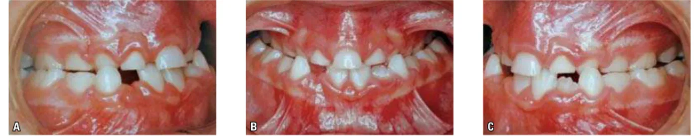
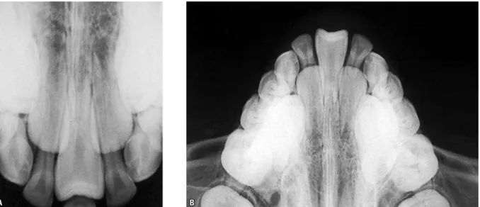
A eight years and three months old patient, fe-male, Caucasian, Brazilian, presented to the Chil-dren’s Dental Integrated Clinic at Federal Univer-sity of Santa Maria/RS (Brazil) for evaluation. After the initial clinical examination, the patient was se-lected and referred to the division of Orthodon-tics at the Children’s Dental Clinic. Once accepted at the Division of Orthodontics of this clinic, the patient was well attended, her clinical history and the records of physical-clinical examinations were obtained and the orthodontic records necessary for diagnosis and treatment planning were requested. During clinical examination a very significant alter-ation was observed, the presence of a single central incisor, compatible with SMMCI, and maxillary atresia, as shown in figures 1, 2 and 3.
Regarding the presence of systemic changes,
FIGURE 1 - A) Right lateral view of the clinical status at diagnosis. B) Initial clinical aspect of the case, with the presence of a solitary maxillary central incisor.
C) Left lateral view of the clinical status at diagnosis.
FIGURE 2 - A) Dental cast study models showing a single central incisor. B) Upper arch study model showing maxillary atresia and a solitary
A B
the patient’s parent reported no involvement. This evaluation is important, since SMMCI may be associated with other developmental problems such as congenital nasal abnormali-ties,1,4,11,15,16,17 growth deficiencies,8,22 holopros-encephaly,6,28 format changes and craniofa-cial morphology,25 congenital heart disease,8,10 among other local and systemic changes. How-ever, there are studies that found no relation-ship between SMMCI and systemic changes.5,27
Some authors also found associations between SMMCI and SHH gene muta-tions9,10,12,14,19,20 and deletions in parts of chro-mosome 18p2,7 and/or of chromosome 7q.10,18,26 Thus, an evaluation of a geneticist can find some association between SMMCI and chro-mosomal abnormalities.
The orthodontic treatment plan comprised a Phase I, which consisted of rapid maxillary expansion (RME), as well as support and in-teraction with Prosthetic Dentistry, Pediatric Dentistry and Oral and Maxillofacial Surgery specialties. At the end a Phase II was scheduled with fixed orthodontic treatment. Furthermore, the patient was referred to a multidisciplinary
health team, including pediatricians, geneti-cists, speech therapists and psychologists, since this anomaly may be associated with other de-velopmental problems.
DISCUSSION
The involvement of SMMCI was initially re-ported by Scott23 who described a girl with the presence of a solitary median maxillary central incisor, as an isolated finding. Another case of SMMCI was verified by Fulstow,8 but the patient showed apart from the single central incisor, short stature, congenital heart disease, microcephaly and scoliosis. Some factors that may be associated with SMMCI are the pituitary gland dysfunction and short stature, whereas in a study involving patients with SMMCI, 7 subjects had short stat-ure and 5 were deficient in growth hormone pro-duction.22 However, Wesley et al27 reported two cases of SMMCI in subjects with normal stature, while Cho and Drummond5 reported three cases of SMMCI in three Chinese girls with no growth deficiencies or systemic involvement.
According to DiBiase and Cobourne,6 the most common cause of a missing maxillary FIGURE 3 - A) Periapical radiograph, which confirms the presence of a solitary maxillary central incisor. B) Occlusal radiograph, confirming the presence of
central incisor is trauma, or more rarely hy-podontia. When dental absence has no explana-tion in the patient’s clinical history, a genetic analysis can show results. It is important to rec-ognize the SMMCI when in an unknown eti-ology, because it may indicate a risk factor for holoprosencephaly. Thus, the role of the ortho-dontist is extremely important in the diagnosis of this condition, which must refer the patient for genetic testing to investigate other possible developmental disorders.
The SMMCI may be associated with vari-ous congenital nasal anomalies such as choanal atresia, intra-nasal stenosis and nasal pyriform aperture stenosis. Choanal atresia consists in a bone or membranous obstruction of the pos-terior nasal aperture caused by a failure in the oronasal disintegration. The intra-nasal stenosis is a bony narrowing of the nasal cavity between the pyriform aperture and the posterior choa-nae, whereas the nasal pyriform aperture steno-sis is an anterior nasal obstruction secondary to the bone growth of the nasal processes of the maxilla. It is important to note that the clinical aspects of the above changes are similar, and often a computed tomography is required for definitive diagnosis,4,17 being that prenatal diag-nosis of SMMCI can be done through magnetic resonance imaging.13
Thus, several studies have looked at the as-sociation of nasal obstructions and SMMCI. Ar-lis and Ward1 evaluated six patients with con-genital stenosis of the nasal pyriform aperture and found that of these, 4 had SMMCI. Lo et al17 found in their results that 63% of patients with congenital stenosis in the nasal pyriform aperture also presented SMMCI, while Hall et al11 found that among 21 patients with SMMCI, all had a positive relationship with a history of nasal congenital obstruction, whereas choanal atresia and intra-nasal stenosis were found in 7 and 8 patients respectively. Already, Levison et al16 reported two cases of neonatal children
with nasal obstruction due to stenosis of the choanae, which had an association with single maxillary central incisor, a fact verified by com-puted tomography.
The presence of chromosomal defects was observed in some children who had SMMCI. Dolan et al7 found chromosomal abnormalities in children with a single central incisor, with deletion of parts of chromosome 18 (18p), which was also reported by Aughton et al.2 Nonetheless, Masuno et al18 reported deletion in the terminal portion of chromosome 7q, which was also found by Hall10 and Tubbs and Oakes.26 Another factor that seems to be as-sociated with the SMMCI is a mutation of the SHH gene.9,10,12,14,19,20
For Yassin and El-Tal,28 the appearance of a solitary incisor in place of the two central incisors may occur due to fusion of two neigh-boring teeth or to agenesis of a tooth germ. However, this can be associated with other sys-temic disorders such as autosomal dominant holoprosencephaly, growth retardation and midline developmental defects. Becktor et al3 evaluated the intermaxillary suture, the erup-tion pattern of the single central incisor and growth of the maxilla in a group of patients with SMMCI. The sample consisted of 11 pa-tients with SMMCI, who underwent orthopan-tomographs, dental and lateral cephalometric radiographs. The X-rays showed that the inter-maxillary suture was abnormal anterior to the incisive foramen, however, the horizontal and vertical growth of the maxilla was normal.
parameters, showed a short anterior cranial base, a short, retrognathic and posteriorly inclined max-illa, and a retrognathic and posteriorly inclined mandible, and morphological changes in the sella turcica were found in five patients examined. Moreover, this group of patients had character-istics such as: nasal obstruction, septal deviation, absence of the fraenum of the upper lip, and a complete or incomplete mid-palatal ridge. Thus, the presence of SMMCI should not be consid-ered as a simple dental anomaly, because it may be associated with other clinical characteristics and craniofacial malformations.
Tabatabaie et al25 evaluated the neurocranial and craniofacial morphology of children with SMMCI using profile radiographs and cepha-lometric analysis. The sample comprised 13 children (12 girls and 1 boy) aged between 7 and 17 years. Cephalometric evaluations were compared with standard measures. The study results showed that the size of the neurocra-nium, the maxillary prognathism and tion, the mandibular prognathism and inclina-tion of lower incisors are significantly decreased in patients with SMMCI. But, the mandibular inclination, vertical jaw relationship and man-dibular angle are significantly increased in pa-tients affected by SMMCI. The data from this study showed that the occurrence of SMMCI is a sign of anomaly development, associated with deviations in neurocranial size and shape and in craniofacial morphology.
According to Hall,10 the etiology of SMMCI is uncertain and may be associated with muta-tions in SHH gene (I111F) in chromosome 7q, with a positive correlation with congenital nasal malformations. These teeth erupt and develop in the midline of the maxillary arch, both in pri-mary and permanent dentitions. The presence of SMMCI may be associated with some com-mon congenital abnormalities such as moderate to severe intellectual disability, congenital heart disease, cleft lip and/or palate and less frequently,
microcephaly, hypopituitarism, strabismus, duo-denal atresia, scoliosis, hypothyroidism, absent kidney, micropenis and ambiguous genitalia. Short stature can be found in children. The di-agnosis of SMMCI should be performed at 8 months of age, but can be done at birth and pos-sibly prenatal, between the 18th and 22nd week of gestation by ultrasound examination. In patients with SMMCI rehabilitation should be undertak-en in accordance with the anomalies presundertak-ented by individuals: choanal stenosis requires surgical treatment, short stature should be approached with growth hormone therapy, and the presence of single maxillary central incisor should be a requirement for an multidisciplinary treatment involving the specialties of Orthodontics, Pros-thetic Dentistry and Oral Surgery.
Cho and Drummond5 suggest that early di-agnosis of SMMCI is extremely important, be-cause it is a sign that the patient may present with other severe congenital malformations. If they are pediatric patients they should be seen together with the pediatrician. In three patients evaluated by these authors,5 all were female and had no growth deficiencies or any systemic involvement. The dental management consisted in preventive and orthodontic ap-proaches, and in two cases expansion of the upper arch was performed, moving the solitary central incisor to one side and obtaining space for placement of osseointegrated implant or prosthesis on the other side.
CONCLUSIONS
Dental procedures for patients with SMMCI vary with the degree of commitment that it causes. Orthodontic procedures are extremely important for the return of function and aes-thetics to the patient, requiring an interdisci-plinary approach with other dental specialties for optimizing clinical outcomes. Moreover,
it is important that the patient should be at-tended by a multidisciplinary health team, in-cluding pediatricians and other medical pro-fessionals, geneticists, speech therapists and psychologists, since this anomaly may be as-sociated with other developmental problems and systemic changes.
1. Arlis H, Ward RF. Congenital nasal pyriform aperture stenosis- isolated abnormality vs developmental ield defect. Arch Otolaryngol Head Neck Surg. 1992 Sep;118(9):989-91. 2. Aughton DJ, AlSaadi AA, Transue DJ. Single maxillary
central incisor in a girl with del(18p) syndrome. J Med Genet. 1991 Aug;28(8):530-2.
3. Becktor KB, Sverrild L, Pallisgaard C, Burhoj J, Kjaer I. Eruption of the central incisor, the intermaxillary suture, and maxillary growth in patients with a single median maxillary central incisor. Acta Odontol Scand. 2001 Dec;59(6):361-6. 4. Brown OE, Manning SC, Myer CM. Congenital nasal
pyriform aperture stenosis. Laryngos. 1989 Jan;99(1):86-91. 5. Cho SY, Drummond BK. Solitary median maxillary central
incisor and normal stature: a report of three cases. Int J Paediatr Dent. 2006 Mar;16(2):128-34.
6. DiBiase AT, Cobourne MT. Beware the solitary maxillary median central incisor. J Orthod. 2008 Mar;35(1):16-9. 7. Dolan LM, Willson K, Wilson WG. 18p-syndrome with a single
central maxillary incisor. J Med Genet. 1981 Oct;18(5):396-8. 8. Fulstow ED. The congenital absence of an upper central
incisor: report of a case. Br Dent J. 1968 Feb 20;124(4):186-8. 9. Gavelli L, Zanacca C, Caselli G, Banchini G, Dubourg C, David
V, et al. Solitary median maxillary central incisor syndrome: clinical case with a novel mutation of sonic hedgehog. Am J Med Genet A. 2004 May 15;127A(1):93-5.
REFERENCES
10. Hall RK. Solitary median maxillary central incisor (SMMCI) syndrome. Orphanet J Rare Dis. 2006 Apr 9;1:12. 11. Hall RK, Bankier A, Aldred MJ, Kan K, Lucas JO, Perks AG.
Solitary median maxillary central incisor, short stature, choanal atresia/midnasal stenosis (SMMCI) syndrome. Oral Surg Oral Med Oral Pathol Oral Radiol Endod. 1997 Dec;84(6):651-62. 12. Hehr U, Gross C, Diebold U, Wahl D, Beudt U, Heidemann P, et al.
Wide phenotypic variability in families with holoprosencephaly and a sonic hedgehog mutation. Eur J Pediatr. 2004 Jul;163(7):347-52. 13. Johnson N, Windrim R, Chong K, Viero S, Thompson M, Blaser
S. Prenatal diagnosis of solitary median maxillary central incisor syndrome by magnetic resonance imaging. Ultrasound Obstet Gynecol. 2008 Jul;32(1):120-2.
14. Kjaer I, Becktor KB, Russell B. Single median maxillary central incisor, SMMCI. Pathogenesis and phenotypic characteristics. In: IADR/AADR/CADR 82nd General Session; 2004 March 10-13; Hawaii: International Association for Dental Research; 2004. abstract 2639. [cited 2010 June 12]. Available from: http://iadr. confex.com/iadr/2004Hawaii/techprogram/abstract_43524.htm. 15. Kjaer I, Becktor KB, Lisson J, Gormsen C, Russell BG. Face, palate,
and craniofacial morphology in patients with a solitary median maxillary central incisor. Eur J Orthod. 2001 Feb; 23(1):63-73. 16. Levison J, Neas K, Wilson M, Cooper P, Wojtulewicz J.
23. Scott DC. Absence of upper central incisors. Br Dent J. 1958; 104:247-8.
24. Simon AR, Roberts MW. Solitary incisor syndrome and holoprosencephaly. J Clin Pediatr Dent. 1993;17(3):175-7. 25. Tabatabaie F, Sonnesen L, Kjaer I. The neurocranial and
craniofacial morphology in children with solitary median maxillary central incisor (SMMCI). Orthod Craniofac Res. 2008 May;11(2):96-104.
26. Tubbs RS, Oakes WJ. Lumbosacral agenesis and anteroposterior split cord malformation in a patient with single central maxillary incisor: case report and review of the literature. J Child Neurol. 2004 Jul;19(7):544-7.
27. Wesley RK, Hoffman WH, Perrin J, Delaney JR Jr. Solitary maxillary central incisor and normal stature. Oral Surg Oral Med Oral Pathol. 1978 Dec;46(6):837-42.
28. Yassin OM, El-Tal YM. Solitary maxillary central incisor in the midline associated with systemic disorders. Oral Surg Oral Med Oral Pathol Oral Radiol Endod. 1998 May;85(5):548-51.
Contact address
Eduardo Machado
Rua Francisco Trevisan, nº 20, Bairro Nossa Sra. de Lourdes CEP: 97.050-230 - Santa Maria / RS, Brazil
E-mail: [email protected]
Submitted: August 2008 Revised and accepted: October 2008
17. Lo FS, Lee YJ, Lin SP, Shen EY, Huang JK, Lee KS. Solitary maxillary central incisor and congenital nasal pyriform aperture stenosis. Eur J Pediatr. 1998 Jan;157(1):39-44.
18. Masuno M, Fukushima Y, Sugio Y, Ikeda M, Kuroki Y. Two unrelated cases of single maxillary incisor with 7q terminal deletion. Jinrui Idengaku Zasshi. 1990 Dec;35(4):311-7. 19. Nanni L, Ming JE, Du Y, Hall RK, Aldred M, Bankier A, et al.
SHH mutation is associated with solitary median maxillary central incisor: a study of 13 patients and review of the literature. Am J Med Genet. 2001 Jul 22;102(1):1-10. 20. Nieuwenhuis E, Hui CC. Hedgehog signaling and congenital
malformations. Clin Genet. 2005 Mar;67(3):193-208. 21. Nordgarden H, Jensen JL, Storhaug K. Reported prevalence
of congenitally missing teeth in two Norwegian counties. Community Dent Health. 2002 Dec;19(4):258-61.