introduction
Orthodontic treatment in patients with trau-matized teeth is not contraindicated, but clini-cal and radiographic repair and/or post-trauma
complications must be established before treat-ment onset.17
Dental injuries occur more frequently in children between ages eight and nine, involving
Orthodontic treatment in patients with
reimplanted teeth after traumatic avulsion:
A case report
Simone Requião Thá Rocha*, Alexandre Moro**, Ricardo César Moresca***, Gilson Sydney****, Fabian Fraiz*****, Flares Baratto Filho******
introduction: The high prevalence of individuals with dental trauma prior to orthodontic treatment justifies the precautions that should be followed before and during treatment, considering all possible effects of orthodontic movement on traumatized teeth. Among the major traumatic dental injuries, avulsion with subsequent tooth reimplantation entails a higher than average risk of complications, such as pulp necrosis, root resorption and ankylosis. Therefore, it gives orthodontists several reasons for concern. objective: This case report sought to analyze the implications of tooth reimplantation after traumatic avulsion in patients requiring orthodontic treatment. conclusions: Tooth movement of a reimplanted tooth after traumatic avulsion is viable provided no signs of abnormality are present. Ankylosed teeth, however, are not amenable to orthodontic movement but should be preserved as space maintainers until root resorption is completed, provided that the teeth do not present with severe infraocclusion. Should an ankylosed tooth be in severe infraocclusion, crown amputation and root burial are indicated as a means to preserve the alveolar bone in the region, since resorption will occur by replacement of the buried root, as was the case in this report.
Abstract
Keywords: Tooth movement. Dental ankylosis. Dental trauma.
* MSc student in Clinical Dentistry, Positivo University. Professor of the Specialization Course in Orthodontics, Positivo University.
** PhD in Orthodontics, FOB-USP. Associate Professor, Department of Orthodontics, Federal University of Paraná (UFPR). Head Professor in the MSc Pro-gram of Clinical Dentistry, Positivo University.
*** PhD in Orthodontics, FO-USP. Associate Professor, Department of Orthodontics, UFPR. Head Professor in the MSc Program of Clinical Dentistry, Positivo University.
**** PhD in Endodontics, FO-USP. Head Professor of Endodontics, UFPR.
***** PhD in Pediatric Dentistry, FO-USP. Associate Professor of Pediatric Dentistry, UFPR.
mostly the upper central incisors.1,21 Individuals who present with overjet in excess of 3 mm are about twice as likely to suffer damage to the anterior teeth, compared to children with fewer than 3 mm overjet.20,22
The high prevalence of individuals with den-tal trauma prior to orthodontic treatment—ap-proximately 10.7%23—justifies the precautions that should be followed before and during treat-ment, taking into account all possible effects of orthodontic movement on traumatized teeth, as well as how these should be monitored. Thus, during patient history and clinical examination one should seek information on prior dental trauma, as well as clinical signs and symptoms of possible sequelae. A thorough assessment of periapical radiographs and/or CT scans is also indicated. If there is history of dental trauma, the general dental practitioner (GP) and/or en-dodontist should be consulted since a multidisci-plinary approach is the most suitable in this case. The purpose of this case report is to analyze the implications of tooth reimplantation after traumatic avulsion in patients requiring orth-odontic treatment.
LitErAturE rEViEW
Preserving an injured tooth in the oral cav-ity depends not only on adequate emergency treatment but also on a long and appropriate follow-up period. During this period, clinical-radiographic monitoring is essential. However, treatment of traumatized teeth is not always performed appropriately.9 Hamilton, Hill and
Holloway10 found an incidence of 34% of
chil-dren with clinical signs of trauma in a popu-lation of 2,022 children. After radiographic analysis of these cases it was found that only 47% of traumatized teeth showed signs of pre-vious treatment. Among these, however, only 41.5% were considered adequate. Some stud-ies conducted in Brazil8,11 indicate that GPs, in particular, when compared with endodontists,
have little knowledge as regards the best treat-ment for patients with traumatized teeth. This is a disquieting reality which must be reversed by developing strategies to enhance the compe-tence of these professionals.
The prognosis for various types of periodontal tissue injuries seems to depend on the type and severity of the damage, as measured by its ex-tent on the periodontal ligament, i.e., if there was compression or rupture of ligaments. It is true that trauma to the periodontium causes exten-sive tissue injury, with the death of cementoblasts in large areas of the cementum surface. Hemor-rhage and necrosis areas occur in the periodontal ligament, which gradually give way to exudates and inflammatory infiltrates, essential for tissue repair. During the repair process several events occur simultaneously, such as the vital phenom-ena of differentiation and cell migration.6 Studies
in monkeys2 showed that when trauma involves
small areas of the periodontal ligament, charac-terized by a moderate amount of surviving cells near the root surface, this is repaired through the intact regions of the periodontal ligament. Dur-ing this repair process, a surface layer of the root can be resorbed. If this is a superficial resorption, the resorbed cavity is repaired by deposition of new cementum. The cementoblasts located in the neighborhood of the cementoblasts that have succumbed, as well as the pcementoblasts, re-position themselves and begin to cover the ce-mentum surface. Thus, if resorption occurs, it will be so minimal that it will not be perceived clinically nor radiographically.6
infected necrotic tissue or a leukocyte infection area, inflammatory root resorption will occur. On the other hand, if the root canal contains normal or inflamed tissue, there will be cavity
repair with the deposition of new cementum.2
Orthodontic treatment can cause root re-sorption. Teeth that have suffered traumatic in-juries are more likely to develop root resorption than non-traumatized teeth.17 Studies, however, are not conclusive because they are based on a small number of patients with different types of injury, appliances and operators.13 Malmgren et al18 found no significant differences in prognosis between teeth with intact periodontal ligament that had suffered minor to moderate injuries, and non-traumatized teeth, which had been moved orthodontically.
Tooth avulsion with subsequent reimplan-tation is the traumatic injury that involves the greatest risk of complications due to a high like-lihood of bacterial infection through both the pulp and the periodontium. Incidence is greater among children due to incomplete root devel-opment and immaturity of the periodontal liga-ment.19 The initial treatment in such cases is often carried out by parents or school teachers, so the community at large should be taught the basics of how to act in the presence of a tooth avulsion. The need to take quick and proper ac-tion, namely, transporting the tooth in humid conditions (in milk or saliva, under the tongue) and taking the patient to a GP is fundamental.21
A poor prognosis in cases of tooth reimplan-tation is directly related to the amount of time that the tooth remains in the extra-alveolar
medium and in a dry environment.4,21 When an
avulsed tooth is immediately and appropriately reimplanted, it ensures the continued vitality of the periodontal ligament, which is still attached to the tooth.23 When a tooth is avulsed, the periodontal fibers are fractured exactly in the center, where the collagen fibers stemming from the alveolar bone and the fibers that originate
from the cementum are intertwined. Hence, part of the fibers remain adhered to the bone and another part to the cementum. It should be underscored that the fibers that remain at-tached to the bone can be regenerated, but the fibers connected to the root surface have a re-duced capacity for regeneration. Therefore, in cases of avulsion, preserving the fibers that re-main attached to the cementum is a decisive factor determining prognosis.19
Other factors affecting the prognosis of re-implanted teeth are: Patient age, the nature of the accident and subsequent long-term treat-ment of the tooth,14 including adequate end-odontic treatment, if indicated.12
Pulp necrosis occurs in most cases, except when root formation is still incomplete and a pre-served dental papilla helps to maintain pulp vital-ity.6 In teeth with an open apex, periodontal heal-ing occurs more frequently than in teeth whose apex is closed.5 The wider—or more open—is the foramen, the better the prognosis, since pulp re-vascularization is facilitated. However, bacterial contamination of the pulp and periodontal liga-ment reduces the likelihood of revascularization.3 The worst possible scenario occurs in chil-dren and adolescents whose root formation is complete because in these cases pulp revascular-ization seldom occurs. The result is ankylosis, im-mediately followed by replacement resorption.7 In adults, tooth reimplantation in the presence of a necrotic periodontal ligament solely enables the maintenance of alveolar anatomy and sym-metry. The reimplantation procedure, however, is worthwhile in all cases, even if only temporarily, since it allows patients, especially children and adolescents, to keep their natural tooth until it is replaced by a prosthesis and/or implant.14
FIGURE 1 - Tooth crown of tooth 11 elongated with composite resin, due to ankylosis. Note height difference between teeth 11 and 21, cervically.
resorption occurs during the first year post-trau-ma. Boyd, Kinirons and Gregg4 found that a time
period ranging between 102 and 997 days14 had
elapsed before root resorption was detected, sug-gesting the need for a longer follow-up period before starting orthodontic treatment.
When the periodontal ligament experiences extensive damage, a small amount of surviving cells near the root surface triggers a repair pro-cess through rapid osteogenesis, leading to an-kylosis of the tooth2 and its subsequent loss by replacement. Dentoalveolar ankylosis involves fusion of the alveolar bone with the root sub-stance and the consequent disappearance of the periodontal space, which loses its structure and function. The close contact between dental tis-sues and alveolar bone structure furthers the bone remodeling process. This results in resorp-tion of part of the bone tissue and tooth tissue, which will be partially or totally replaced by
new bone formation6.
Replacement resorption increases if the avulsed tooth is allowed to remain outside the oral cavity for extended periods of time. It rang-es from only 9.5% in short periods (lrang-ess than fifteen minutes) to 100% if periods exceed sixty
minutes, in a dry medium.5
Ankylosed teeth are not amenable to orth-odontic movement due to the absence of peri-odontal ligament. Once ankylosis has been di-agnosed, professionals are left with two alter-natives only, extraction, or preservation of the tooth in the arch as a space maintainer until root resorption is completed. In children and adolescents the alveolar bone undergoes consid-erable vertical growth, which is not the case in the region of the ankylosed tooth, causing in-creased tooth infraposition and tipping of adja-cent teeth (Fig 1).17 Infraposition evolves differ-ently in different individuals. There is a high risk of severity when ankylosis is established before pubertal growth, at around 10 years of age.15
Extraction is recommended in cases of
in-clined adjacent teeth or extensive infraposi-tion.17 In other cases, teeth should be examined at intervals of six months until root resorption ceases and the tooth crown either comes loose or can be removed with forceps, after most of the root has been replaced by bone.16
Clinical experience has shown that extraction of ankylosed teeth involves substantial bone loss both horizontally and vertically, which affects, in particular, the thin buccal bone wall in the max-illa.16 To prevent this loss, Malmgren et al16,17 de-scribed a technique that involves removal of the tooth crown with subsequent closure of the alve-olus with the root inside it. When root resorption by replacement takes place it preserves or even enhances alveolar bone height in the vertical direction. It also preserves the alveolar bone in the buccolingual direction, which improves the conditions for orthodontic treatment—if neces-sary—and/or subsequent placement of a prosthe-sis and/or implant.
B A
FIGURE 2 - A) Periapical radiograph taken immedi-ately after trauma. B) After reimplantation of tooth 21 and repositioning of tooth 11.
cLinicAL cAsE rEport
Female patient, aged 10 years, was referred by her pedodontist for orthodontic treatment. She presented with a slightly convex profile, good maxillomandibular relationship (Fig 3), Class I malocclusion, constricted upper and lower arches (Fig 4), anterior mandibular and maxillary crowding, and mandibular midline shift (1 mm to the right).
Four years earlier, the patient had suffered a fall with total avulsion of tooth 21 and extru-sion of tooth 11 (Fig 2, A). According to her pe-dodontist, both radiographically and clinically, the teeth had open apices with divergent walls. Tooth 11 was repositioned and tooth 21 was reimplanted (Fig 2, B). A semi-rigid retainer was bonded and fishing line (nylon) was placed around teeth 13, 11, 21 and 23. Amoxicillin 250 mg was prescribed for 7 days, Cataflan drops for three days, and aqueous polyvinyl-pyrrolidone for cleaning the region. Liquid and semi-liquid food was recommended.
Subsequently, tooth 21 underwent endodon-tic treatment with calcium hydroxide for root apexification.
At the time of the initial orthodontic ex-amination, tooth 21 showed signs of resorp-tion (Fig 5), light browning of the crown and a slight step between teeth 11 and 22. Tooth 11 appeared normal both clinically and radio-graphically (Fig 4, B).
The orthodontic plan provided for the use of a standard Bimler appliance for upper and lower arch expansion and a fixed orthodontic appliance in a second stage for tooth alignment and leveling, and occlusion detailing.
After nine months of treatment with the removable appliance, we observed a significant increase in the size of the step between tooth 21 and teeth 11 and 22 due to the ankylosis in tooth 21.
B A
B C
A
B A
FIGURE 3 - Extraoral photographs before orth-odontic treatment.
B A
B C
A
B C
A
D
FIGURE 7 - Finishing phase of orthodontic treatment.
FIGURE 8 - A, B, C) Final intraoral photographs. D) Final occlusal photograph.
FIGURE 6 - A) Periapical radiograph immediately after crown amputation and burial of the root of tooth 21. B) Periapical radiograph 8 months after crown amputation.
The patient will wait until her growth is completed before having an implant and pros-thesis placed in the edentulous area (Fig 10).
06/2007
discussion
B C A
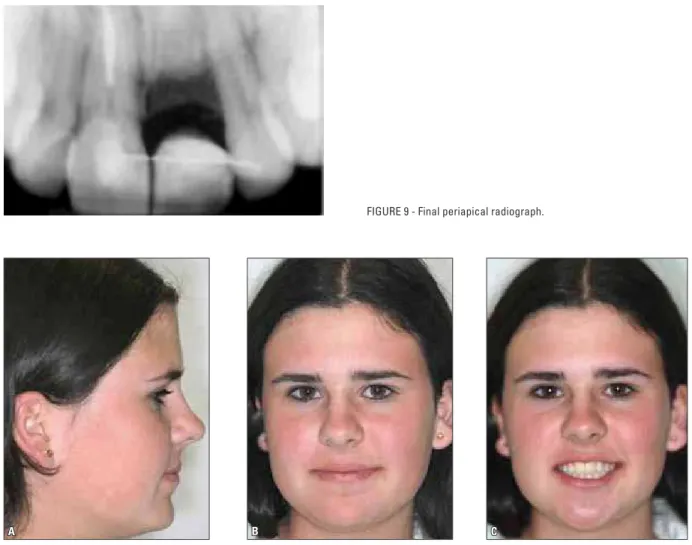
FIGURE 9 - Final periapical radiograph.
FIGURE 10 - Final extraoral photographs.
high prevalence of this condition in patients at an early age and the possibility that these cases do not receive treatment and proper follow-up,9 it is of great importance that orthodontists obtain a comprehensive dental history of their patients. The analysis of periapical radiographs allows the diagnosis of root resorption and ankylosis prior to orthodontic treatment. Regular X-ray control during the entire period of orthodontic move-ment is essential for the diagnosis of root changes. Dental trauma is not a condition that con-traindicates orthodontic treatment, but some studies indicate that it is a predisposing factor
to tooth resorption.6,17 Due to the length of follow-up time required in these cases, a direct relationship between dental trauma and root re-sorption cannot be established at this time.
In this clinical case we diagnosed a cervical root resorption in tooth 21 prior to orthodon-tic treatment, and given the likelihood that this tooth was also ankylosed, no orthodontic forces were ever applied to it.
the initiation of treatment. During orthodontic treatment, tooth 11 exhibited pulp necrosis, which is probably related to the damage suf-fered four years earlier. This scenario is to be ex-pected for trauma cases as sequelae can appear years after an injury. Pulp necrosis is among such sequelae21. Tooth 11 was then subjected to endodontic treatment.
Tooth avulsion, with subsequent reimplanta-tion, displays the most uncertain prognosis of all dental trauma types. A reimplanted tooth can only be moved orthodontically after two years’ follow-up, and only if normal conditions are re-stored.4,6,14,17 The likelihood of root resorption and/or ankylosis is extremely high. Should anky-losis occur, orthodontic treatment becomes limit-ed as an ankyloslimit-ed tooth loses its periodontal liga-ment, therefore rendering orthodontic movement impracticable. Furthermore, the alveolar bone loses its capacity for vertical growth in this region. In younger patients, especially those in the growth phase, ankylosis is accompanied by a growing infraposition of the tooth,17 such as
oc-curred in our clinical case. Given the fact that in this case the degree of infraposition was severe be-cause ankylosis occurred before pubertal growth, we amputated the affected tooth crown and ‘buried’ its root using the technique described by Malmgren et al.16,17 This procedure allows the re-placement of root tissue by alveolar bone, favoring the maintenance of alveolar bone in the region. In some cases it may lead to increased alveolar bone height, eliminating the need for bone grafting for implant and/or prosthesis placement.
concLusions
1. Ankylosed teeth should be preserved as space maintainers until root resorption is com-pleted, provided that the teeth do not present with severe infraposition.
1. Andreasen JO, Andreasen FM. Texto e atlas colorido de trauma-tismo dental. 3ª ed. Porto Alegre: Artmed; 2001.
2. Andreasen JO. Relationship between cell damage in the peri-odontal ligament after replantation and subsequent develop-ment of root resorption. Acta Odontol Scand. 1980;39:15-25. 3. Andreasen JO, Vinding TR, Christensen SS. Predictors for
healing complications in the permanent dentition after dental trauma. Endod Topics. 2006;14:20-7.
4. Boyd DH, Kinirons MJ, Gregg TA. A prospective study of factors affecting survival of replanted permanent incisors in children. Int J Paediatr Dent. 2000 Sep;10(3):200-5.
5. Chappuis V, von Arx T. Replantation of 45 avulsed perma-nent teeth: a 1-year follow-up study. Dent Traumatol. 2005 Oct;21(5):289-96.
6. Consolaro A. Reabsorções dentárias nas especialidades clínicas. Maringá: Dental Press; 2005.
7. Ebeleseder KA, Friehs S, Ruda C, Pertl C, Glockner K, Hulla H. A study of replanted permanent teeth in different age groups. Endod Dent Traumatol. 1998 Dec;14(6):274-8.
8. França RI, Traebert J, Lacerda JT. Brazilian dentist’s knowledge regarding immediate treatment of traumatic dental injuries. Dent Traumatol. 2007 Oct;23(5):287-90.
9. Hamilton FA, Hill FJ, Holloway PJ. An investigation of dento-alveolar trauma and its treatment in an adolescent population. Part 1: the prevalence and incidence of injuries and the extend and adequacy of treatment received. Br Dent J. 1997 Feb 8;182(3):91-5.
10. Hamilton FA, Hill FJ, Holloway PJ. An investigation of dento-alveolar trauma and its treatment in an adolescent population. Part 2: dentists´ knowledge of management methods and their perceptions of barriers to providing care. Br Dent J. 1997 Feb 22;182(4):129-33.
11. Hu LW, Prisco CR, Bombana AC. Knowledge of Brazilian general dentists and endodontists about the emergency management of dento-alveolar trauma. Dent Traumatol. 2006 Jun;22(3):113-7. 12. Keklikoglu N, Asci SK. Histological evaluation of a
re-planted tooth retained for 49 years. Dent Traumatol. 2006 Jun;22(3):157-9.
rEfErEncEs
13. Kindelan SA, Day PF, Kindelan JD, Spencer JR, Duggal MS.
Dental trauma: an overview of its inluence on management of
orthodontic treatment. Part 1. J Orthod. 2008 Jun;35(2):68-78.
14. Kinirons MJ, Boyd DH, Gregg TA. Inlammatory and replace -ment resorption in reimplanted permanent incisor teeth: a study of the characteristics of 84 teeth. Endod Dent Traumatol. 1999 Dec;15(6):269-72.
15. Malmgren B, Malmgren O. Rate of infraposition of reimplanted ankylosed incisors related to age and growth in children and adolescents. Dent Traumatol. 2002 Feb;18(1):28-36. 16. Malmgren B, Cvek M, Lundberg M, Frykholm A. Surgical
treat-ment of ankylosed and infrapositioned reimplanted incisors in adolescents. Scand J Dent Res. 1984 Oct;92(5):391-9. 17. Malmgren O, Malmgren B, Goldson l. Abordagem ortodôntica
da dentição traumatizada. In: Andreasen JO, Andreasen FM. Texto e atlas colorido de traumatismo dental. 3ª ed. Porto Alegre: Artmed; 2001.
18. Malmgren O, Goldson L, Hill C, Orwin A, Petrini L, Lundberg M. Root resorption after orthodontic treatment of traumatized teeth. Am J Orthod. 1982 Dec;82(6):487-91.
19. Melo LL. Traumatismo alvéolo-dentário: etiologia, diagnóstico e tratamento. São Paulo: Artes Médicas: EAP-APCD; 1998. 20. Nguyen QV, Bezemer PD, Habets L, Prahl-Andersen B. A
systematic review of the relationship between overjet size and traumatic dental injuries. Eur J Orthod. 1999 Oct;21(5):503-15. 21. Simões FG, Leonardi DP, Baratto F Filho, Ferreira EL, Fariniuk LF, Sayão SMA. Fatores etiológicos relacionados ao trauma-tismo alvéolo-dentário de pacientes atendidos no pronto socorro odontológico do Hospital Universitário Cajuru. RSBO; 2004; 1(1):50-5.
22. Traebert JICS, Almeida ICS,Garghetti CW, Marcenes W. Preva-lência, necessidade de tratamento e fatores predisponentes do traumatismo na dentição permanente de escolares de 11 a 13 anos de idade. Cad Saúde Pública. 2004 mar-abr;20(2):403-10. 23. Westphalen VP, Martins WD, Deonizio MD, Silva UX Neto,
Cunha CB, Fariniuk LF. Knowledge of general practitioners dentists about the emergency management of dental avulsion in Curitiba, Brazil. Dent Traumatol. 2007 Feb;23(1):6-8.
Submitted: December 2008 Revised and accepted: May 2009
contact address Simone Requião Thá Rocha
Av. Visconde de Guarapuava, 4663, ap. 2301 - Batel CEP: 80.240-010 – Curitiba / PR, Brazil