51 Radiol Bras. 2013 Jan/Fev;46(1):51–55
Magnetic resonance imaging in the differential diagnosis
of infectious and inflammatory conus medullaris lesions
*
Diagnóstico diferencial das lesões inflamatórias e infecciosas do cone medular utilizando a ressonância magnética
Gustavo Balthazar da Silveira Carvalho1, Gabriel Barbosa Sandim1, Luis Antônio Tobaru Tibana1,
Franklin Freitas Tertulino1, Marcos Hideki Idagawa2, Nitamar Abdala3
The conus medullaris is frequently affected by inflammatory and infectious lesions which many times are hardly differentiated because of the similarity of their clinical history and physical examination among the different etiologies. Magnetic resonance imaging presents high sensitivity in the detection of these lesions and plays a relevant role in the diagnosis as well as in the evolutive control of the condition. The present pictorial essay with selected cases from the archives of the authors’ institution is aimed at demonstrating imaging findings which might help in the diagnosis of a specific etiology amongst inflammatory an infectious conditions and in the differentiation with diseases of neoplastic and vascular etiologies, for example. Findings such as enhancement pattern, presence of cysts, edema and involvement of other regions of the central nervous system are important for this differentiation, and may define a specific etiology as associated with clinical and laboratory tests findings.
Keywords: Magnetic resonance imaging; Myelitis; Transverse myelitis; Schistosomiasis.
O cone medular é frequentemente acometido por lesões de etiologia inflamatória e infecciosa, muitas vezes de difícil diferenciação devido a história clínica e exame físico semelhantes entre as diversas entidades. A ressonância magné-tica apresenta alta sensibilidade na detecção de lesões no cone medular e tem importante papel no diagnóstico e controle evolutivo. Este ensaio iconográfico com casos selecionados dos arquivos do nosso serviço tem como objetivo demonstrar achados de imagem que possam auxiliar no diagnóstico de uma etiologia específica entre as doenças inflamatórias e infecciosas e na diferenciação com doenças de outras etiologias como neoplasias e causas vascula-res. Características como padrão de realce, presença de cistos, edema, além do acometimento de outras regiões do sistema nervoso central são importantes para esta diferenciação, podendo definir uma etiologia específica quando associadas ao quadro clínico e laboratorial.
Unitermos: Ressonância magnética; Mielite; Mielite transversa; Esquistossomose. Abstract
Resumo
* Study developed at Department of Imaging Diagnosis, Es-cola Paulista de Medicina – Universidade Federal de São Paulo (EPM-Unifesp), São Paulo, SP, Brazil.
1. MDs, Radiologists, Fellows Master degree, Department of Imaging Diagnosis, Escola Paulista de Medicina – Universidade Federal de São Paulo (EPM-Unifesp), São Paulo, SP, Brazil.
2. MD, Radiologist, Assistant at the Department of Imaging Diagnosis, Escola Paulista de Medicina – Universidade Federal de São Paulo (EPM-Unifesp), São Paulo, SP, Brazil.
3. PhD, Professor of Radiology, Chief of the Department of Imaging Diagnosis, Escola Paulista de Medicina – Universidade Federal de São Paulo (EPM-Unifesp), São Paulo, SP, Brazil.
Mailing Address: Dr. Gustavo Balthazar da Silveira Carvalho. Departamento de Diagnóstico por Imagem – EPM-Unifesp. Rua Napoleão de Barros, 800, Vila Clementino. São Paulo, SP, Bra-zil, 04024-002. E-mail: gustavobalthazar@gmail.com.
Received December 7, 2011. Accepted after revision July 20, 2012.
Carvalho GBS, Sandim GB, Tibana LAT, Tertulino FF, Idagawa MH, Abdala N. Magnetic resonance imaging in the differential diagnosis of infectious and inflammatory conus medullaris lesions. Radiol Bras. 2013 Jan/Fev;46(1):51–55.
cal and laboratory findings may lead to a more accurate diagnosis(1,3). The knowl-edge of such conditions and their presen-tations is fundamental for radiologists.
The present study is aimed at approach-ing the findapproach-ings related to conus medullaris lesions of inflammatory and infectious eti-ologies. For such purpose, the authors have selected images of individual cases in the files of their institution, demonstrating findings which may be useful to differen-tiate among the several etiologies.
MEDULLARY SCHISTOSOMIASIS
Schistosomiasis is endemic in Brazil. Its most frequent ectopic presentation – med-ullary schistosomiasis –, is considered the most common cause of non-traumatic and disseminated through the cerebrospinal
fluid or by meningeal or vertebral exten-sion. Cases of neoplastic and vascular eti-ologies may present similar clinical signs. Many times, the clinical history and physi-cal examination may be not sufficient to determine a specific cause because of in-terposition of symptoms(1).
Magnetic resonance imaging (MRI) plays a relevant role in the diagnosis and differentiation as well as in the evolutive follow-up of such lesions. Such imaging method is highly sensitive and many times allows the differentiation with of other eti-ologies such as neoplasms and vascular diseases(1,2). However the specificity of this method is limited for differentiating be-tween inflammatory etiologies. But some imaging findings in association with
clini-INTRODUCTION
non-neoplastic myelitis(4). It is believed that the involvement of the central nervous sys-tem (CNS) occurs by means of retrograde flow through the Batson venous plexus, which is valveless. Schistosoma mansoni eggs are large and spiculated, impairing their progression through the CNS. For that reason, the conus medullaris is the most frequent presentation site and encephalic involvement is rare with this species(5). The clinical picture is that of acute/subacute myelopathy, characterized by pain and weakness of the lower limbs, loss of bowel and bladder sphincter control, besides changes in sensitive and sexual reflexes(4– 6). MRI generally identifies a conus
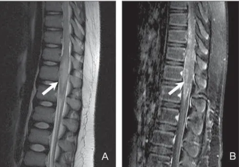
medullaris expansion, with hyposignal on T1-weighted, hypersignal on T2-weighted images and contrast-enhancement at the granuloma site. This finding may simulate a neoplastic lesion. A linear and nodular enhancement pattern with an “arborized” appearance is considered to be character-istic, and although it is not present in all cases, is a strong indication for the diagno-sis of neuroschistosomiadiagno-sis (Figure 1)(6).
NEUROCYSTICERCOSIS
Cysticercosis is the most common para-sitic infection of the CNS. The intracranial presentation is most frequently found. Ver-tebral canal involvement is rare, represent-ing only 2% to 5% of total neurocysticer-cosis cases. Leptomeningeal neurocysti-cercosis is the most common presentation, affecting the subarachnoid space by means of intracranial migration. Intramedullary involvement is rarely observed and occurs generally in the thoracic segment(5,7).
In medullary neurocysticercosis, the clinical picture depends on the compres-sion and edema mechanisms, as well as on the inflammatory response. It may be either asymptomatic or present signs and symp-toms of myelopathy, including radiating pain, flaccid or spastic paresthesia, and neurogenic bladder(5,7,8).
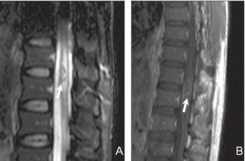
MRI is the method of choice for the study of medullary neurocysticercosis and shows cysts with similar signal to that of the cephalorachidian fluid, with hyposignal on T1-weighted and hypersignal on T2-weighted images (Figure 2). The scolex is represented by an eccentric nodule near the
cyst wall, and its identification is diagnos-tic. In cysts with some degree of degenera-tion, peripheral postcontrast enhancement can be observed(7,8).
TUBERCULOSIS
Involvement of the CNS occurs in ap-proximately 10–15% of all infections by tuberculosis(9). Intramedullary tuberculoma is a rare finding, occurring in approximately
2% of cases of neurotuberculosis, with or without association with pulmonary in-volvement. The clinical presentation is gen-erally subacute, with a variable picture of muscle weakness, paraparesis or quadripa-resis. Constitutional symptoms such as fe-ver and weight loss may not be present(9,10). At the disease onset, the inflammatory pro-cess with ill defined edema predominates. After formation of the granuloma, the le-sions become more defined, generally with
Figure 1. Twelve-year-old child diagnosed with neuroschistosomiasis. Sagittal MRI T2-weighted sequences (A), remarkable, central hypersignal with ill defined limits (arrow), besides volumetric increase of the conus medullaris obliterating the anterior and posterior CSF column. The contrast-enhanced T1-weighted se-quence demonstrates heterogeneous enhancement with an “arborized” pattern (arrow in B).
hyposignal on T1-weighted images and iso- to hypersignal on T2-weighted images. There is postcontrast enhancement, being such enhancement nodular and heteroge-neous or annular and peripheral depending on the presence or not of caseous necrosis (Figure 3)(5,10). In immunocompromised patients, the finding of nodular lesions with nodular or peripheral enhancement may also be observed in cases of toxoplasmo-sis or fungal infections, but such conditions are extremely rare(11–13).
ACUTE TRANSVERSE MYELITIS
Idiopathic acute transverse myelitis gen-erally refers to a single phased, acute or subacute inflammatory process, without defined etiology, which develops with bi-lateral motor and sensitive deficit, some-times with autonomic dysfunction. Ap-proximately one third of the patients re-cover completely, one third remain with moderate sequels, and one third maintain severe dysfunction(5,14).
Several conditions may present similar clinical signs, such as infectious, vascular and demyelinating diseases and actinic le-sions. Therefore, the final diagnosis of id-iopathic acute transverse myelitis depends on the investigation and ruling out of a possible identifiable etiology(1,14). The in-clusion criteria for such diagnosis comprise motor/sensitive deficits or autonomic dys-function attributed to the spinal cord, iden-tifiable level of sensitivity, absence of com-pressive cause, medullary inflammatory process demonstrated by pleocytosis or increase in IgG in the cephalorachidian fluid, or post-gadolinium injection en-hancement at MRI, with worsening peak in 4 hours to 21 days after the symptoms on-set. The exclusion criteria are history of local radiation, evidence of collagen dis-ease, syphilis, Lyme disdis-ease, HIV, HTLV and other viral etiologies, vascular dis-eases, lesions suggestive of multiple scle-rosis or optic neuritis(15).
Findings at MRI are variable, most fre-quently affecting thoracic segments, and least frequently the conus medullaris. A centrally located hypersignal is observed on T2-weighted images, with or without post-contrast enhancement (Figure 4). Increase in the spinal cord caliber, mimicking neo-plastic lesion, may be observed(2,5,16).
In the suspicion of a myelinating etiol-ogy, further investigation should be under-taken in search of other lesions in the
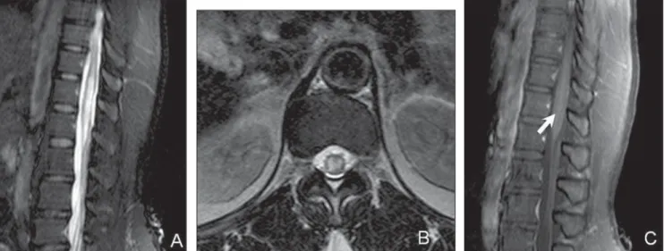
re-Figure 3. Immunocompromised patient diagnosed with neurotuberculosis. Sagittal MRI identify the pres-ence of a nodular lesion with peripheral hyposignal and centrally located hypersignal on T2-weighted (arrow on A), besides adjacent edema presenting peripheral annular post-contrast enhancement (arrow on B).
maining medullary and intracranial seg-ments, besides investigation for optical neuritis, in suspected cases. Approximately 90% of the patients with medullary lesions associated to multiple sclerosis present in-tracranial foci of demyelination(16). Usu-ally, medullary lesions related to multiple sclerosis are peripherally located and with an extent of less than two vertebral bodies, while optic neuromyelitis presents lesions that extend for more than three vertebral bodies, with sharp hyposignal on T1-weighted images and greater association with medullary atrophy(17).
VIRAL MYELITIS
Herpes virus, enterovirus and retrovirus (HIV) are some of the viral agents most frequently causing myelitis. The presenta-tions are very similar to each other and the etiological diagnosis is only confirmed by means of laboratory tests.
The clinical diagnosis of the infection by herpes zoster is many times difficult, as only one third of the patients present the typical skin lesion. Myelitis is a rare mani-festation of the infection by herpes zoster and generally occurs after reactivation of a latent infection. In cases where a con-comitant skin lesion is present, the medul-lary level corresponds to the affected der-matome(2,18).
The presentation at MRI varies, ranging from single or multiple lesions, with or with-out postcontrast enhancement (Figure 5)(18).
Myelitis related to HIV occurs in 5% to 8% of patients with acquired immunode-ficiency syndrome, generally in association with a severe encephalic involvement(5).
The HTLV-1 is associated to a progres-sive presentation of spastic paraparesis. The most affected medullary segment is the thoracic one and there may be association with medullary atrophy, besides lesions in the encephalic white matter(19).
The infection by cytomegalovirus causes polyradiculomyelitis which fre-quently involves the conus medullaris and cauda equina roots, represented at MRI by contrast-enhanced, clumped and thickened nerve roots, a finding compatible with arachnoiditis(20).
SARCOIDOSIS
Neurosarcoidosis is a rare condition which occurs in approximately 5% of the cases of systemic sarcoidosis(21) and may affect any part of the CNS(22). Medullary in-volvement affection is rarely observed, and generally presents a subacute or chronic course. The symptoms are varied and gen-erally are followed by pain, with possible occurrence of motor and sensitive deficits. The diagnosis of neurosarcoidosis repre-sents a challenge in the clinical practice. Correlation with laboratory tests and imag-ing findimag-ings is essential(21,23). At MRI, fre-quently multiple and centrally located, het-erogeneously contrast-enhanced lesions are observed with hypersignal on T2-weighted
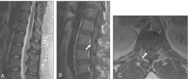
sequences. There may be fusiform medul-lary expansion. For patients with a compat-ible clinical condition, in the presence of pial enhancement beyond the intramedul-lary lesions, medulintramedul-lary sarcoidosis must be included among the differential diagnoses (Figure 6). The lesions tend to regress with the utilization of corticoids, generally af-ter clinical improvement(22,24,25).
CONCLUSION
MRI is the method of choice in the ap-proach to myelitis. In the present study, the authors have attempted to describe some relevant imaging findings of inflammatory conus medullaris diseases which can dif-ferentiate them from neoplastic or vascu-lar lesions, while suggesting a specific etio-logic agent.
The type of contrast-enhancement must be carefully evaluated. The “arborized” pattern is typical of neuroschistosomiasis, while a peripheral annular enhancement may be present in cases of granulomatous lesions such as in tuberculosis and toxo-plasmosis. In the presence of cysts, the di-agnosis of cysticercosis should be consid-ered, and may be confirmed by the identi-fication of the scolex. The differentiation amongst the several viral etiologies by MRI alone is difficult, and association of imag-ing findimag-ings with clinical and laboratory findings is necessary. Finally, a comprehen-sive evaluation of the whole neuroaxis may be useful in the definition of a specific
ology, since in many cases there is con-comitant intracranial involvement.
REFERENCES
1. Mendonça RA. Spinal infections and inflamma-tory disorders In: Atlas SW, editor. Magnetic reso-nance imaging of the brain and spine. 4th ed. Philadelphia, PA: Lippincott Williams & Wilkins; 2009. p. 1647–737.
2. Choi KH, Lee KS, Chung SO, et al. Idiopathic transverse myelitis: MR characteristics. AJNR Am J Neuroradiol. 1996;17:1151–60. 3. Tumors of the spine [Book review]. AJNR Am J
Neuroradiol. 2009;30:E137.
4. Silva LC, Maciel PE, Ribas JGR, et al. Schisto-somal myeloradiculopathy. Rev Soc Bras Med Trop. 2004;37:261–72.
5. DeSanto J, Ross JS. Spine infection/inflamma-tion. Radiol Clin North Am. 2011;49:105–27. 6. Sanelli PC, Lev MH, Gonzalez RG, et al. Unique
linear and nodular MR enhancement pattern in schistosomiasis of the central nervous system: report of three patients. AJR Am J Roentgenol. 2001;177:1471–4.
7. Gonçalves FG, Neves PO, Jovem CL, et al. Chronic myelopathy associated to intramedullary cysticer-cosis. Spine (Phila Pa 1976). 2010;35:E159–62. 8. Homans J, Khoo L, Chen T, et al. Spinal intramed-ullary cysticercosis in a five-year-old child: case report and review of the literature. Pediatr Infect Dis J. 2001;20:904–8.
9. Prabhakar S, Thussu A. Central nervous system tuberculosis. Neurol India. 1997;45:132–40. 10. Lu M. Imaging diagnosis of spinal intramedullary
tuberculoma: case reports and literature review. J Spinal Cord Med. 2010;33:159–62. 11. Elias J Jr, dos Santos AC, Carlotti CG Jr, et al.
Central nervous system paracoccidioidomycosis: diagnosis and treatment. Surg Neurol. 2005;63 Suppl 1:S13–21.
12. GültaÕli NZ, Ercan K, Orhun S, et al. MRI find-ings of intramedullary spinal cryptococcoma. Diagn Interv Radiol. 2007;13:64–7.
13. Poon TP, Tchertkoff V, Pares GF, et al. Spinal cord toxoplasma lesion in AIDS: MR findings. J Comput Assist Tomogr. 1992;16:817–9. 14. de Seze J, Lanctin C, Lebrun C, et al. Idiopathic
acute transverse myelitis: application of the recent diagnostic criteria. Neurology. 2005;65:1950–3. 15. Transverse Myelitis Consortium Working Group. Proposed diagnostic criteria and nosology of acute transverse myelitis. Neurology. 2002;59: 499–505.
16. Tartaglino LM, Friedman DP, Flanders AE, et al. Multiple sclerosis in the spinal cord: MR appear-ance and correlation with clinical parameters. Radiology. 1995;195:725–32.
17. Filippi M, Rocca MA, Moiola L, et al. MRI and magnetization transfer imaging changes in the brain and cervical cord of patients with Devic’s neuromyelitis optica. Neurology. 1999;53:1705– 10.
Figure 6. MRI study of a patient diagnosed with neurosarcoidosis, showing a slight increase of the conus medullaris on sagittal (A) T2-weighted sequence, with no alteration in central signal, with post-contrast pial and radicular enhancement (arrows on B and C).
18. Gilden DH, Beinlich BR, Rubinstien EM, et al. Varicella-zoster virus myelitis: an expanding spectrum. Neurology. 1994;44:1818–23. 19. Ferraz AC, Gabbai AA, Abdala N, et al. Magnetic
resonance in HTL-I associated myelopathy. Leu-koencephalopathy and spinal cord atrophy. Arq Neuropsiquiatr. 1997;55:728–36.
20. Whiteman ML, Dandapani BK, Shebert RT, et al. MRI of AIDS-related polyradiculomyelitis. J Comput Assist Tomogr. 1994;18:7–11. 21. Baughman RP, Teirstein AS, Judson MA, et al.
Clinical characteristics of patients in a case con-trol study of sarcoidosis. Am J Respir Crit Care Med. 2001;164(10 Pt 1):1885–9.
22. Zajicek JP, Scolding NJ, Foster O, et al. Central nervous system sarcoidosis – diagnosis and man-agement. QJM. 1999;92:103–17.
23. Bradley DA, Lower EE, Baughman RP. Diagno-sis and management of spinal cord sarcoidoDiagno-sis. Sarcoidosis Vasc Diffuse Lung Dis. 2006;23:58– 65.
24. Sakushima K, Yabe I, Nakano F, et al. Clinical features of spinal cord sarcoidosis: analysis of 17 neurosarcoidosis patients. J Neurol. 2011;258: 2163–7.