Arq Neuropsiquiatr 2006;64(2-B):507-510
Headache Clinic, Department of Neuro l o g y, Hospital das Clínicas, University of Sao Paulo, Sao Paulo, Brazil:1Doutorando em
Neurologia;2Médico-Assistente;3Professor Assistente-Doutor; 4Professor Livre-Docente.
Received 13 October 2005, received in final form 22 February 2006. Accepted 9 March 2006.
Dr. Antonio Cezar R. Galvão - Rua Apinages 761/111 - 05017-000 São Paulo SP - Brasil. E-mail: [email protected]
SUNCT SYNDROME ASSOCIATED
WITH PITUITARY TUMOR
Case report
Pedro A.S. Rocha Filho
1, Antonio Cezar R. Galvão
3, Manoel J. Teixeira
4,
Getulio D. Rabello
3, Ida Fortini
2, Marcelo Calderaro
2, Dalva Carrocini
2ABSTRACT - For twelve years, the subject of this report, a 38-year-old man, presented a clinical condition compatible with the SUNCT (short-lasting unilateral neuralgiform headache attacks with conjunctival injec-tion and tearing) syndrome. He presented a stabbing and intense daily pain located in the left pre-auric-ular and temporal regions. Each of these intense pain attacks lasted around one minute and presented a f requency of two to eight times per day. The pain was associated with ipsilateral lacrimation, conjunctival injection and rhinorrhea. MRI revealed a pituitary tumor with little suprasellar extent. The subject’s serial assays of prolactin, GH, TSH and ACTH were within normal levels. Following transsphenoidal hypophysec-t o m y, wihypophysec-th complehypophysec-te removal of hypophysec-the hypophysec-tumor, hypophysec-the subjechypophysec-t no more presenhypophysec-ted pain. The pahypophysec-thological diagno-sis was non-secreting adenoma. Fourteen months after the surgery, he remains symptom-free.
KEY WORDS: SUNCT, pituitary tumor, surgery, ultra- shorting headaches.
Síndrome SUNCT associada a tumor de hipófise: relato de caso
RESUMO - O paciente relatado neste artigo apresentou uma condição clínica compatível com síndro m e SUNCT (cefaléia de curta duração, unilateral, neuralgiforme com hiperemia conjuntival e lacrimejamen-to). Ele referia dor diária, intensa, em facada, localizada na região pré-auricular e temporal esquerd a s . Cada ataque de dor permanecia por cerca de um minuto, com freqüência de duas a oito vezes por dia. A dor se acompanhava de lacrimejamento ipsolateral, congestão conjuntival e rinorréia. A RM mostrou um tumor de hipófise com pouca extensão suprasselar. Dosagens de prolactina, GH, TSH e ACTH estavam em níveis normais. Foi então submetido a hipofisectomia transesfenoidal com remoção completa do tumor após o que a dor cessou completamente. O diagnóstico anátomo-patológico foi adenoma não secre t o r. Quatorze meses após a cirurgia, o paciente permanecia livre de dor.
PALAVRAS-CHAVE: SUNCT, tumor de hipófise, cirurgia, cefaléia de curta duração.
s y n d rome is the poor response to pharm a c o l o g i c a l t re a t m e n t s3 , 4. There have been re p o rts of impro v
e-ment upon the use of amitriptyline, carbamazepine, gabapentin, prednisone, topiramate5, lamotrigine6,
nifedipine and sumatriptan4. Anesthetic blockades
do not work very well, although there have been re p o rts of improvement following the local opioid blockade of the superior cervical ganglion7. There
have also been re p o rts of surgical pro c e d u res that worked eff e c t i v e l y5while others re p o rts have
indi-cated a lack of response to such procedures8.
This syndrome has been listed in the second edi-tion of The Internaedi-tional Classificaedi-tion of Headache D i s o rders together with trigeminal autonomic cepha-The SUNCT syndrome (short-lasting unilateral
neu-r a l g i f o neu-rm headache attacks with conjunctival injec-tion and tearing) is an uncommon headache charac-terized by moderate to severe pain. Usualy, the loca-tions with the most severe pain are the ocular/peri-ocular regions and the frontal region. The latter is the main location for irr a d i a t i o n1. The mean
dura-tion of the attack lasts around 40 seconds, with a fre-quency ranging from two episodes per day up to 10 to 30 episodes, predominantly during the daytime2.
508 Arq Neuropsiquiatr 2006;64(2-B)
l a l g i a s9. This group of headaches presents a
trigemi-nal autonomic reflex that consists of a brainstem con-nection between the trigeminal nerve (responsible for the pain) and the VIIth cranial nerve (responsible for the autonomics symptoms)1 0. According to this new
classification system, which was published by the IHS in 20049, the SUNCT syndrome is defined according to
the following characteristics: A. At least 20 attacks ful-filling criteria B-D; B. Attacks of unilateral orbital, supraorbital or temporal stabbing or pulsating pain lasting between 5 and 240 seconds; C. Pain is accom-panied by ipsilateral conjunctival injection and lacrima-tion; D. Attacks occur with a frequency from 3 to 200 per day; E. Not attributed to another disord e r.
This paper presents the description of a case of SUNCT syndrome associated with pituitary adenoma.
CASE
Over a twelve-year period the patient, a 38-year- o l d male, presented daily attacks of severe, short - d u r a t i o n stabbing pain located in the pre-auricular and left tempo-ral regions. These attacks lasted for about one minute each, taking place between two and eight times per day. At the same time, he presented, along w ith the pain, ipsilateral conjunctival injection, lacrimation and rh i n o rrhea. During this period he was diagnosed elsewhere as having trigem-inal neuralgia. The patient was treated for a few years with carbamazepine (even 600 mg/day). He presented intoler-ance to carbamazepine and no clinical im provement was re p o rted. In the end he was only taking 50 mg of this dru g per day, due to sleepiness that evolved as a side effect of bigger dosages. In addition, the use of amitriptyline and m e t h y s e rgide was prescribed to this patient, without clin-ical response. He also sought hom eopathic and acupunc-t u re acupunc-treaacupunc-tmenacupunc-ts wiacupunc-th no resulacupunc-ts. Aacupunc-t acupunc-the beginni ng of acupunc-the
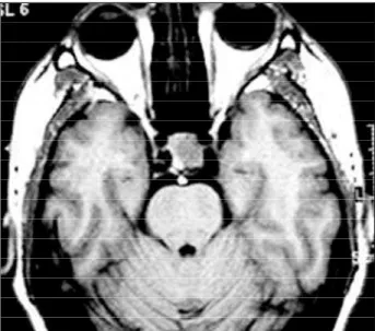
year 2002, he underwent a cranial MRI. This pro c e d u re revealed a pituitary tumor with little suprasellar extent (Fig 1, 2 and 3). Serial assays of prolactin, GH, TSH and ACTH w e re within normal levels (ACTH= 42,3 pg/ mL; GH= 3,10 ng/ mL; prolactin= 16 ng/ mL; hTSH= 0,747µUI/ mL; T3= 0,82 ng/ mL; T4= 7,99 µg/ mL).
He was submitted to transsphenoidal hypophysectomy with complete removal of the tumor, which was a pituitary adenoma (the pathological diagnosis was non-secre t i n g adenoma). He took prednisone for a few weeks following the surgical pro c e d u re as usual in this kind of surg e ry. Following the surgery, he evolved toward a complete dis-appearance of pain. Over the past 14 months his condition has remained stable and he has not taken any pre s c r i p t i o n medicines or presented any pain during this period.
Fig 1. MRI showing a pituitary tumor. Fig 2. MRI pituitary adenoma do not invade cavernous sinus.
Arq Neuropsiquiatr 2006;64(2-B) 509
DISCUSSION
C h ronic paroxysmal hemicrania, cluster headache, trigeminal neuralgia of the first nerve division, pri-m a ry stabbing headache5and cluster-tic syndro m e
a re among the main diff e rential diagnoses for the SUNCT syndrome. Regardless of the fact that the majority of cases of SUNCT syndrome are primary, secondary cases cannot be ruled out.
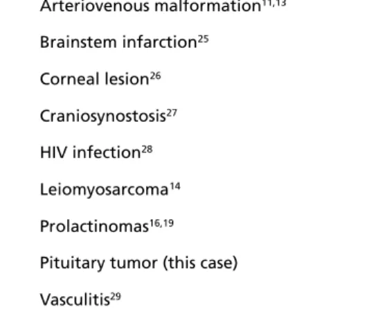
Table shows the conditions to which the SUNCT s y n d rome may be associated with. In two cases the a rteriovenous malformation was located at the cere-bellopontine angle1 1 , 1 2. This location suggested irr
i-tative compression of the facial, trigeminal or gre a t e r p e t rous superficial nerves. In another case, cavern o u s angioma of the brainstem1 3may have triggered irr
tation of the trigeminal nerve and greater superf i-cial petrous nerve, which would explain the symp-toms. The leiomyosarcoma case occurred in a patient who had undergone kidney transplantion. This patient was using immunosuppressive therapy, and because of the location of the tumor in the cavern o u s sinus, it is possible that the trigeminal nerve may have been involved14.
B e f o re the SUNCT syndrome was first described and defined by Sjaastad et al.1 5, the case of a patient
with a pituitary tumor and bro m o c r i p t i n e - i n d u c e d headaches compatible with the syndrome had been re p o rt e d1 6. Later on two more cases were described1 7.
These two cases presented pain induced by dopamine agonists. In the first of these, the prolactinoma had invaded the cavernous sinus that was on the same side as the pain. The resulting pain may have been due to trigeminal activation. There was a significant d e c rease in the frequency of the pain following radio-t h e r a p y. The second of radio-these radio-two cases did noradio-t pre s-ent any invasion of the cavernous sinus.
R e c e n t l y, another two trigeminal autonomic headache cases linked with pituitary micro a d e n o m a s have been re p o rt e d1 8. The first of these re p o rted
cas-es was clinically compatible with the SUNCT syn-drome. The second case was compatible with hemi-crania continua. In both cases there was an incre a s e in pain due to the use of dopamine agonists. In the first case, which was compatible with the SUNCT syn-drome, the patient’s pain was eliminated following the surgical removal of the adenoma. It is import a n t to note that there was an exacerbation of the pain at the time when the tumor re c u rred. Thus these facts suggest a causal relationship. Additionally, there has been a description of headaches caused by pro l a c t i n-oma, that were compatible with SUNCT1 9. There was
i m p rovement in the condition with the use of b romocriptine and cabergoline, and the headache was resolved within three months.
The pathophysiology of the headache associated with pituitary tumors is not completely clear. Dural s t re t c h2 0 , 2 1, invasion of the cavernous sinus1 7and local
p re s s u re eff e c t s2 2have been suggested as mechanisms.
It has been found that diff e rences in tumor size were not apparent between those who pre s e n t e d headaches and those who did not2 3. Also, there were
no clear correlations between the pituitary volume and headache score2 4. The extent of cavernous sinus
invasion was not associated with the pre s e n c e / e x t e n t of headache2 4. One explanation given for why
headaches get worse after the use of dopamine ago-nists is that the growth of the tumor is transitory1 8o r
that a neurohumoral mechanism is possible1 9.
In the case of the 38-year-old male patient in the p resent study, the pituitary adenoma did not invade the cavernous sinus. The pain can there f o re not be explained by a mechanism of invasion of the cav-e rnous sinus. A non-functioning pituitary adcav-enoma can stay asymptomatic for many years since it will not compress the neighboring structures. It is possi-ble that this patient had already the tumor during the previous twelve years when he presented the attacks of headache without others signs or symp-toms. There was complete remission of the pain fol-lowing surg e ry. The patient was treated for a few weeks after the surgery with prednisone. The pred-nisone could have some positive effect in patients with SUNCT syndro m e5, but the patient had alre a d y
no pain when this drug were prescribed. This patient had used prednisone for a few weeks and, despite of its interruption, the pain did not relapse. The patient has presented no headache symptoms for 14
Table. Diseases associated with SUNCT syndrome.
Arteriovenous malformation11,13
Brainstem infarction25
Corneal lesion26
Craniosynostosis27
HIV infection28
Leiomyosarcoma14
Prolactinomas16,19
510 Arq Neuropsiquiatr 2006;64(2-B)
14. Kaphan E, Eusebio A, Donnet A, Witjas T, Ali Cherif A. Shortlasting, unilateral, neuralgiform headache attacks with conjunctival injection and tearing (SUNCT syndrome) and tumour of the cavernous sinus. Cephalalgia 2003;23:395-397.
15. Sjaastad O, Saunte C, Salvesen R, et al. Shortlasting, unilateral, neural-giform headache attacks with conjunctival injection, tearing, sweating, and rhinorrhea. Cephalalgia 1989;9:147-156.
16. Ferrari MD, Haan J, van Seters A P. Bromocriptine-induced trigeminal neuralgia attacks in a patient with pituitary tumor. Neuro l o g y 1988;38:1482-1484.
17. Massiou H, Launay JM, Levy C, El Amrani M, Emperauger B, Bousser MG. SUNCT syndrome in two patients with prolactinomas and bromocriptine-induced attacks. Neurology 2002;58:1698-1699. 18. Levy MJ, Matharu MS, Goadsby PJ. Prolactinomas, dopamine agonists
and headache: two case reports. Eur J Neurol 2003;10:169-173. 1 9 . M a t h a ru MS, Levy MJ, Merry RT, Goadsby PJ. SUNCT syndrome
second-ary to prolactinoma. J Neurol Neuro s u rg Psychiatry 2003;74:1590-1592. 20. Forsyth PA ,Posner JB. Headaches in patients with brain tumors: a study
of 111 patients. Neurology 1993;43:1678-1683.
21. Suwanwela N, Phanthumchinda K, Kaoropthum S. Headache in brain tumor: a cross-sectional study. Headache 1994;34:435-438.
22. Arafah BM, Prunty D, Ybarra J, Hlavin ML, Selman WR. The dominant role of increased intrasellar pre s s u re in the pathogenesis of hypopitu-itarism, hyperprolactinemia, and headaches in patients with pituitary adenomas. J Clin Endocrinol Metab 2000;85:1789-1793.
23. Abe T, Matsumoto K, Kuwazawa J, Toyoda I, Sasaki K. Headache asso-ciated with pituitary adenomas. Headache 1998;38:782-786. 24. Levy MJ, Jager HR, Powell M, Matharu MS , Meeran K, Goadsby PJ.
Pituitary volume and headache: size is not everything. A rch Neuro l 2004;61:721-725.
2 5 . Penart A, Firth M, Bowen JR. Short-lasting unilateral neuralgiform headache with conjunctival injection and tearing (SUNCT) following pre-sumed dorsolateral brainstem infarction. Cephalalgia 2001;21:236-239. 26. Piovesan EJ, Kowacs PA, Werneck LC. SUNCT syndrome: report of a
case preceded by ocular trauma. A rq Neuropsiquiatr 1996;54:494-497. 27. Moris G, Ribacoba R, Solar DN, Vidal JA. SUNCT syndrome and seb-o r rheic dermatitis assseb-ociated with craneseb-osynseb-ostseb-osis. Cephalalgia 2001;21:157-159.
28. B a rea LM ,Forcelini CM. Onset of short-lasting unilateral, neuralgi-forme headache with conjunctival injection and tearing (SUNCT) after acquiring human immunodeficiency virus (HIV): more than a coinci-dence? Cephalalgia 2001; 21: 518.
29. Hannerz J, Greitz D, Hansson P, Ericson K. SUNCT may be another manifestation of orbital venous vasculitis. Headache 1992;32:384-389.
months to date. These facts suggest a causal re l a t i o n-ship between the adenoma and the headache in this patient. It is important to emphasize the value of ru l-ing out secondary SUNCT by appropriately obtainl-ing the patient’s history and placing emphasis on pitu-i t a ry - related symptoms, neuropitu-imagpitu-ing pitu-investpitu-igatpitu-ions and also hormonal assaying.
REFERENCES
1 . P a reja JA ,Sjaastad O. SUNCT syndrome: a clinical re v i e w. Headache 1997;37:195-202.
2. P a reja JA, Shen JM, Kruszewski P, Caballero V, Pamo M, Sjaastad O. SUNCT syndrome: duration, fre q u e n c y, and temporal distribution of attacks. Headache 1996;36:161-165.
3. P a reja J, Caminero A, Sjaastad O. SUNCT syndrome: diagnosis and treatment. Headache 2003;43:306.
4. P a reja JA, Kruszewski P, Sjaastad O. SUNCT syndrome: trials of dru g s and anesthetic blockades. Headache 1995;35:138-142.
5. P a reja JA, Caminero AB, Sjaastad O. SUNCT syndrome: diagnosis and treatment. CNS Drugs 2002;16:373-383.
6. Piovesan EJ, Siow C, Kowacs PA, Werneck LC. Influence of lamotrig-ine over the SUNCT syndrome: one patient follow-up for two years. Arq Neuropsiquiatr 2003;61:691-694.
7. Sabatowski R, Huber M, Meuser T, Radbruch L. SUNCT syndrome: a t reatment option with local opioid blockade of the superior cervical ganglion? A case report. Cephalalgia 2001;21:154-166.
8. Black DF ,Dodick DW. Two cases of medically and surgically intractable S U N C T: a reason for caution and an argument for a central mechanism. Cephalalgia 2002;22:201-204.
9. The International Classification of Headache Disorders: 2nd edition. Cephalalgia 2004;24(Suppl 1):S9-S160.
10. Goadsby PJ ,Lipton RB. A review of paroxysmal hemicranias, SUNCT syndrome and other short-lasting headaches with autonomic feature, including new cases. Brain 1997;120:193-209.
11. Bussone G, Leone M, Volta G, Strada L, Gasparotti R, Di Monda V. Short-lasting unilateral neuralgiform headache attacks with tearing and conjunctival injection: the first “symptomatic” case? Cephalalgia 1991;11:123-127.
12. Morales F, Mostacero E, Marta J, Sanchez S. Vascular malformation of the cerebellopontine angle associated with “SUNCT” syndro m e . Cephalalgia 1994;14:301-302.