Serviço de Cirurgia de Epilepsia, Hospital Brigadeiro, São Paulo SP, Brazil.
Received 4 October 2005, received in final form 16 January 2006. Accepted 15 March 2006.
Dr. Arthur Cukiert - Rua Dr. Alceu Campos Rodrigues 247 / 121 - 04544-000 São Paulo SP - Brasil. E-mail: acukiert@uol.com.br
SURGICAL OUTCOME IN PATIENTS WITH
REFRACTORY EPILEPSY ASSOCIATED TO
MRI-DEFINED UNILATERAL MESIAL
TEMPORAL SCLEROSIS
Cristine Mella Baldauf, Arthur Cukiert, Meire Argentoni, Carla Baise-Zung,
Cássio Roberto Forster, Valeria Antakli Mello, José Augusto Burattini,
Pedro Paulo Mariani, Ródio Brandão Câmara, Lauro Seda
ABSTRACT - I n t roduction: Several pre-operative work-up protocols have been used while selecting epilep-tic patients for surg e ry among diff e rent centers. The relative value of the diff e rent available pre - o p e r a-tive tests is still under discussion. Objective: We re p o rt on the surgical outcome obtained in patients with re f r a c t o ry temporal lobe epilepsy associated to mesial temporal sclerosis (MTS) and who were evaluated p re-operatively by interictal EEG and MRI alone. Method: F o rty one patients with re f r a c t o ry unilateral temporal lobe epilepsy were evaluated using interictal EEG and MRI. MRI disclosed unilateral MTS in all patients. All patients had at least 4 interictal EEG re c o rdings. All patients were submitted to cort i c o - a m y g-dalo-hippocampectomy at the side determined by MRI. Results: Interictal EEG showed unilateral epilep-t i f o rm discharges compaepilep-tible wiepilep-th MRI findings in 37 paepilep-tienepilep-ts; in four ouepilep-t of epilep-the 41 paepilep-tienepilep-ts, bilaepilep-teral dis-charges were found. Mean follow-up time was 4.3±1.1 years. Thirty-nine patients (95.1%) were classified as Engel’s Class I (70.6% Engel I-A). Two patients (4.9%) were rated as Engel's Class II. All patients in whom bilateral discharges were found were in Engel’s Class I. Pathological examination showed MTS in all patients. Conclusion: It is possible to adequately select good surgical candidates for temporal lobe resection using MRI and interictal EEG alone. In patients with MRI-defined MTS we should expect a postoperative remis-sion rate higher then 90%. The finding of MTS on MRI is the most important good prognostic factor after temporal lobe resection.
KEY WORDS: re f r a c t o ry temporal lobe epilepsy, hippocampal sclerosis, surg e ry, electro e n c e p h a l o g r a p h y, magnetic resonance image.
Resultados cirúrgicos em pacientes com epilepsia refratária associada a esclerose mesial tem-poral unilateral definida por ressonância magnética
Temporal lobe epilepsy (TLE) is the commonest epileptic syndrome and re p resents up to 40% of the epileptic patients. It is the most frequently re f r a c t o-ry epileptic syndro m e1 - 3. Mesial temporal epilepsy
(MTE) is the most frequent re f r a c t o ry epileptic syndro-me in adults and its treatsyndro-ment re p resents two-third s of the epilepsy surg e ry pro c e d u re s4. Ve ry good surg
i-cal outcome has been re p o rted in patients with MTE. E n g e l5a rgued that MTE was the best prototype of a
“ s u rgically remediable epileptic syndrome”. In this patient population, a 70% to 90% postoperative sei-zure remission rate might be expected6-7.
M o re re c e n t l y, many centers have reevaluated the relative value of each exam included in the pre o p e r-ative workup of epileptic patients8 - 1 0. The need for
video-EEG seizure ’s re c o rding in all patients has been extensively studied1 1 , 1 2. Many authors emphasized
the value of MRI and interictal EEG findings in pati-ents with TLE who were considered candidates for s u rg e ry1 3 - 1 5. Better surgical outcome related to
seizu-res was found in patients with concordant MRI and interictal EEG findings, especially when MRI disclosed unilateral mesial temporal sclerosis (MTS)12,16,17.
We studied the surgical outcome of patients with TLE and MTS selected based on the anatomical find-ings provided by MRI and that were submitted to c o rtico-amygdalo-hippocampectomy (CAH) at the side shown by imaging.
METHOD
F o rty-one consecutive patients (23 women and 18 men) with re f r a c t o ry TLE and unilateral MTS that were submit-ted to surgery at the Hospital Brigadeiro Epilepsy Surgery P rogram from 1997 to 1999 were studied. The study pro-tocol was approved by Hospital Brigadeiro IRB.
Mean age at surg e ry was 32.7±8.8 years (range: 11-51 years) and all patients had TLE and unilateral MTS as shown by MRI.
All patients had clinical and semiological findings com-patible with TLE. All had re f r a c t o ry epilepsy and had been p reviously unsuccessfully treated by at least two adequate antiepileptic drug (AED) regimens. All patients had unilat-eral MTS on MRI and at least 4 inter ictal EEGs perf o rm e d over the last 2 years showing unilateral or bilateral tempo-ral lobe spiking. All patients were submitted to CAH at the Hospital Brigadeiro Epilepsy Surgery Program.
Patients that had been previously operated , had nor-mal MRI, other extra-hippocampal lesions or pseudo-seizu-res were excluded from the study.
The clinical diagnosis was based on the Intern a t i o n a l Classification of Seizure s1 8and Epileptic Syndro m e s1 9. The
following clinical characteristics were considered as diag-nostic for TLE: simple partial seizures of the déjà vu or j a
-mais vu type, or including epigastric or psychic
manifesta-tions (p.e., fear) followed by complex partial seizures
char-acterized by staring and masticatory automatisms, accom-panied or not by superior limb automatisms or contralat-eral superior limb distonia.
The following clinical variables were analyzed: sex, age at onset of seizures, weekly seizure fre q u e n c y, type of sei-z u re, pre- and post-operative AED regimen and the pre s e n-ce or not of febrile seizures in childhood.
All patients had 32-channels (Medelec, Profile) interic-tal EEG (10-20 system) re c o rdings including zygomatic elec-t rodes. The presence of elec-temporal lobe inelec-tericelec-tal spiking and absence of extratemporal discharges were considere d findings related to TLE1 2 , 1 6. The finding of at least 90% of
the discharges at one side was considered a lateralizing sign in patients with bilateral EEG findings.
All patients had high resolution MRI which showed MTS in all of them. MRI examinations included sequences for the adequate study of the hippocampal formation: 3 mm thick (0.3 mm interval) FLAIR, T2 and IR coronal slices; 6 mm thick T1, T2, gradient echo, FLAIR and IR axial slices and T1 sagittal slices. MTS was diagnosed when there was c l e a r-cut volumetric reduction of the hippocampus as seen on T1 coronal s lices and increased hippocampal signal in T2 and FLAIR coronal slices as noted by two independent observers.
All patients were submitted to CAH at the side deter-mined by MRI. The procedure was carried out under gen-eral anesthesia and without intraoperative electro c o rt i c o g-raphy.
S u rg e ry consisted of cortical resection that included the s u p e r i o r, middle, and inferior temporal, parahippocampal and fusiform gyri (with its posterior border at the level of the central art e ry), total hippocampectomy and re s e c t i o n of the intratemporal portion of the amygdala. All patients were operated by the same surgeon (Arthur Cukiert, Hos-pital Brigadeiro Epilepsy Surgery Program).
All surgical specimens were analyzed and MTS was found in all of them.
E n g e l ’s scale was used to rate post-operative outcome, and could be summarized as follows20: Class I: No seizures
or simple partial seizures (SPS) only; Class II: 90% or more s e i z u re frequency reduction; Class III: Seizure fre q u e n c y reduction from 50-90%; Classe IV: Seizure frequency re d u c-tions lower then 50% or no worthwhile reduction.
RESULTS
Men were significantly younger than women ( m e a n = 2 8 . 4 4±9.17 versus 36.04±7.07 years) by the time of surg e ry (p=0.005). No other gender diff e r-ence was noted when analyzing age at seizure onset, weekly seizure frequency or seizure type. Mean age at the onset of seizures was 9.60±8.24 years for wo-men and 8.46±8.44 years for men. Mean weekly sei-zure frequency was 3.02±3.23 for women and 3.47±
2.34 for men. There was no significant relationship b e t-ween age of seizure onset and its weekly fre q u e n c y.
vegeta-tive (20 patients; 48.7%) or psychic (12 patients; 29.2%). Complex partial seizures (CPS) occurred in all patients. In 1 patient (2.5%), CPS occurred isolately, while in 6 patients (14.6%) they were associated to generalized tonic-clonic seizures (GTC). No patient p resented with isolated GTC. Secondarily general-ized seizures occurred in 29 patients (70.7%).
Febrile seizures occurred in 10 patients (24.39%); t h e re was a latency of a mean of 7.61±9.25 years for the appearance of non-febrile seizures. There was no statistically significant relationship between the p resence or not of febrile convulsion and age of non-febrile seizure onset.
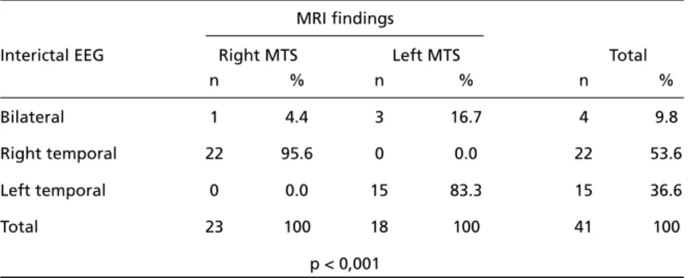
Bilateral interictal EEG temporal lobe spiking was noted in 4 patients (9.8%, all female). Right interic-tal EEG findings were noted in 22 patients (53.6%) and in 15 (36.6%) left temporal lobe spiking was not-ed. There was no significant gender difference.
MRI showed right MTS in 23 (56.1%) and left MTS in 18 (43.9%) patients. There was no significant gen-der difference.
Among the patients with bilateral interictal EEG findings, 1 (4.4%) had right and 3 (16.7%) had left MTS on MRI. There was a significant re l a t i o n s h i p between interictal EEG and MRI findings (p<0.001) (Table 1).
Table 1. Distribution of patients according to interictal EEG and MRI findings.
MRI findings
Interictal EEG Right MTS Left MTS Total
n % n % n %
Bilateral 1 4.4 3 16.7 4 9.8
Right temporal 22 95.6 0 0.0 22 53.6
Left temporal 0 0.0 15 83.3 15 36.6
Total 23 100 18 100 41 100
p < 0,001 MTS, Mesial temporal sclerosis.
Table 2. Distribution of Engel I patients according to the presence or not of postoperative SPS and preoperative seizure type.
Postoperative outcome (ENGEL I)
Preoperative No seizure With SPS Total
seizure type n % n % n %
CPS 0 0.0 1 10.0 1 2.6
CPS – GTC 6 20.7 0 0.0 6 15.4
SPSp – CPS 4 13.7 0 0.0 4 10.3
SPSp- CPS - GTC 5 17.2 2 20.0 7 17.9
SPSs- CPS - GTC 1 3.4 0 0.0 1 2.6
SPSv – CPS 6 20.7 0 0.0 6 15.4
SPSv- CPS - GTC 7 24.3 6 60.0 13 33.2
SPSvi – CPS 0 0.0 1 10.0 1 2.6
Total 29 100 10 100 39 100
p=0.034
At late follow-up (mean: 4.3±1.1 years; range: 9 months - 6 years and 10 months), 29 patients (70.6%) had no seizure during the last follow-up year; 10 (24.5%) presented 1 to 6 SPS; 1 (2.5%) presented 8 p a rtial seizures (5 SPS and 3 CPC) and 1 GTC and 1 patient (2.5%) presented 1 GTC. Thus, 95.1% of the patients were rated as Engel’s Class I and the re m a i n-ing (4.9%) as Class II.
We noted that 5 patients had seizures during their postoperative follow-up that were related to re d u c-tion or withdrawal of AED; 4 of these patients had no seizures over the last follow-up year.
We were not able to analyze the data derived from Engel’s Class II patients from a statistical point of view since the sample was very small (n=2). The-re f o The-re, we analyzed the data from Engel’s Class I pa-tients, according to the presence or not of SPS dur-ing follow-up.
No statistically significant results were found while analyzing the surgical outcome and any clinical vari-able such as sex, age of seizure onset, weekly seizure f re q u e n c y, type of seizure or presence of febrile sei-zure in childhood (Table 2).
Unilateral EEG findings did not correlate with bet-ter surgical outcome; all 4 patients (9.8%) with bilat-eral EEG findings were seizure-free after surgery.
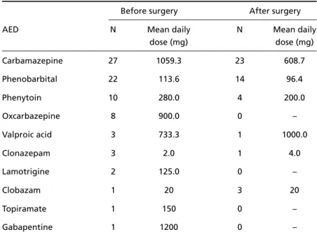
We noted a relevant reduction on the amount of AED postoperatively. Table 3 summarizes these find-ings. Before surg e ry, 14 (34.1%) patients were re c e i v-ing 1 AED and the remainv-ing 27 (65.9%) were under 2 or more AED. At the last follow-up visit after surg e-ry, 11 patients (26.8%) were receiving no medication,
15 (36.6%) were receiving 1 AED and 15 (36.6%), 2 or more AED.
DISCUSSION
R i g o rous pre s u rgical evaluation protocols are es-sential for adequate focus localization and good sur-gical outcome in patients with re f r a c t o ry temporal lobe epilepsy. The introduction of MRI into clinical practice and the better knowledge of the pathophys-iology of epilepsy have led many centers to re e v a l u-ate the relative role of each exam or technique used in the pre s u rgical evaluation of epileptic pati-ents4,12,13,17,21.
Jeong et al.1 0 studied patients with MTS and
showed postoperative seizure remission in 84% of them; these authors considered age at surg e ry (26.9 ±7.3 years in the seizure - f ree group versus 35.4±9 . 4 years in the non-seizure - f ree group) and duration of epilepsy (14.5±6.8 years in the seizure - f ree group ver-sus 19.5±8.7 years in the non-seizure - f ree group) as p rognostic indicators of postoperative outcome. On the other hand, McIntosh et al.22showed in a
meta-analysis study that gender, age of seizure onset and p reoperative weekly seizure frequency were not post-operative prognostic factors.
SPS occurred preoperatively in 82.9% of our pa-tients, as was noted by others, who re p o rted that up to 90% of patients with MTS had SPS4 , 2 3. The most
f requently found SPS were vegetative, also in agre e-ment with other findings7. CPS occurred in all our
pa-tients and secondarily generalized GTC in 70.7% of
Table 3. Pré- and postoperative AED regimens.
Before surgery After surgery
AED N Mean daily N Mean daily
dose (mg) dose (mg)
Carbamazepine 27 1059.3 23 608.7
Phenobarbital 22 113.6 14 96.4
Phenytoin 10 280.0 4 200.0
Oxcarbazepine 8 900.0 0 –
Valproic acid 3 733.3 1 1000.0
Clonazepam 3 2.0 1 4.0
Lamotrigine 2 125.0 0 –
Clobazam 1 20 3 20
Topiramate 1 150 0 –
them. GTC was not the most usual seizure type in pa-tients with MTS, although 50% of the papa-tients might have one GTC at some point7. Our data, which were
in agreement with those from Kilpatrick et al.2 4, did
not suggest that the presence of GTC would be a bad p rognostic factor after surg e ry. On the other hand, Spencer et al.8and Henessy et al.2 5suggested that
the absence of GTC would be a good prognostic fac-tor after CAH.
F rench et al.7found that febrile seizures were
pre-sent in 77% of the patients with MTS and that the mean latent period for the appearance of non-febrile seizures was 7.5 years. Although febrile seizures oc-c u rred in only 25% of our patients, the latenoc-cy for the appearance of non-febrile seizures (7.61 years) was the same as found by others. Prolonged febrile s e i z u res had been implicated in the development of MTS. Although some authors considered the pre s e n-ce of febrile seizures in childhood as a poor surgical outcome indicator2 6, our data and those from
Kilpa-trick et al.2 4and Henessy et al.2 5did not support this
idea.
The surgical results re p o rted for patients with MTS varied among centers, and seemed to be better when t h e re was agreement among the clinical, EEG and imaging preoperative findings. The presence of MTS on MRI had been related to a better surgical out-come after CAH2 7. Radhakrishnan et al.9found
excel-lent outcome in 95% of the patients with MRI-defi-ned MTS submitted to CAH and only 65% of good results in patients without MTS. In this study, they al-so pointed out that exclusively unilateral interictal EEG discharges were as important as the MR findings as positive prognostic factor; only 60% of the patients without that interictal EEG finding got good surg i-cal outcome. Gilliam et al.1 6re p o rted good surg i c a l
outcome in 77% of the patients with concordant MRI and interictal EEG findings, in contrast to 53% of good results in patients with concordant or non-l o c a non-l i z a t o ry findings. In our sampnon-le, patient senon-lection was essentially based on anatomical MRI data, and the majority of them got excellent results (95%). In our series, the patients with bilateral interictal EEG findings did not have a worst postoperative pro g n o-sis. This finding suggests that the presence of MTS on MRI would be the most important positive post-operative prognostic factor after CAH1 7. We found
that all patients with MTS on MRI had favorable post-operative outcome. We did not analyze the impact of other (non-MTS) temporal lobe MRI findings.
EEG re c o rdings have always been included as part
of the pre s u rgical evaluation of epileptic patients. On the other hand, the introduction of new techno-logical tools in the diagnosis of epilepsy led to the need to reevaluate its role in this setting. All patients w e re submitted to invasive re c o rdings by the time epilepsy surg e ry was introduced for adequate focus localization, which is clearly not the actual situation. Williamson et al.2 8, in a re t rospective analysis of 67
patients, showed that 96% of the patients had inter-ictal EEG abnormalities and that 94% of them were localized over the anterior temporal region. In their s t u d y, independent bilateral EEG discharges were found in 42% of the patients; in 50% of them the d i s c h a rges prevailed over the ictal-onset side. In our s t u d y, anterior temporal lobe spikes were present in all patients and bilateral interictal findings were ra-rely seen. Cascino et al.29evaluated the relative role
of routine interictal EEG, vídeo-EEG and MRI find-ings in the postoperative prognosis of patients with TLE. They found that interictal EEG abnorm a l i t i e s w e re re c o rded in 81.1% of the patients and were unilateral in 77.3% of them. Bilateral findings with unilateral predominance occurred in 5.6% of the pa-tients and bilateral EEG findings with no lateraliza-tion occurred in 6.9% of them, which is in agre e m e n t with our data. There was a statistically significant re-lationship between the MRI and interictal EEG find-ings.
Pataraia et al.30studied 118 seizures from 24
pa-tients with clinical history compatible with TLE, uni-lateral MTS on MRI and uniuni-lateral interictal EEG find-ings. They concluded that these findings are highly sensitive for focus localization and that ictal re c o rd-ings did not bring additional useful information in this patient population.
Cendes et al.1 2evaluated MRI and ictal and
inter-ictal EEG findings in 184 consecutive patients with MTS. They concluded that all patients with unilater-al MTS had congruent ictunilater-al and interictunilater-al EEG find-ings and in only 3% of them interictal and ictal EEG w e re not concordant. They suggested that, in this patient population, serial routine interictal EEG would be sufficient for focus localization. Our findings are in agreement with the latter and, additionally, sug-gest that MRI findings might be isolately eff i c a c i o u s for focus localization and good postoperative out-come forecast.
We noted that 5 of our patients had GTC after s u rg e ry that was related to reduction or withdraw-al of AED. Williamson et withdraw-al.2 8also re p o rted that GTC
events, that they usually occurred during the first 2 years of follow-up and were related to inadequate management of AED.
In this study, we re p o rted a significant re d u c t i o n on the amount of AED during the postoperative peri-od. It is our policy to initiate clobazam postopera-tively in Class II patients; thus, clobazan was the only AED which was added postoperatively. The main objective after epilepsy surg e ry would be to have a s e i z u re - f ree patient under no AED2 9. On the other
hand, improvement in quality of life could be easily documented in those patients who were not AED-f ree but were seizure - AED-f ree under a more modest AED regimen. These patients showed less side effects and lower AED-related costs.
We found persistent SPS (auras) in 10 patients, as re p o rted by others. SPS are often thought as “benign” by most authors. On the other hand, this is not true in some patient’s perception, although the majority of them feel comfortable about them in the long term follow-up.
Our study included only patients with unilateral MTS who presumably had the best surgical outcome p rognosis. The number of patients who were not re n-d e ren-d seizure - f ree was very low. This fact man-de it impossible to further analyze predictive factors in Class II patients. Radhakrishnan et al.9found that
uni-lateral MTS on MRI was a strong positive pre d i c t i v e factor for good surgical outcome after CAH, and sug-gested that other clinical findings did not influence s u rgical outcome. The finding of unilateral MTS on MRI is the single most important positive predictive factor for good surgical outcome after CAH10,11.
REFERENCES
1. Mathern GW, Babb TL, Vi c k rey BG, Melendez M, Petrorius JK. The clinical-pathogenic mechanisms of hippocampal neuron loss and sur-gical outcomes in temporal lobe epilepsy. Brain 1995;118:105-118. 2. Kuzniecky R, de la Sayette V, Ethier R, et al. Magnetic resonance
imag-ing in temporal lobe epilepsy: pathological correlations. Ann Neuro l 1987;22:341-347.
3. Hauser WA, Annegers JF, Kurland LT. Prevalence of epilepsy in Roches-ter, Minnesota, 1940-80. Epilepsia 1991;32:429-445.
4. Engel J Jr. Mesial temporal lobe epilepsy: what have we learned? Neu-roscientist 2001b; 7:340-352.
5. Engel J Jr. Surgery for seizures. N Engl J Med 1996; 334:647-652. 6. Wieser HG, Engel J Jr, Williamson PD, Babb TL, Gloor P. Surg i c a l l y
remediable temporal lobe syndromes. In: Engel J Jr, (ed). Surgical tre a t-ment of the epilepsies. New York: Raven Press, 1993:49-63. 7. F rench JA, Williamson PD, Thadani VM, et al. Characteristics of
medi-al tempormedi-al lobe epilepsy: I. Results of history and physicmedi-al examina-tion. Ann Neurol 1993;34:774-780.
8. Spencer SS. The relative contributions of MRI, SPECT, and PET imag-ing in epilepsy. Epilepsia 1994;35(Suppl 6):S72-S79.
9. Radhakrishnan K, So EL, Silbert PL, et al. Predictors of outcome of ante-rior temporal lobectomy for intractable epilepsy: a multivariate study. Neurology 1998;51:465-471.
10. Jeong SW, Lee SK, Kim KK, Kim JY, Chung CK. Prognostic factors in anterior temporal lobe ressection for mesial temporal lobe epilepsy: multivariate analysis. Epilepsia 1999;40:1735-1739.
11. Engel J Jr. When is imaging enough? Epileptic Disord 1999;1:249-253. 12. Cendes F, Li LM, Watson C, Andermann E, Dubeau F, Arnold DL. Is ictal re c o rding mandatory in temporal lobe epilepsy? Not when the interictal eletroencephalogram and hipocampal atrophy coincide. A rc h Neurol 2000;57:497-500.
13. Cascino GD. Clinical correlations with hippocampal atro p h y. Magn Reson Imaging 1995;13:1133-1136.
14. Chee MW, Morris HH 3rd, Antar MA, et al. Pre s u rgical evaluation of
temporal lobe epilepsy using interictal temporal spikes and positro n emission tomography. Arch Neurol 1993;50:45-48.
15. Bronen RA, Fulbright RK, et al. Qualitative MR imaging of refractory temporal lobe epilepsy requiring surgery: correlation with pathology and seizure outcome after surg e r y. AJR Am J Roentgenol 1997;169: 875-882.
16. Gilliam F, Bowling S, Bilir E, et al. Association of combined MRI, inter-ictal EEG and inter-ictal EEG results with outcome and pathology after tem-poral lobectomy. Epilepsia 1997;38:1315-1320.
17. Cukiert A, Buratini JA, Machado E, et al. Seizure - related outcome after corticoamygdalohippocampectomy in patients with refractory tempo-ral lobe epilepsy and mesial tempotempo-ral sclerosis evaluated by magnet-ic resonance imaging alone. Neurosurg Focus 2002;13:ecp2. 18. ILAE - International League Against Epilepsy, Comission on
Classifi-cation and Te r m i n o l o g y. Proposal for revised clinical and electro g r a p h-ic classifh-ication of epilepth-ic seizures. Epilepsia 1981;22:489-501. 19. ILAE - International League Against Epilepsy, Comission on
Classifi-cation and Te r m i n o l o g y. Proposal for revised classifiClassifi-cation of epilep-sies and epileptic syndromes. Epilepsia 1989;30:389-399.
20. Engel J Jr, van Ness PC, Rasmussen TB, Ojemann L. Outcome with re s-pect to epileptic seizures. In: Engel J Jr (ed). Surgical treatment of epilep-sies. New York: Raven Press, 1993:609-621.
21. Kuzniecky R, Burg a rd S, Faught E, Morawetz R, Bartolucci A. Pre d i c t i v e value of magnetic resonance imaging in temporal lobe epilepsy sur-gery. Arch Neurol 1993;50:65-69.
22. McIntosh AM, Wilson SJ, Berkovic SF. Seizure outcome after temporal lobectomy: current re s e a rch practice and findings. Epilepsia 2001;42: 1288-1307.
23. Wieser HG, Williamson P. Ictal semiology. In: Engel J Jr (ed). Surg i c a l treatment of epilepsies. New York: Raven Press, 1993:161-171. 24. Kilpatrick C, Cook M, Matkovic Z, O'Brien T, Kaye A, Murphy M.
Sei-z u re frequency and duration of epilepsy are not risk factors for post-operative seizure outcome in patients with hippocampal sclero s i s . Epilepsia 1999;40:899-903.
25. Hennessy MJ, Elwes RD, Rabe-Hesketh S, Binnie CD, Polkey CE. Pro g-nostic factors in surgical treatment of medically intractable epilepsy associated with mesial temporal sclerosis. Acta Neurol Scand 2001; 103:344-350.
26. Kim W-J, Park S-C, Lee S-J, et al. The prognosis for control of seizures with medications in patients with MRI evidence for mesial temporal sclerosis. Epilepsia 1999;40:290-293.
27. Jack CR Jr, Sharbrough FW, Cascino GD, Hirschorn KA, O’Brien PC, Marsh WR. Magnetic resonance image-based hippocampal volume-try: correlation with outcome after temporal lobectomy. Ann Neuro l 1992;31:138-146.
28. Williamson PD, French JA, Thadani VM, et al. Characteristics of medi-al tempormedi-al lobe epilepsy: II. Interictmedi-al and ictmedi-al scmedi-alp electro e n c e p h a l o g-r a p h y, neug-ropsychological testing, neug-roimaging, sug-rgical g-results, and pathology. Ann Neurol 1993;34:781-787.
29. Cascino GD, Trenerry MR, So EL, et al. Routine EEG and temporal lobe epilepsy: relation to long term EEG monitoring, quantitative MRI, and operative outcome. Epilepsia 1996;37:651-656.