Arq Neuropsiquiatr 2006;64(2-B):359-362
D e p a rtamento de Neurologia, Universidade Estadual de Campinas (Unicamp) e Laboratório de Neurofisiologia Clínica, Instituto de Psiquiatria, Universidade de São Paulo (USP), Brazil: 1Pós-Graduanda; 2Professora Doutora; 3Professor Associado.
Received 21 November 2005. Accepted 14 March 2006.
Dra. Marilisa M. Guerre i ro - Departmento de Neurologia - FCM - Unicamp / Caixa Postal 6111 - 13083-970 Campinas SP - Brasil. E-mail: [email protected]
INTERICTAL ELECTROENCEPHALOGRAPHIC
FINDINGS IN CHILDREN AND ADULTS WITH
TEMPORAL LOBE TUMORS
Renata C. Franzon
1, Maria Augusta Montenegro
2, Clarissa Lin Yasuda
1,
Catarina A. Guimarães
1, Carlos A.M. Guerreiro
3, Fernando Cendes
3,
Kette D. Valente
2, Marilisa M. Guerreiro
3ABSTRACT - Objective: To characterize clinical and interictal electroencephalographic aspects of childre n and adults with temporal lobe epilepsy (TLE) due to tumoral lesions. Method: We perf o rmed a re t ro s p e c-tive analysis of the clinical and interictal electroencephalographic aspects of 16 children (64 exams) and 12 adults (78 exams) with lesions in the temporal lobe. Results: The most frequent etiologies were gangli-ogliomas, DNETs, followed by astrocytomas. Auras occurred in both groups, the most common being epi-gastric sensation. Other findings such as myoclonias, behavioral arrest and vomiting were more frequent in children. Temporal epileptiform and nonepileptiform activities, mostly unilateral, were found in both g roups. Extratemporal epileptiform activities (frontal, parietal, central, occipital and generalized) were also found equally in both groups. Conclusion: Our data show that children and adults with TLE due to expansive lesions present with similar EEG findings.
KEY WORDS: epilepsy, childhood, temporal lobe, EEG, tumors.
Eletrencefalograma interictal em crianças e adultos com tumores de lobo temporal
RESUMO - Objetivo: Avaliar os aspectos eletrencefalográficos interictais e clínicos de crianças e adultos com epilepsia do lobo temporal secundária a lesões tumorais. Método: Análise re t rospectiva dos aspectos clínicos e eletrencefalográficos interictais de 16 crianças (64 exames) e 12 adultos (78 exames) com lesões tumorais no lobo temporal. Resultados: As etiologias mais freqüentes foram gangliogliomas e DNETs , seguidos por astrocitomas. As auras ocorreram em ambos os grupos, sendo a sensação epigástrica a mais comum. Outros achados tais como mioclonias, parada comportamental e vômitos foram mais freqüentes em crianças. Atividade epileptiforme e não epileptiforme temporal, principalmente unilateral, foi encon-trada nos dois grupos. Atividade epileptiforme extra temporal (frontal, parietal, central, occipital e gene-ralizada) foi também igualmente detectada em ambos os gru p o s . Conclusão: Crianças e adultos com epilepsia do lobo temporal secundária a lesões tumorais apresentam padrão eletrencefalográfico seme-lhante.
PALAVRAS-CHAVE: epilepsia, lobo temporal, EEG, tumores.
Due to a wider use of magnetic resonance imag-ing (MRI), neurodevelopmental tumors have been diagnosed with increasing frequency as a cause of re f r a c t o ry temporal lobe epilepsy (TLE), especially in c h i l d ren. In some studies, they are considered the main cause of re f r a c t o ry TLE in childre n1 , 2. The tumors
most frequently found are neuronal or glioneural tu-mors (gangliogliomas, gangliogliocytoma, ganglioneuroma, dysembryoplastic neuroepithelial tumor -DNET), oligodendrogliomas and astrocytomas,
includ-ing pleomorphic xanthoastrocytoma. Gangliogliomas are the main tumors responsible for the occurrence of epileptic seizures which are difficult control with medications, and which have an onset before the age of fifteen3. They are tumors of slow growth and
t h e re is an absence of clinical signs of localization and also the absence of signs of intracranial hyper-tension4.
gangli-360 Arq Neuropsiquiatr 2006;64(2-B)
ogliomas and DNET, might either originate from a c o rtical malformation or be the final end of the spec-t rum of spec-the corspec-tical dysplasias5 - 9. In children with TLE
of diff e rent etiologies there is a wide clinico-electro-encephalographic diversity2,10-13.
Knowing that tumoral lesions lead to a variable e l e c t roencephalographic pattern, particularly in chil-d ren, we aimechil-d to compare interictal EEG finchil-dings of children with those of adults with TLE due to tu-moral lesions.
METHOD
We perf o rmed a re t rospective analysis of 16 patients (9M:7F) younger than 17 years of age (mean age: 10 years) with an expansive lesion in the temporal lobe, who were followed at the childhood epilepsy clinic of the HC/Unicamp and of the Institute of Psychiatry / U S P, from 1998 to 2005 (Group 1).
The interi ctal electroencephalographic fi ndings were c o m p a red to those of 12 adult patients ( 6M:6F), from 21 to 51 years of age, followed at the ambulatory clinic of epilepsy of difficult medical control of the HC/Unicamp (Group 2).
Two to 11 routine EEGs were re c o rded in each patient, following the international rules for electrodes placement: “system 10-20”. Two EEG devices were used, one w ith 14 channels (analog) and another with 32 channels (digital), both from Nihon Kohden Company. The montages were in a c c o rdance with the American EEG Society, using montages with zygomatic electrodes besides bipolar montages (lon-gitudinal and transverse) and re f e rential with the vert e x . The minimal duration of the tracings was 20 minutes. The examinations were perf o rmed during sleep, somnolence and while awake. Methods of activation (hyperv e n t i l a t i o n and intermittent photic stimulation) were routinely used. In younger children and in those less collaborative, when-ever necess ary, the sleep phase was induced with chloral hydrate.
Te l e m e t ry was perf o rmed in nine children and six adults. We used the t-student and Fisher tests for statistical analysis.
RESULTS
The mean age of onset of the epileptic seizure s was 16.1 years in the adult patients and 6.4 years in the children, which is statistically significant (p=0.01).
Simple partial seizures occurred in nine childre n and in 11 adults, being predominantly epigastric and visual aura. There were complex manual automatisms in nine patients of the groups 1 and 2 (56% and 75%, respectively). Other findings, such as myoclonias (two c h i l d ren), behavioral arrest at the beginning of the ictus (six children and two adults), and vomiting (four
Table 1. Etiology-neuropathological data.
Neuropathological findings Children Adults
Ganglioglioma 6 3
DNET 4 0
Astrocytoma 0 1
Pilocytic astrocytoma 1 0
Cavernoma 1 1
Ruptured epidermic cyst 0 1
Without definition 4 6
Table 2. Interictal EEGs with temporal discharges (in numbers).
Children Adults p
Temporal EA 15 9 0.285
Contralateral temporal EA 3 5 0.230
Bilateral temporal EA 6 4 1
Temporal NEA 13 7 0.230
Bilateral t NEA 5 5 0.697
Contralateral temporal NEA 0 3 0.067
Normal 4 9 0.020
EA, epileptiform activity; NEA, nonepileptiform activity.
Table 3. Interictal EEGs with extratemporal discharges (in numbers).
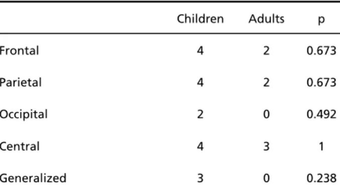
Children Adults p
Frontal 4 2 0.673
Parietal 4 2 0.673
Occipital 2 0 0.492
Central 4 3 1
Arq Neuropsiquiatr 2006;64(2-B) 361
c h i l d ren and two adults) were more frequent in chil-d ren, anchil-d there was no significant chil-diff e rence bet-ween the two groups in signs and symptoms.
The neuropathological data of the patients who had already undergone surg e ry are shown in Ta b l e 1 (12 children and six adults).
The most frequent etiologies were ganglioglioma, D N E T, followed by astrocytoma. We found six tempo-ral mesial lesions in the children and six in the adults, and there were 10 lateral lesions in the children and six in the adults. The lesions occurred most fre q u e n tl y in the right hemisphere: 10 children and eight adults. Table 2 shows the temporal EEG findings of gro u p s 1 and 2. The childhood group (Group 1) underwent 64 exams (mean: 4 exams per patient) and the adult g roup (Group 2) had 78 exams (mean: 6.5 exams per patient).
Table 3 shows the extratemporal EEG findings of groups 1 and 2.
DISCUSSION
Our data show that temporal interictal epilepti-f o rm and nonepileptiepilepti-form activity occur equally in all age ranges. Normal EEGs were significantly more f requent in the adults in this study. However, accord-ing to Harvey et al.1 4, in the pediatric age range,
15.9% of the patients with TLE may have persistent-ly normal EEGs. Interictal extratemporal epileptiform activity was also found in both groups, which was unexpected, as TLE in childhood usually presents with a greater clinico-electroencephalographic diversity1.
P re-operative studies perf o rmed in adults with temporal lobe tumors have demonstrated electro-physiological variability: temporal focal discharg e s , bilateral temporal activity, contralateral activity, extratemporal activity and the presence of “mirro r focus”15-22.
Interictal EEGs of children with TLE may show ex-tratemporal (especially frontal) discharges more fre-quently than those seen in adults with mesial sclero-s i sclero-s1 2. In the present study we observed that the EEGs
of adult patients with tumoral lesions also displayed extratemporal discharges. That suggests that this n e u rophysiological characteristic might be related to the etiology, independent of the age range. In oth-er words, thoth-ere was no significant diff e rence between the extratemporal findings of children and adults be-cause both age groups seem to have a similar neuro-physiological behavior when the etiology is an
expan-sive lesion of the temporal lobe. In patients with TLE caused by hippocampal sclerosis, however, there is a difference between the age ranges12,23.
Our data show that seizure onset was significant-ly earlier in children than in adults, despite the same pathology and similar EEG findings. This may re i n-f o rce that EEG n-features in temporal lobe tumors are not age-dependent.
The clinical features of adults with tumoral lesions in the temporal lobe can be diff e rentiated from those with mesial temporal sclerosis by the initial ictal pat-t e rn, by pat-the behavioral sequence and by pat-the pat-time of its appearance during the seizure2 4. Other authors
found that seizures of neocortical origin are signifi-cantly shorter in duration25. In spite of these clinical
d i ff e rences, there is difficulty in making a distinction in an individual patient26. Our clinical findings were
suggestive of mesial TLE with the presence of typi-cal auras and automatisms, even in the lateral lesions. These findings may either reflect a rapid pro p a g a-tion to the mesial stru c t u res or simply occur by the activation of cortical areas distant from the epilep-tic focus. Few patients presented auras with sugges-tive of neocortical involvement.
To conclude, this study suggests that interictal dis-c h a rges in dis-children and adults with TLE due to expasive lesions present with a polymorphic electro e n-cephalographic pattern. Although children with TLE have frequent extratemporal epileptiform discharg e s , independent of the etiology, there is no significant d i ff e rence when one compares children and adults with tumoral lesions. In TLE due to mesial sclero s i s , t h e re seems to be a diff e rence between the two age groups.
REFERENCES
1. Duchowny M, Levin B, Jayakar P, et al. Temporal lobectomy in early childhood. Epilepsia 1992;33:298-303.
2. Wyllie E, Chee M, Granstrom ML, et al. Temporal lobe epilepsy in ear-ly childhood. Epilepsy 1993;34:859-868.
3. Sutton LN, Roger JP, Rorke LB, Derek AB, Schut L. Cerebral gangli-ogliomas during childhood. Neurosurgery 1983;13:124-128. 4. Johannsson HJ, Rekate HL, Roessmann U. Gangliogliomas:
patholog-ical and clinpatholog-ical correlation. J Neurosurg 1981;54:58-63.
5. Lach B, Duggal N, DaSilva VF, Benoit BG. Association of pleomorphic x a n t h o a s t rocytoma with cortical dysplasia and neuronal tumors: a report of three cases. Cancer 1996;78:2551-2563.
6. Madsen JR, Vallat AV, Poussaint TY, Scott RM, De Girolami U, A n t h o n y DC. Focal cortical dysplasia with glioproliferative changes causing seizures: report of three cases. Pediatr Neurosurg 1998;28:261-266. 7. Prayson RA, Estes ML. Dysembryoplastic neuroepithelial tumor. Am
J Clin Pathol 1992;97:398-401.
362 Arq Neuropsiquiatr 2006;64(2-B)
9. Shimbo Y, Takahashi H, Hayano M, Kumagai T, Kameyama S. Te m p o r a l lobe lesion demonstrating features of dysembryoplastic neuroepithe-lial tumor and ganglioglioma: a transitional form? Clin Neuro p a t h o l 1997;16:65-68.
10. B rockhaus A, Elger CE. Complex partial seizures of temporal lobe ori-gin in children of different age groups. Epilepsia 1995;36:1173-1181. 11. Fogarasi A, Jokeit H, Faveret E, Janszky J, Tuxhorn I. The effect of age
on seizure semiology in childhood temporal lobe epilepsy. Epilepsia 2002;43:638-643.
12. Franzon RC, Montenegro MA, Guimarães CA, Guerreiro CA, Cendes F, Guerre i ro MM. Clinical, electroencephalographic, and behavioral f e a t u res of temporal lobe epilepsy in childhood. J Child Neurol 2004; 19:418-423.
13. N o rdli DR, Kuroda MM, Hirsch LJ. The ontogeny of partial seizure s in infants and young children. Epilepsia 2001;42:986-990.
14. Harvey AS, Berkovic SF, Wrennall JA, et al. Temporal lobe epilepsy in childhood: clinical, EEG, and neuroimaging findings and syndro m e classification in a cohort with new-onset seizures. Neurology 1997; 49:960-968.
15. M o r rell F, Rasmussen T, Gloor P, De To l e d o - M o r rell L. Secondary epilep-togenic foci in patients with verified temporal lobe tumors. Eletro-encephalogr Clin Neurophysiol 1983;54:26.
16. Morris HH, Maltkovic Z, Estes ML, et al. Ganglioglioma and intractable epilepsy: clinical and neurophysiologic features and predictors of out-come after surgery. Epilepsia 1998;39:307-313.
17. Hamer HM, Najm I, Mohamed A, Wyllie E. Interictal epileptiform dis-c h a rges in temporal lobe epilepsy due to hippodis-campal sdis-clerosis ver-sus medial temporal lobe tumors. Epilepsia 1999;40:1261-1268.
18. J o rge CL, Nagahashi-Marie SK, Pedreira CC, et al. Clinical character-istics and surgical outcome of patients with temporal lobe tumors and epilepsy. Arq Neuropsiquiatr 2000;58:1002-1008.
19. Iannelli, Guzzetta F, Battaglia D, Iuvone L, Di Rocco C. Surgical treat-ment of temporal tumors associated with epilepsy in children. Pediatr Neurosurg 2000;32:248-254.
20. Z a a t reh MM, Firlik KS, Spencer DD, Spencer SS. Temporal lobe tumoral epilepsy: characteristics and predictors of surgical outcome. Neuro l o g y 2003;61:636-641.
21. Labate A, Briellmann RS, Harvey AS, et al. Temporal lobe dysembry-oplastic neuroepithelial tumour: significance of discordant interictal spikes. Epileptic Disord 2004;6:107-114.
22. Sampaio L, Yacubian EM, Manreza ML. The role of mirror focus in the s u rgical outcome of patients with indolent temporal lobe tumors. A rq Neuropsiquiatr 2004;62:9-14.
23. Blume WT, Girvin JP, Mclachlan RS, Gilmore BE. Effective temporal lobectomy in childhood without invasive EEG. Epilepsia 1997;38: 164-167.
24. Saygi S, Spencer SS, Scheyer R, Katz A, Mattson R, Spencer DD. Diff e-rentiation of temporal lobe ictal behavior associated with hippocam-pal sclerosis and tumors of temporal lobe. Epilepsia 1994;35:737-742. 25. Foldvary N, Lee N, Thwaites G, et al. Clinical and electrographic
man-ifestations of lesional neocortical temporal lobe epilepsy. Neuro l o g y 1997;49:757-763.