Po ly-D L-lactide -co -glyco lide
micro sphe re s as a co ntro lle d
re le ase antige n de live ry syste m
Laboratório de Tecnologia Farmacêutica, Departamento de Produtos Farmacêuticos, Universidade Federal de Minas Gerais, Belo Horizonte, MG, Brasil
K.M. Lima and J.M. Rodrigues Júnior
Abstract
Successful vaccine application means maximum protection with mini-mal number of administrations. A rational development of vaccines involves studies of the nature of the antigen as well as of the adjuvant to be used to improve the immune responses. This has provided the impetus for studies to design the degradable devices and for different approaches to antigen delivery by different routes of administration. The development of controlled release systems based on polymeric devices that permit a sustained or pulsed release of encapsulated antigens has attracted much interest. Polymeric delivery systems consist of polymers that release their content continuously in a con-trolled manner over a period of time. The development of a biocompatible delivery system for parenteral administration offers several advantages in terms of immunoadjuvanticity over other com-pounds. It was found that, in contrast to other carriers, microspheres are more stable, thus permitting administration by the oral or parenter-al route. In the present study, we describe the main characteristics and potentialities of this new immunoadjuvant for oral and parenteral administration.
Co rre spo nde nce
J.M. Rodrigues Júnior Departamento de Produtos Farmacêuticos, UFMG Av. O legário Maciel, 2360 30180-112 Belo Horizonte, MG Brasil
Fax: + 55-31-291-9769
E-mail: rodrigue@ dedalus.lcc.ufmg.br
Presented at the International Symposium “The Third Revolution on Vaccines: DNA Vaccines”, Belo Horizonte, MG, Brasil, November 3-7, 1997.
Research supported by FAPEMIG and CNPq.
Received November 19, 1998 Accepted December 8, 1998
Ke y words
·Vaccine ·Adjuvant ·Microspheres ·Microencapsulation ·Biomaterial
Intro ductio n
The investigation of formulations for the controlled release in vaccine delivery is a top priority because of their potential for reduc-ing the number of administrations required to induce protection (1-4). A rational devel-opment of vaccines involves studies of the nature of the antigen as well as the adjuvant used to improve the immune responses. With the progress in the development of new anti-gens resulting from biotechnology studies, the search for new adjuvants has become the main research subject for many groups who
study their role and safety for therapeutical use (5-8).
The ideal and safe adjuvant should not gen-erate local or systemic reactions after admin-istration. It should elicit an early, high and long-lasting immunoresponse and should be stable and chemically defined to permit re-producible manufacturing (5,8). A good ad-juvant needs to be able to give signals to the immunological system so that lymphocytes and phagocytes migrate to the vaccinated area (10).
However, after many decades of research for adjuvants, only aluminum and liposomes (lipid vesicles) have been approved for use in humans (9). Although many advantages may be considered for alum as adjuvant, after years of safe history, the use of alumi-num-based vaccines has declined consider-ably due to variations in the production of alum-precipitated vaccines (8). Other limi-tations are their ineffectiveness for certain antigens and their inability to elicit cell-mediated immune responses, particularly cytotoxic T-cell responses, which limit their application against intracellular parasites and viral infections. Also, aluminum adjuvants cannot be frozen or lyophilized (8).
Other adjuvants have been developed and a few have been evaluated in clinical trials, but most of them were never accepted for routine vaccines due to their adverse side effects. The use of liposomes as immunoad-juvants is also controversial. However, en-capsulated antigens have the potential ad-vantage of providing a higher antigen-carry-ing capacity. Both humoral and cell-medi-ated immune responses have been elicited by these systems. With the continuous hard effort of the past two decades, a considerable number of issues may yet be overcome such as shelf life and targeting specific cells.
Controlle d de live ry syste ms
The need for new adjuvants has led to the development of controlled release systems based on polymeric devices, which could permit a sustained or pulsed release of
en-capsulated or entrapped antigens (11-13). This has provided the impetus for active studies to design biodegradable materials and approaches for delivery by different routes of administration (14-17). Benefits of controlled release systems are delivery to a specific site, protection of the antigen from degradation, better patient compliance and more efficient antigen dosing, which may eliminate the need for boosters.
Polymeric delivery systems consist of polymers that release their content in a con-trolled manner continuously over time. The development of a biocompatible delivery system for parenteral administration offers several advantages in terms of immunoadju-vanticity over other compounds. In contrast to other carriers, polymeric microspheres are stable enough to permit their administra-tion by the topical, oral or parenteral route. This is particularly advantageous when a mucosal immune response is needed. Fur-thermore, the association of antigens with microspheres has been able to elicit both cellular and humoral immune response (18). The stability of this formulation under stor-age conditions is related to hydrolytic degra-dation. However, the formulations can be freeze-dried and stored as a dry powder, which can be easily reconstituted immedi-ately before use. The main goal of polymeric microencapsulation is to coat the antigenic material with a biodegradable polymer which will protect and control the antigen delivery. The possibility of obtaining free-flowing par-ticles with well-defined diameters from 1 to 100 micrometers offers many advantages for its manufacture and interaction with immu-nocompetent cells.
Po ly-lactide -co -glyco lide micro sphe re s
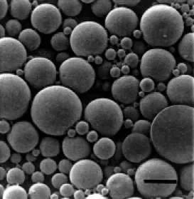
immuno-logical studies as a controlled delivery sys-tem of peptides, native and synthetic pro-teins and lately, nucleic acids (13-27). PLGA microspheres are composed of a spherical-shaped polymeric matrix ranging in diam-eter from 1 to 250 µm (Figure 1). Many factors are important to formulate these sys-tems: i) ability to release the entrapped sub-stance in a controlled way, which is influ-enced by polymer molecular weight, mono-mer ratio and morphology (28-30); ii) par-ticle size, which is important in terms of interaction with phagocytic cells (12,13,31); iii) safety, which is related to the in vivo
polymer degradation (32,33), and iv) stabil-ity, both in storage and in the biological fluids (34,35).
The antigen is physically entrapped into microspheres inside an injectable solid poly-meric matrix. The combination of diffusion through pores and of polymer matrix biodeg-radation allows the control of antigen re-lease rates. The biodegradation rate of the polymer depends on its molecular weight and it is well established that shorter chains are degraded faster (30,36). After their ter-minal chain hydrolysis, the resulting degraded products are monomers of lactide and glycolide, which are innocuous to the body. These degraded products are eliminated by the Krebs cycle as carbon dioxide and in the urine (37). During biodegradation, the en-capsulated antigen is released. Since these polymers are chemically defined, the anti-gen release is uniform and reproducible. This release can vary from hours to months de-pending on the polymer combinations.
Microsphere diameter plays an impor-tant role in the interactions with phagocytic cells. Particles smaller than 10 µm may be phagocytosed faster by macrophages, which are recruited to the site of administration after subcutaneous injection, and particles larger than 10 µm act as a depot releasing the antigens in a second step (13,16). We also have to consider the fate of these particles across the digestive tract to be taken up by
the M cells of the Peyers patches, when administered orally, since this could play a role by facilitating the antigen presentation to the immunocompetent cells and conse-quently the elicitation of an effective mu-cosal immune response. Enhancement of the stability of macromolecules may also be at-tained since they are protected from enzymes or other factors that could compromise the stability of the antigens.
Another important factor in favor of this new immunoadjuvant is related to its safety. These polymers have a long history as a safe material used in the composition of sutures and implants. This fact provided the basis for injectable delivery systems now used by 300,000 patients annually for treating ad-vanced prostate cancer, endometriosis or precocious puberty (38).
Polymer selection is the most important step in the manufacture of microspheres be-cause it critically influences their rate of biodegradation and hence devices with short-or long-lasting action can be constructed. Nowadays, several biocompatible polymers are commercially available. By changing the homopolymer ratio, different physicochemi-cal compositions can influence its bility and permeability, resulting in degrada-tion times ranging from weeks up to several months depending on the shape, size and porosity of the device (Table 1). These
rameters can be easily monitored using dif-ferent polymer and copolymer compositions and controlling the variables during the manu-facture process. Since the amorphous poly-mers are more permeable, poly-DL-lactide acid (DL-PLA), which is less amorphous than PLGA copolymers, has a slower degra-dation rate, usually over several months. The combination of particles with different di-ameters and polymer composition in the same formulation has permitted to establish the concept of the single-dose vaccine (1), which can be programmed to mimic the reinforce-ment dosages, inducing protection with a minimal number of administrations (Figure 2). Finally, PLA and its copolymers have
proved to be biocompatible and produce little or no local or systemic toxicity (39,40). PLGA subcutaneously administrated acts by means of three mechanisms: i) a depot formation at the site of administration; ii) targeting antigens to antigen-presenting cells, since it has been well described that macro-phages are recruited to the site of the injec-tion, or adherence to the Peyers patches, which could facilitate the antigen presenta-tion (41,42), and iii) protecpresenta-tion of the anti-gen by storage, with a slow release of the remaining antigens that could eliminate the need for a booster (43,47).
Microsphe re pre paration
PLGA microspheres can be obtained by two main methods: simple emulsification (O/W) or multiple water-in-oil-in-water (W/ O/W) emulsion followed by solvent elimina-tion. The choice of method depends on the physical and chemical characteristics of the antigen, permitting the matrix entrapment of both lipophilic and hydrophilic molecules.
In the first case, an emulsion is formed by dissolving (or dispersing) the antigen in an organic solvent immiscible in water (meth-ylene chloride, chloroform or ethyl acetate) containing the polymer, under strong me-chanical agitation. This emulsion may be stabilized by a surfactant added to the aque-ous phase. The solvent is eliminated by evaporation at room temperature, followed by washing and freeze-drying.
The multiple emulsion method involves water-in-oil-in-water emulsification (16,31). The inner aqueous phase containing the hy-drophilic substances is obtained after emul-sification with the immiscible organic sol-vent-containing polymer under strong me-chanical agitation. This first emulsion is sta-bilized by the addition of an aqueous solu-tion containing a surfactant (e.g., poly vinyl alcohol) and is further homogenized to pro-duce a W/O/W double emulsion. This double emulsion is gently stirred with a
homog-Table 1 – Physicochemical characteristics and biodegradation time of poly-lactide and co-polymers.
* Biodegradation time depends on the formulation, porosity, surface area and polymer molecular w eight. Adapted from Ref. 35.
Polymer Crystallinity Glass transition Biodegradation
time (months)*
Poly (L-lactide) Crystalline 45-60oC 18-24
Poly (D,L-lactide) Amorphous 50-65oC 12-16
50:50 Poly (D,L-lactide-co-glycolide) Amorphous 40-55oC 2 85:15 Poly (D,L-lactide-co-glycolide) Amorphous 45-60oC 5
Antigen release time (after injection)
Days
Weeks
M onths Figure 2 - The single-dose
enizer at room temperature for solvent evapo-ration. The microspheres are collected by centrifugation, washed with water and freeze-dried. The composition of the aqueous phase as well as the surfactant applied plays an important role in the pattern of antigen re-lease since porosity and water permeability will be dependent on these factors. A mix-ture of hydrophilic and lipophilic molecules can also be successfully entrapped by using the W/O/W method (48).
Both of these methods allow high levels of antigen entrapment. The particles are char-acterized by determining the antigen entrap-ment rate, their average diameter, porosity and in vitro antigen release kinetics in appro-priate medium. The maintenance of immu-nogenicity after antigen contact with organic solvents is also an important aspect to con-sider. Many studies have indicated that the antigenic properties and the structure of the proteins can be preserved during entrapment (26,44,49-53). Another important aspect is related to the possibility of scaling up the process under aseptic conditions, allowing the preparation of a pyrogen-free and sterile product (54).
Conside rations about PLGA microsphe re biocompatibility
Polymeric microspheres have attracted much attention because of their biocompa-tible characteristics. The phagocytosis of bio-degradable and nonbiobio-degradable particles has been reported to depend on their size, surface charge and hydrophobicity (55,56). After subcutaneous administration, PLGA microspheres ranging in diameter from 1 to 10 µm are readily phagocytosed by macro-phages recruited to the site of injection, thereby providing an intracellular delivery of the antigen. This mechanism may en-hance antibody responses and consequently decrease the required antigen dose. On the other hand, particles larger than 10 µm in diameter would remain as a depot at the site
of injection providing a sustained release of antigen. Improving their hydrophobicity can increase phagocytosis of the particles, whereas microsphere preparations with dif-ferent compositions of PLA and PLGA do not alter the extent of phagocytosis. How-ever, precoating microspheres with opsonins can enhance phagocytosis (55). The intra-cellular fate of these particles is dependent on their composition. The rate of micro-sphere degradation inside cells could be con-trolled by changing the molecular weight and the monomer composition. PLGA mi-crospheres are readily degraded releasing the entrapped molecules. Small particles of 200 nm in diameter are readily phagocy-tosed by Kupffer cells and reach the lysoso-mial compartment in liver tissues after intra-venous administration, as demonstrated by transmission electron microscopy (56).
The relationship between the maximal dose of polymer and its implications in the intracellular residential time is not yet clear. In some reports of immunization studies, the entrapped protein:polymer ratio is very low, requiring administration of a huge amount of microspheres to attain the desirable antigen dose. It is well documented that the antigen and adjuvant doses interfere with the im-mune responses. Studies conducted with sub-cutaneous implants indicate that the physi-cochemical characteristics of PLA and PLGA polymers play a decisive role in the in vivo
degradation time and also in the local in-flammatory reaction (32,33,39). These re-sults highlight an important aspect to be considered in the development of micro-spheres as carriers, i.e., the dose of polymer. We focused our studies on the clarification of the local responses after subcutaneous administration of microspheres, using an air pouch model in rats. We observed that 6 h after the administration of a single dose of 125 mg or a higher dose of 625 mg of poly-mer per kg rat body weight, 14.3 x 10³
and 31.5 x 10³
injec-tion, the number of cells decreased con-spicuously without a significant difference between the treated groups and the control group treated with saline only. Histological analysis allowed us to identify an organic reactivity in the injected area characteristic of macrophage infiltration. Using soluble and particulate Leishmania antigens as a model, we found that the mononuclear infil-trate obtained at the injection site was larger with antigen-loaded microspheres as com-pared to unloaded ones. No granulocytic cell infiltration was observed. Higher doses (250 and 625 mg/kg) provoked the formation of a fibrovascular capsule surrounding the mi-crospheres. In contrast, after administration of lower doses this reaction was not ob-served and PLGA microspheres were found to be completely degraded one month after injection. We selected the maximal polymer dose of 150 mg/kg to design our experimen-tal protocols.
Syste mic immunization
The diameter of the particles plays an important role in the induction of protection by parenteral immunization. In fact, when ovalbumin was adsorbed to or entrapped in PLGA microspheres of different diameters, drastic differences were observed in the anti-body responses (12,13). Two weeks after immunization with a single dose by the sub-cutaneous route, the serum IgG antibody re-sponse to ovalbumin was significantly greater than the response to soluble ovalbumin. This response was more significant for particles of 1.5 µm than for particles larger than 73 µm. Interestingly, the level of antibodies remained high even one year after injection (16). Uchida et al. (31) reported similar results showing the superior efficacy of microspheres over alum. Vaccine formulations prepared by simple mixing of blank microspheres (with-out antigens) and ovalbumin exhibited low primary immune responses, which were only elevated by boosting (45).
Three batches of PLA and PLGA micro-spheres were used to investigate the single dose vaccine delivery by incorporating teta-nus toxoid (18). Its comparison with alumi-num hydroxide as immunoadjuvant provid-ed data to support the superior efficacy of PLGA microspheres to elicit T cell and anti-body responses after subcutaneous adminis-tration. These results were confirmed by Walker et al. (57). The cellular and antibody responses of mice to tetanus toxoid were compared after subcutaneous administration of the toxoid alone or incorporated into alu-minum hydroxide and PLGA microspheres. The safety of a similar vaccine was demon-strated in rats (40).
The entrapment of a synthetic peptide in PLGA microspheres was evaluated by Partidos et al. (23). Intraperitoneal immuni-zation with particles smaller than 10 µm containing a chimeric peptide constructed by synthesis of a potential B cell site induced anti-peptide antibody responses after a single dose. This single intraperitoneal administra-tion protocol was also applied to induce pro-tection against Bordetella pertussis respira-tory infection in mice (26). Although, PLGA microspheres containing pertussis fimbrae elicited lower levels of serum antibody than those produced by alhydrogel-containing an-tigen, both formulations protected against intranasal infection by B. pertussis.
The magnitude of the immune response induced by peptides is comparable or supe-rior to that induced by peptide emulsified in complete Freunds adjuvant (58). In vitro, peptides (carrying an immunodominant T-helper cell epitope delineated from the ra-bies virus nucleoprotein) incorporated into PLGA microspheres induced the prolifera-tion of a peptide-specific T cell line. After subcutaneous immunization of mice, the immune response induction was related to the nature of the polymer.
adju-vants in a birth control vaccine (25), HIV-1 vaccine (52,54), rabies virus nucleoprotein (58), malaria antigen (59) and type A botu-lism (60).
Mucosal immunization
New approaches to vaccine development have become possible after a common mu-cosal defense system was recognized where an antigen interacting with localized lym-phoid tissue could stimulate IgA precursor cells that could migrate to other mucosal surfaces (12,13,61). The mucosal immune system displays a unique ability to respond to an array of immunogens presented by the respiratory and oral routes. Oral vaccines are often more desirable than other routes be-cause of easy administration to large popula-tions and a reduced number of side effects (62). Since PLGA microspheres are readily absorbed by the Peyers patches, they have considerable potential as a vehicle for oral immunization. They are taken up by modi-fied epithelial cells (M cells) and transported to the lymphoid tissue where they encounter antigen-presenting cells including macro-phages, dendritic cells and B cells. So, the use of microspheres has two advantages: i) protection from low pH and from proteases (since the antigen and/or adjuvant are en-trapped in the polymer matrix, they are pro-tected from both gastric and gastrointestinal proteolysis), and ii) targeting to IgA induc-tive sites.
It was found that peroral immunizations with a toxoid vaccine of staphylococcal en-terotoxin B encapsulated in 1-10 µm PLGA microspheres stimulated circulating IgM, IgG and IgA antitoxin antibodies in mice. A dis-seminated mucosal IgA response was also observed. Systemic immunization was ef-fectively primed for a mucosal IgA response when a booster was administered by the oral or intratracheal route, indicating that under these experimental conditions the systemic immunization did not induce tolerance to or
down-regulation of a subsequent mucosal antibody response. Nonencapsulated con-trols did not elicit any significant response. Microspheres less than 5 µm in diameter were effectively taken up by Peyers patches and carried by macrophages to the mesenter-ic lymph nodes and spleen. On the other hand, microspheres in the range of 5-10 µm remained in the Peyers patches throughout the 35 days of the experiment. Microspheres larger than 10 µm in diameter were not adsorbed at any point in the gastrointestinal tract (13).
The entrapment of a branched peptide immunogen representing a portion of the principal neutralizing determinant of HIV-1 has been developed for oral and combined oral and subcutaneous immunization against HIV. Under aseptic conditions a pyrogen-free and sterile preparation was obtained. The system showed high levels of both se-rum IgG and neutralizing antibodies against HIV in guinea pigs in both protocols. The authors proposed the formulation for clini-cal trials (52,54). Recently, it was demon-strated that immunization with a recombi-nant HIV envelope protein entrapped in PLGA microspheres induced consistent HIV-specific CD4+ and CD8+ T-cell responses in mice. Cytotoxic T lymphocyte (CTL) re-sponses were detected after a single sys-temic immunization with gp120 entrapped in microparticles; when given by the intrana-sal route these microparticles induced HIV-specific CD8+ CTL and secretory IgA. The induction of Th1 cells was observed after generation of CD4+ T cells that secreted moderate to high levels of interferon-g fol-lowing immunization with gp120 entrapped in microparticles (63).
sta-bility and stimulated the secretion of IgA, IgG and IgM after oral administration. On the other hand, only the IgM and IgG re-sponses were significant after intraperito-neal administration.
Concluding re marks
PLGA microspheres with an incorporated antigen represent a good antigen delivery system for both cellular and humoral re-sponses. Further efforts are needed to elabo-rate an effective controlled release system
for antigens. The promising use of PLA and PLGA microspheres has been confirmed by experimental and clinical trials. The ques-tion whether pulsed or continuous antigen release provides the better immune responses remains to be clarified. The easy manufac-ture of microspheres and the possibility of administration by different routes offer the additional advantage of their use as a phar-maceutically acceptable adjuvant for vac-cines. These findings may have major impli-cations for the design of novel vaccine deliv-ery strategies.
Re fe re nce s
1. Aguado M T (1993). Future approaches to vaccine development: single-dose vac-cines using controlled-release delivery systems. Vaccine, 11: 596-597.
2. Khan M ZI, Opdebeeck JP & Tucker IG (1994). Immunopotentiation and delivery systems for antigens for single-step im-munization: recent trends and progress.
Pharmaceutical Research, 1: 2-11. 3. Pow ell M F & New man M J (1994).
Vac-cine Design: The Subunit and Adjuvant Approach. Plenum Publishing Corpora-tion, New York.
4. Cleland JL (1995). Design and production of single-immunization vaccines using polylactide polyglycolide microsphere sys-tems. In: Pow ell M F & New man M J (Edi-tors), Vaccine Design: The Subunit and Adjuvant Approach. Plenum Publishing Corporation, New York, 439-462. 5. Bomford R (1989). Adjuvants for
anti-para-site vaccines. Parasitology Today, 5: 41-46.
6. Allison AC & Byars NE (1991). Immuno-logical adjuvants: desirable properties and side effects. M olecular Immunology, 28: 279-284.
7. Audibert FM & Lise LC (1993). Adjuvants: current status, clinical perspectives and future prospects. ImmunologyToday, 14: 281-284.
8. Gupta RK & Siber GR (1995). Adjuvants for human vaccines - current status, prob-lems and future prospects. Vaccine, 13: 1263-1276.
9. Gregoriadis G (1998). Genetic vaccines: strategies for optimization. Pharmaceuti-cal Research, 15: 661-669.
10. Gizurarson S (1996). Optimal delivery of vaccines - clinical pharmacokinetic
con-siderations. Clinical Pharmacokinetics, 30: 1-15.
11. Partidos CD, Vohra P, Jones D, Farrar G & Stew ard M W (1997). CTL responses in-duced by a single immunization w ith pep-tide encapsulated in biodegradable micro-particles. Journal of Immunological M eth-ods, 7: 143-151.
12. Eldridge JH, Staas JK, M eulbroek JA, Tice TR & Gilley RM (1991). Biodegradable mi-crospheres as a vaccine delivery system.
M olecular Immunology, 28: 287-294. 13. Eldridge JH, Staas JK, Dexiang C, Preston
AM , Tice TR & Gilley RM (1993). New advances in vaccine delivery systems.
Seminars in Hematology, 4: 16-25. 14. M estecky J & Eldridge JH (1991).
Target-ing and controlled release of antigens for the effective induction of secretory anti-body responses. Current Opinion in Im-munology, 3: 492-495.
15. Allaoui-Attarki K, Fattal E, Pecquet S, Trolle S, Chachat y E, Couvreur P & Andremont A (1998). M ucosal immuno-genicity elicited in mice by oral vaccina-tion w ith phosphorylcholine encapsulated in poly (D,L-lactide-co-glycolide) micro-spheres. Vaccine, 16: 685-691.
16. O’Hagan DT, Jeffery H & Davis SS (1993). Long-term antibody responses in mice fol-low ing subcutaneous immunization w ith ovalbumin entrapped in biodegradable mi-croparticles. Vaccine, 11: 965-969. 17. O’Hagan DT, Rafferty D, Wharton S &
Illum L (1993). Intravaginal immunization in sheep using a bioadhesive microsphere antigen delivery system. Vaccine, 11: 660-664.
18. M en Y, Thomasin C, M erkle HP, Gander B & Corradin G (1995). A single
adminis-tration of tetanus toxoid in biodegradable microspheres elicits T cell and antibody response similar or superior to those ob-tained w ith aluminum hydroxide. Vaccine, 7: 683-689.
19. Santiago N, M ilstein S, Rivera T, Garcia E, Zaidi T, Hong H & Bucher D (1993). Oral immunization of rats w ith proteinoid mi-crospheres encapsulating influenza virus antigens. Pharmaceutical Research, 8: 1243-1247.
20. Gander B, Thomasin C, M erkle HP, M en Y & Corradin G (1993). Pulsed tetanus tox-oid release from PLGA-microspheres and its relevance for immunogenicity in mice. In: Proceedings of the International Sym-posium of Controlled Release Bioactive M aterials. Washington DC, 20: 65-66. 21. Hazrati AM , Lew is DH, Atkins TJ, Stohrer
RC, M cPhilips CA & Little JE (1993). Sal-monella enteritidis vaccine utilizing biode-gradable microspheres. Proceedings of the International Sym posium of Con-trolled Release Bioactive M aterials. Wash-ington DC, 20: 101-102.
22. Alm eida AJ, Alpar HO & Brow n RW (1993). Immune response to nasal deliv-ery of antigenically intact tetanus toxoid associated w ith poly(L-lactic acid) micro-spheres in rats, rabbits and guinea-pigs.
Journal of Pharmacy and Pharmacology, 45: 198-203.
23. Partidos CD, Shaw DM , Gander B, M erkle HP, How ard CR & Stew ard M W (1994). Induction of antibody responses to a syn-thetic peptide entrapped in biodegradable microparticles. Vaccine Research, 4: 203-209.
Singh M (1995). Synthetic delivery sys-tem for tuberculosis vaccines: immuno-logical evaluation of the M . tuberculosis
38 kDa protein entrapped in biodegrad-able PLG microparticles. Vaccine, 16: 1576-1582.
25. Singh M , Singh O & Talw ar GP (1995). Biodegradable delivery system for a birth control vaccine: Immunogenicity studies in rats and monkeys. Pharmaceutical Re-search, 12: 1796-1800.
26. Jones DH, M cBride BW , Jef f ery H, O’Hagan DT, Robinson A & Farrar GH (1995). Protection of mice from Bordetella pertussis respiratory infection using mi-croencapsulated pertussis fimbriae. Vac-cine, 7: 675-681.
27. Jones DH, Corris S, M cDonald S, Clegg JC & Farrar GH (1997). Poly(DL-lactide-co-glycolide)-encapsulated plasmid DNA elic-its systemic and mucosal antibody re-sponses to encode protein after oral ad-ministration. Vaccine, 15: 814-817. 28. Pistner H, Gutw ald R, Ordung R, Reuther
J & M uhling J (1993). Poly(l-lactide): a long-term degradation study in vivo. I. Bio-logical results. Biomaterials, 14: 671-677. 29. Dorta M J, M unguía O & Llabrés M (1993). Effects of polymerization variables on PLGA properties: molecular w eight, com-position and chain structure. International Journal ofPharmaceutics, 100: 9-14. 30. Park TG (1994). Degradation of
poly(D,L-lactic acid) microspheres: effect of molec-ular w eight. Journal of Controlled Re-lease, 30: 161-173.
31. Uchida T, Goto S & Foster T (1994). Par-ticle size studies for subcutaneous deliv-ery of poly(lactide-co-glycolide) micro-spheres containing ovalbumin as vaccine formulation. Journal of Pharmacy and Pharmacology, 47: 556-560.
32. Pekarek KJ, Dyrud M J, Ferrer K, Jong YS & M athiow itz E (1996). In vitro and in vivo
degradation of double-w alled polymer mi-crospheres. Journal of Controlled Re-lease, 40: 169-178.
33. Spenlehauer G, Vert M , Benoit JP & Boddaert A (1989). In vitro and in vivo
degradation of poly(D,L-lactide/glycolide) t ype m icrospheres m ade by solvent evaporation method. Biomaterials, 10: 557-563.
34. Langer R (1996). Controlled release of a therapeutic protein. Nature M edicine, 7: 742-743.
35. Lew is DH (1990). Controlled release of bioactive agents from lactide/glycolide polymers. In: Chasin M & Langer R (Edi-tor), Biodegradable Polymers as Drug
De-livery Systems. M arcel Dekker, New York, 1-43.
36. Park TG, Lu W & Crotts G (1995). Impor-tance of in vitro experimental conditions on protein release kinetics, stability and polymer degradation in protein encapsu-lated poly (D,L-lactic acid-co-glycolic acid) microspheres. Journal of Controlled Re-lease, 33: 211-222.
37. Bazile DV, Ropert C, Huve P, Verrecchia T, M arland M , Frydman A, Veillard M & Spenlehauer G (1992). Body distribution of fully biodegradable 14C-poly(lactic acid) nanoparticles coated w ith albumin after parenteral administration to rats. Bioma-terials, 13: 1039-1102.
38. Langer R (1998). Drug delivery and target-ing. Nature, 392 (Suppl): 5-10.
39. Visscher GE, Robison RL, M aulding HV, Fong JW, Pearson JE & Argentieri GJ (1986). Biodegradation of and tissue reac-tion to poly(D,L-lactide) microcapsules.
Journal of Biomedical M aterials Research, 20: 667-676.
40. Chaudhury M R, Sharm a K & Giri DK (1996). Poly(D,L-lactice) glycolide polymer microsphere entrapped tetanus toxoid: safety evaluation in Wistar rats. Human and Experimental Toxicology, 15: 205-207.
41. O’Hagan DT (1996). The intestinal uptake of particles and the implications for drug and antigen delivery. Journal of Anatomy, 19: 477-482.
42. Beier R & Gebert A (1998). Kinetics of particle uptake in the domes of Peyer’s patches. American Journal of Physiology, 275: G130-G137.
43. An-Cheng C & Gupta RK (1996). Stabiliza-tion of tetanus toxoid in poly(DL-lactic-co-glycolic acid) microspheres for the con-trolled release of antigen. Journal of Phar-maceutical Sciences, 85: 129-132. 44. Schw endesm an SP, Const ant ino HR,
Gupta RK, Tobio M , Chang AC, Alonso M J, Siber GR & Langer R (1996). Strate-gies for stabilising tetanus toxoid tow ard the development of a single-dose tetanus vaccine. Developments in Biological Stan-dardization, 87: 293-306.
45. Coombes AG, Lavelle EC, Jenkins PG & Davis SS (1996). Single dose, polymeric, microparticle-based vaccines: the influ-ence of formulation conditions on the magnitude and duration of the immune response to a protein antigen. Vaccine, 14: 1429-1438.
46. Crotts G & Park TG (1997). Stability and release of bovine serum albumin encap-sulated w ithin
poly(D,L-lactide-co-glyco-lide) microparticles. Journal of Controlled Release, 44: 123-134.
47. Johansen P, M en Y, Audran R, Corradin G, M erkle HP & Gander B (1998). Improv-ing stability and release kinetics of mi-croencapsulated tetanus toxoid by co-en-capsulation of additives. Pharmaceutical Research, 15: 1103-1110
48. Lima KM , Chiarini-Garcia H, Bambirra EA, Af onso LCC & Rodrigues Júnior JM (1998). Development and characterization of Leishm ania m ajor ant igens-loaded PLGA microspheres. Proceedings of the III PHARM ATECH: New Perspectives in Drug Delivery Systems, July 26-29, Belo Horizonte, M G, Brasil, 103-104. 49. M cGee JP, Davis SS & O’Hagan DT
(1994). Zero order release of protein from poly(D,L-lactide-co-glycolide) m icropar-ticles prepared using a modified phase separation technique. Journal of Con-trolled Release, 34: 77-86.
50. Yeh M -K, Coombes AGA, Jenkins PG & Davis SS (1995). A novel emulsification-solvent extraction technique for produc-tion of protein loaded biodegradable mi-croparticles for vaccine and drug delivery.
Journal of Controlled Release, 33: 437-445.
51. Kofler N, Ruedl C, Klima J, Recheis H, Bock G, Wick G & Wolf H (1996). Prepara-tion and characterizaPrepara-tion of poly-(D,L-lactide-co-glycolide) and poly-(L-lactic acid) microspheres w ith entrapped pneumotro-pic bacteria antigens. Journal of M icrobio-logical M ethods, 192: 25-35.
52. Cleland JL, Lim A, Barrón L, Duenas ET & Pow ell M F (1997). Development of a single-shot subunit vaccine for HIV-1: part 4. Optimizing microencapsulation and pul-satile release of M N rgp120 from biode-gradable microspheres. Journal of Con-trolled Release, 47: 135-150.
53. Allaoui-Attarki K, Pecquet S, Fattal E, Trolle S, Chachat y E, Couvreur P & Andremont A (1997). Protective immunity against Salmonella typhimurium elicited in mice by oral vaccination w ith phospho-rylcholine encapsulated in poly(DL-lactide-co-glycolide) microspheres. Infection and Immunity, 65: 853-857.
phagocytosis of biodegradable m icro-spheres composed of L-lactic acid/glycolic acid homo- and copolymers. Journal of Biomedical M aterials Research, 22: 837-858.
56. Rodrigues Jr JM , Croft SL, Fessi H, Bories C & Devissaguet J Ph (1994). The activity and ultrastructural localization of prima-quine-loaded poly(D,L-lactide) nanopar-ticles in Leishmania donovani-infected mice. Tropical M edicine and Parasitology, 45: 223-228.
57. Walker KB, Xing DK, Sesardic D & Corbel M J (1998). M odulation of the immune re-sponse to tetanus toxoid by polylactide-polyglycolide m icrospheres. Develop-ments in Biological Standardization, 92: 259-267.
58. Ertl HC, Varga I, Xiang ZQ, Kaiser K,
Stephens L & Otvos Jr L (1996). Poly(DL-lactide-co-glycolide) microspheres as car-riers for peptide vaccine. Vaccine, 14: 879-885.
59. M en Y, Tamber H, Audran R, Gander B & Corradin G (1997). Induction of a cytotoxic T lymphocytes response by immunization w ith a malaria specific CTL peptide en-trapped in biodegradable polymer micro-spheres. Vaccine, 15: 1405-1412. 60. Whalen RL, Dempsey DJ, Thompson LM ,
Bucknell K, Kunitomo R, Okazaki Y & Harasaki H (1996). M icroencapsulated vaccines to provide prolonged immunity w ith a single administration. American Society for Artificial Internal Organs Trans-actions, 46: M 649-M 654.
61. Santiago N, Hass S & Baughman RA (1994). Vehicles for Oral Immunization.
In: Pow ell M F & New man M J (Editors),
Vaccine Design: The Subunit and Adju-vant Approach. Plenum Publishing Corpo-ration, New York, 413-438.
62. Shalaby WSW (1995). Development of oral vaccines to stimulate mucosal and systemic immunity: barriers and novel strategies. Clinical Immunology and Im-munopathology, 74: 127-134.