129
Imagen s em Hematologi a Rev. bras. hematol. hemoter. 2003;25(2):125-127
P53 gen e del eti on i n m ul ti pl e m yel om a
Deleção da gene P53 no mi eloma múlti plo
Man oela M. Ortega
1Môn i ca B. Melo
2Cár mi n o A. de Sou za
1Ir en e Lor an d-Metze
1Fer n an do F. Costa
1Car men S. P. Li ma
21
Departmen t of In tern al Medi ci n e.
2
Haematology an d Haemotherapy Cen tre,
State Un i versi ty of Campi n as, Campi n as, SP, Brazi l.
The role of
P53gene abnormalities in the
pathogenesis of multiple myeloma (MM) and their
p otenti al use as p rognosti c i ndi cator remai ns
uncertain.
1-4To further define this question, we studied
genomic DNA from 50 MM and one plasma cell
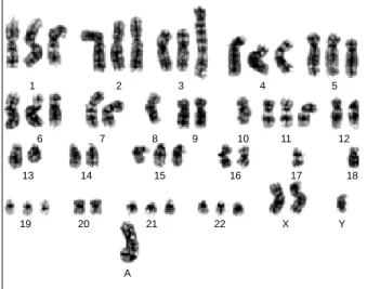
Fig. 1 –G-banded multiple myeloma patient karyotype: 55, XY, + X, + 1, + 2, + 3, + 4, + 5, + 6, del(6)q21, add(7)q36, -8, -10, + 11, del(13)q14, + 15, -17, -18, + 19, + 21, + 22, + mar (A)
1 2 3 4 5
6 7 8 9 10 11 12
13 14 15 16 17 18
A
19 20 21 22 X Y
Cor r espondence to:Car men S. P. Li ma
Hemocen tr o – UNICAMP – Ci dade Un i versi tári a Zeferi n o Vaz Cai xa postal 6198 – Cep: 13083-970 – Campi n as-SP – Brazi l
Tel: (+55 19) 3788-8729 – fax: (+55 19) 3788-8600 – e-mai l: carmen l@u n i camp.br
leukaemia patients by polymerise chain reaction single
strand conformation polymorphism and sequencing, and
fluorescence in situ hybridisation in order to detect
P53mutations and deletions, respectively. Kaplan-Meier
survival curves and the log-rank test w ere used to
analyse the survival data.
Fig. 2 –Interphase fluorescence in situ hybridisation analysis in multiple myeloma patient, using specific probes for chromosome 17 centromere ( ·) and P53 locus (· ) (Vysis, Downers Grove, IL, USA). (A): two hybridisation signals for each probe in normal interphasenuclei and, (B) abnormal nuclei exhibiting a monoallelic (left) and biallelic (right) deletions of P53 locus
130
Rev. bras. hematol. hemoter. 2003;25(2):125-127 Imagen sem Hematologi a
No
P53mutation was detected in our patients. In
contrast,
P53deletion, predominantly monoallelic, was
detected in 8 out of 51 (15.7%) patients. Patients w ith
P53
deletions had significantly shorter median overall
survival compared w ith those w ithout a deletion (7.4
vs.
139.0 months,
P<0.0001). In univariate regression
analysis,
P53deletion w as a parameter for predicting
shortened survival (
P=0.02).
We concluded that
P53mutation might be seen
as a prognostic indicator of limited value in MM. In
contrast,
P53deletion might be seen as a prognostic
indicator of poor outcome. These results have already
been accepted for publication in Annals of Hematology
and has been submitted to Rev. Bras. Hematol.
Hemoter., w ith the purpose of presenting the images
obtained by conventional and molecular cytogenetics
analyses performed in study (Figures 1 and 2).
Refer ên ci as Bi bl i ogr áfi cas
1. Avet-Loiseau H, Li JY, Godon C et al. P53 deletions is not a frequent event in multiple myeloma. Br JHaematol 1999;106:717-9.
2. Corradini P, Inghirami G, Astolfi M, et al. Inactivation of tumor suppressor genes, P53 and Rb1 in plasma cell dyscrasias. Leukemia 1994;8:758-67.
3. Drach J, Ackerman J, Fritz E et al. Presence of a P53 gene deletion in patients with multiple myeloma predicts for short survival after conventional-dose chemo-therapy. Blood 1998;92:802-9.
4. Ow en RG, Davis SAA, Randerson J et al. P53 gene mutations in multiple myeloma. JClin Pathol Mol Pathol 1997;50:18-20.
Recebi do: 15/04/03 Acei to: 02/05/03