A
rt
ic
le
0103 - 5053 $6.00+0.00
#Present address: Instituto de Física de São Carlos, Universidade de São
Paulo, Av. do Trabalhador São Carlense, 400 13566-590 São Carlos-SP, Brazil
*e-mail: [email protected]
Chemical Constituents of
Habenaria petalodes
Lindl. (Orchidaceae)
Betania B. Cota,a Alviclér Magalhães,b,# Adriano M. C. Pimenta,c Ezequias P. Siqueira,a
Tânia M. A. Alvesa and Carlos L. Zani*,a
a Laboratório de Química de Produtos Naturais, Av. Augusto de Lima, 1715,
30190-002 Belo Horizonte-MG, Brazil
b Instituto Tecnológico de Fármacos, Rua Sizenando Nabuco, 100, 21041-250 Rio de Janeiro-RJ, Brazil
c Departamento de Bioquímica e Imunologia, Av. Antônio Carlos, 6.627,
31270-901 Belo Horizonte-MG, Brazil
Uma nova substância, denominada habenariosídeo {[(2R)-2-[(2,3,4,6-tetra-O-acetil-b-D -glicopiranosil)oxi]-2-(2-metilpropil)-1,4-dioxo-1,4-butanodiil]bis(oximetileno-4,1-fenileno) bis-b-D-glicopiranosídeo}, foi isolada das frações polares do extrato etanólico de Habenaria petalodes Lindl. (Orchidaceae), juntamente com duas substâncias conhecidas e estruturalmente relacionadas, a loroglossina e a militarina. Os flavonóides: isoquercitrina, isorramnetina 3-O-b
-D-glicopiranosídeo e isorramnetina 3,7-di-O-b-D-glicopiranosídeo, também foram isolados. As estruturas do habenariosídeo e das demais substâncias foram determinadas pela análise dos dados de EM, IV, UV e RMN mono e bidimensional e comparação com os dados publicados na literatura.
Habenarioside, a new natural product identified as [(2R)-2-[(2,3,4,6-tetra-O-acetyl-b-D - glucopyranosyl)oxy]-2-(2-methylpropyl)-1,4-dioxo-1,4-butanediyl]bis(oxymethylene-4,1-phenylene) bis-b-D-glucopyranoside, along with two known related metabolites, loroglossin and militarin, were isolated from the ethanol extract of the whole plant Habenaria petalodes
Lindl. (Orchidaceae). The flavonoids isoquercitrin, isorhamnetin 3-O-b-D-glucopyranoside, and isorhamnetin 3,7-di-O-b-D-glucopyranoside were also isolated. The structures of all compounds were established by analysis of their MS, IR, UV, 1D and 2D NMR spectra, and comparison with published data.
Keywords: orchid, loroglossin, militarin, habenarioside, flavonoids
Introduction
The Brazilian flora is exceedingly rich and comprises more than 56,000 endemic plant species. Among the 22,000 members of the family Orchidaceae that have been described worldwide, approximately 1,800 grow endemically in Brazil.1 In spite of its size and diversity when compared with other plant families, a relatively small number of orchid species have been investigated regarding their chemical composition, with about 300 secondary metabolites reported so far. These included, among others, flavonoids, terpenoids, quinones, phenanthrenes, stilbenes, lignans, and glucosylated benzyl
esters of succinic, malic, tartaric and citric acids.2-6 The phytochemical investigations of Brazilian Orchidaceae are also restricted to few published works: a report concerning the presence of methylated C-glycosylflavones as taxonomic markers in fifteen orchids,7 and the investigation of the floral composition of other twenty-one species.8
Cota et al. 1099 Vol. 19, No. 6, 2008
Results and Discussion
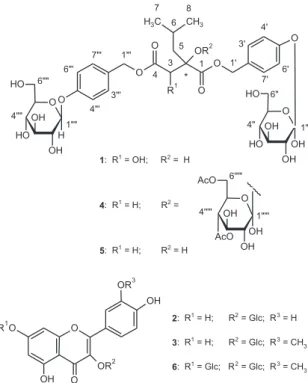
The whole plant was extracted by maceration with ethanol and the extract was subjected to liquid-liquid partition between CH2Cl2 and MeOH:H2O (1:1) to afford an organic and a hydroalcoholic fraction. The latter was subjected to solid phase extraction (SFE) using RP-18 cartridges followed by semi-preparative RP-HPLC to yield six compounds (Figure 1-2).
The ESI-MS of compounds 1 and 5 exhibited quasi -molecular ionpeaks ([M + Na]+) at m/z 765 and m/z 749, respectively. Their molecular weight, together with their UV, 1H and 13C NMR data were in accordance with those reported for loroglossin (1) and militarin (5).5,14
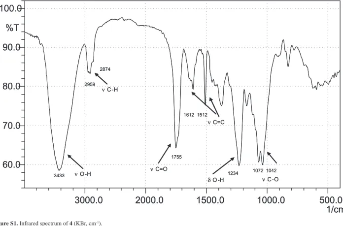
Habenarioside (4) was isolated as a resinous material with negative specific rotation ([a]D25 –30.0°, c 0.16 in MeOH). The positive HRESIMS spectrum showed a quasi-molecular ionpeak [M + Na]+ at m/z 1079.3578, consistent with the molecular formula C48H64O26 (calc. 1079.3584). The ESI-MS in positive ion mode showed neutral losses of 60 Da, attributed to the presence of acetate groups. Its IR spectrum exhibited absorption bands at 1755 cm-1 (C=O), 1612 and 1512 (C=C), 1234 (C-O) due to ester carbonyl functions and
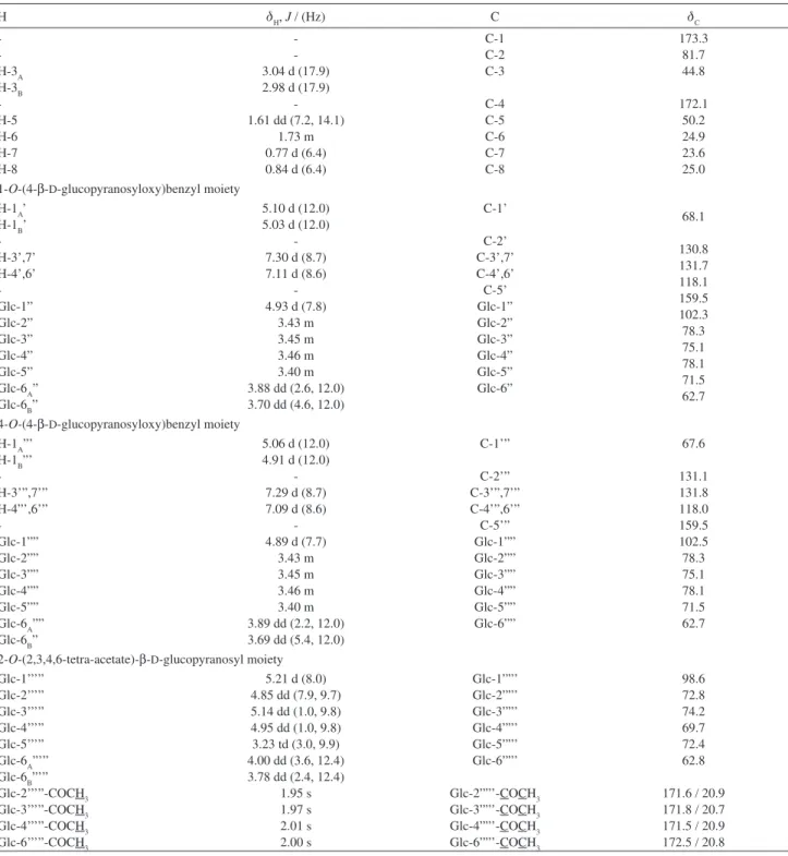
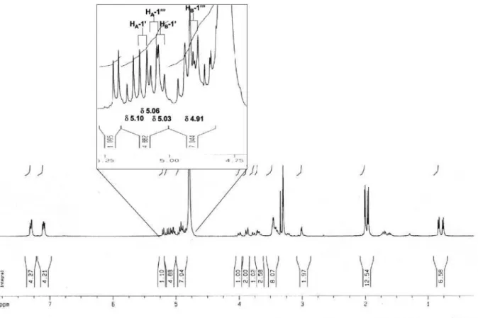
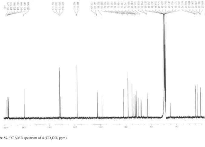
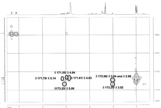
aromatic rings, and strong absorptions at 3433 (OH) and 1072 (C-O) indicating the presence sugar moieties. Acid hydrolysis of 4 followed by reduction and acetylation, and GC-MS analysis of the resulting alditol acetate, confirmed the presence of D-glucose by comparison with an authentic sample. In the UV spectrum of 4, absorptions were similar to those of compounds 1 and 5 (Figure 1), with maxima observed at 224 nm (log ε 3.76) and 271 nm (3.28). NMR spectra (Table 1) showed signals attributed to four acetyl groups at dC (C=O/Me) 171.6 /20.9, 171.8/20.7, 171.5/20.9, and 172.5/20.8, and dH (Me) 1.95, 1.97, 2.01, and 2.00. On the basis of DEPT spectrum other carbon resonances were ascribed to two methyls (dC 23.6, 25.0), four methylenes (dC 44.8, 50.2, 67.6, 68.1), a methine (dC 24.9), seven quaternary carbons (dC 81.7, 130.8, 131.1, 2 × 159.5, 172.1, 173.3), and two sets of four aromatic carbons (dC 118.1, 131.7 and 118.0, 131.8). Also, signals due to the presence of three glucose units were evident in the HMQC experiment which showed correlations between pairs of anomeric protons and carbons with the following chemical shifts dH/dC: 5.21/98.6, 4.93/102.3, and 4.89/102.5. The coupling constants between the anomeric protons and their respective H-2 neighbor in the sugar moieties (3J 7.7 Hz) indicate that they are all in b configuration. The above data appeared closely related to those of malic acid derivatives, gymnosides (J 7.6 Hz)23 and dactylorhins (J 7.5 Hz)5. The HMBC experiments showed a clear correlation between the C-2 (dC 81.7) in the malic acid skeleton and H-1’’’’’ of a glucose moiety (dH 5.21). The Figure 1. HPLC chromatogram of H. petalodes ethanol extract (UV
detection at 230 nm), and UV spectra (200-400 nm) of loroglossin (1), isoquercitrin (2), isorhamnetin 3-O-b-D-glucopyranoside (3), habenarioside (4), militarin (5), and isorhamnetin 3,7-di-O-b-D -glucopyranoside (6). Column, 250 mm × 4.6 mm i.d.; mobile phase, MeOH:H2O (10:90 →70: 30 in 30 min; 70:30 →100: in 5 min), flow rate 1 mL min-1. Mass data was obtained on an ion-trap mass spectrometer
equipped with an ESI ion source operating in the positive ion mode.
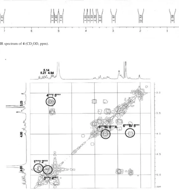
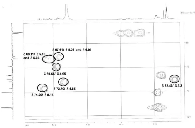
later form a complete hexose spin system, deducible from the 1H-1H COSY NMR experiment, in which correlations between the following protons pairs could be observed: H-1’’’’’ and H-2’’’’’ (dH 5.21/4.85), H-2’’’’’ and H-3’’’’’ (dH 4.85/5.14), H-3’’’’’ and H-4’’’’’ (dH 5.14/4.95), H-4’’’’’ and H-5’’’’’ (dH 4.95/3.23), H-5’’’’’ and H-6B’’’’’ (dH 3.23/4.00), and H-5’’’’’ and H-6A’’’’’ (dH 3.23/3.78). This sugar unit is completely acetylated as can be inferred on the basis of the following HMBC correlations (dC/dH) between the acetate carbonyls and the above mentioned sugar protons: 171.6/4.85 (C=O/H-2’’’’’); 171.8/5.14 (C=O/H-3’’’’’); 171.6/4.95 (C=O/H-4’’’’’); 172.5/3.78 (C=O/H-6A’’’’’); and, 172.5 /4.00 (C=O/H-6B’’’’’). Carbons at C-5’ and C-5’” (dC 159.5) showed long range correlations with the anomeric protons at dH 4.93 (H-1”) and dH 4.89 (H-1””), indicating the position of the other two sugar units. On the basis of these evidences, compound 4 was identified as [2-[(2,3,4,6-tetra-O-acetyl-b-D-glucopyranosyl) oxy]-2-(2-methylpropyl)-1,4-dioxo-1,4-butanediyl]bis (oxymethylene-4,1-phenylene) bis-b-D-glucopyranoside, which was given the trivial name habenarioside as it is a mono-glucosylated derivative of habenariol. This compound can also be regarded as a tetra-acetate derivative of dactylorhin A, a natural product previously isolated from
Dactylorhiza hatagirea D. Don.5 and from Gymnadenia
conopsea.23 From the latter species it was also described the presence of acetic and cinnamic esters of dactylorhin A, all at the glucose connected to the C-2 position of the malic acid. These findings indicate that further esters of dactylorhin A may be produced by Orchidaceae species.
Gitterman and co-workers13 demonstrated that in
Cephalotaxus (Cephalotaxaceae) 2-isobutyl-malic acid is biosynthesized from L-leucine, and have R configuration at C-2. Assuming that this is also the case in Orchidaceae, and based on structures of compounds already isolated from this family, a biosynthetic route is suggested for habenarioside (Scheme S1 - Supplementary Material). Accordingly, 2R-isobutyl-malic acid could be subjected to a sequence of esterification and glucosylation reactions to afford habenariol, militarin, dactylorhin A, and gymnoside III, as intermediates in the biosynthesis of habenarioside. This hypothetical sequence assumes that all intermediates have 2R configuration in order to avoid the unlikely inversion/reversion of the C-2 configuration. However, habenariol was reported to have [a]D25 + 12° and 2S configuration.12 On the other hand, catalytic hydrogenation of militarin14, and comparison of the resulting 2-isobutyl malic acid with a synthetic standard, established its absolute stereochemistry as 2R.14 This data rises doubt about the published configuration for habenariol. Even though, it is possible to devise a pathway bypassing this compound,
and use only 2R intermediates. In such pathway, militarin would be produced via esterification of 2R-isobutyl-malic with gastrodin (Scheme S1) keeping the R configuration at C-2. Based on the above discussion we propose that habenarioside have 2R configuration.
Malic acid derivatives 1 and 5 were previously isolated from Orchis militaris L.,13,21 Dactylorhiza hatagirea D. Don,5 and Coeloglossum viride (L.) Hartm. var. bracteatum (Willd.),22 whilst 5, together with Gymnoside III (Scheme S1), were recently found in tubers of Gymnadenia conopsea R. Br.23 Zhang et al. 24,25 reported that a fraction from the rhizomes of Coeloglossum viride rich in 1 and 5
significantly improved the memory of mice treated with scopolamine, cycloheximide or alcohol. These authors also demonstrated that this fraction protected neurons against injury by b-amyloid or H2O2, and suggested that these components could be of value in the treatment of Alzheimer’s disease and other dementia.
Compounds 2, 3 and 6 were isolated as yellow powders, and their UV spectra in MeOH exhibited absorptions consistent with those of flavonoids.15 The ESI-MS in the negative ion mode of compound 2 showed a [M - H]– quasi -molecular ion peak at m/z 463,while the ESI-MS in the positive ion mode of compounds 3 and 6 showed [M + Na]+
quasi-molecular ion peaks at m/z 501 and 663, respectively. Their 1H NMR spectra showed signals characteristic of glycosylated flavonoids, and comparison with published data allowed 2 to be identified as isoquercitrin16, 3 as isorhamnetin 3-O-b-D-glucopyranoside,16 and 6 as isorhamnetin 3,7-di-O-b-D-glucopyranoside.17 Although isoquercitrin and isorhamnetin 3-O-b-D-glucoside have been previously isolated from some Orchidaceae species,18-20 this is the first report on the occurrence of isorhamnetin 3,7-di-O-b-D-glucopyranoside in this family.
The present study contributes to the phytochemical characterization of Orchidaceae, disclosing the presence of a new compound and the occurrence of two known compounds reported for the first time in this family.
Experimental
General experimental procedures
Cota et al. 1101 Vol. 19, No. 6, 2008
a Beckman DU Series 600 spectrophotometer, whilst infrared spectra were measured in KBr pellets on a Shimadzu FTIR-8400 instrument. 1H NMR (400 MHz), 13C NMR (100 MHz), DEPT, HMQC and HMBC
experiments were carried out using a Bruker DRX 400 spectrometer. ESI-MS were recorded on a Thermo Finnigan
LCQ-Advantage spectrometer, whilst high resolution mass measurements were obtained using a Micromass Q-TOF Micro™ instrument equipped with an ESI source operated in the positive ion mode. Extract, fractions and compounds were analyzed by HPLC using a Shim-pack C18 column (5 µm, 250 mm × 4.6 mm i.d.) in a Shimadzu Table 1. 1H and 13C NMR data for compound 4 (CD
3OD, 400 and 100 MHz, respectively)
H dH, J / (Hz) C dC
-H-3A H-3B -H-5 H-6 H-7 H-8 -3.04 d (17.9) 2.98 d (17.9)
-1.61 dd (7.2, 14.1)
1.73 m 0.77 d (6.4) 0.84 d (6.4)
C-1 C-2 C-3 C-4 C-5 C-6 C-7 C-8 173.3 81.7 44.8 172.1 50.2 24.9 23.6 25.0 1-O-(4-b-D-glucopyranosyloxy)benzyl moiety
H-1A’ H-1B’ -H-3’,7’ H-4’,6’ -Glc-1” Glc-2” Glc-3” Glc-4” Glc-5” Glc-6A” Glc-6B”
5.10 d (12.0) 5.03 d (12.0)
-7.30 d (8.7) 7.11 d (8.6)
-4.93 d (7.8)
3.43 m 3.45 m 3.46 m 3.40 m 3.88 dd (2.6, 12.0) 3.70 dd (4.6, 12.0)
C-1’ C-2’ C-3’,7’ C-4’,6’ C-5’ Glc-1” Glc-2” Glc-3” Glc-4” Glc-5” Glc-6” 68.1 130.8 131.7 118.1 159.5 102.3 78.3 75.1 78.1 71.5 62.7
4-O-(4-b-D-glucopyranosyloxy)benzyl moiety H-1A”’
H-1B”’ -H-3’”,7’” H-4”’,6’” -Glc-1”” Glc-2”” Glc-3”” Glc-4”” Glc-5”” Glc-6A”” Glc-6B”
5.06 d (12.0) 4.91 d (12.0)
-7.29 d (8.7) 7.09 d (8.6)
-4.89 d (7.7)
3.43 m 3.45 m 3.46 m 3.40 m 3.89 dd (2.2, 12.0) 3.69 dd (5.4, 12.0)
C-1’” C-2’” C-3’”,7’” C-4’”,6’” C-5’” Glc-1”” Glc-2”” Glc-3”” Glc-4”” Glc-5”” Glc-6”” 67.6 131.1 131.8 118.0 159.5 102.5 78.3 75.1 78.1 71.5 62.7
2-O-(2,3,4,6-tetra-acetate)-b-D-glucopyranosyl moiety Glc-1’’’”
Glc-2’’’” Glc-3’’’” Glc-4’’’” Glc-5’’’” Glc-6A”’” Glc-6B”’” Glc-2’’’”-COCH3 Glc-3’’’”-COCH3 Glc-4’’’”-COCH3 Glc-6’’’”-COCH3
5.21 d (8.0) 4.85 dd (7.9, 9.7) 5.14 dd (1.0, 9.8) 4.95 dd (1.0, 9.8) 3.23 td (3.0, 9.9) 4.00 dd (3.6, 12.4) 3.78 dd (2.4, 12.4)
1.95 s 1.97 s 2.01 s 2.00 s Glc-1””’ Glc-2””’ Glc-3””’ Glc-4””’ Glc-5””’ Glc-6””’ Glc-2””’-COCH3 Glc-3””’-COCH3 Glc-4””’-COCH3 Glc-6””’-COCH3 98.6 72.8 74.2 69.7 72.4 62.8
171.6 / 20.9 171.8 / 20.7 171.5 / 20.9 172.5 / 20.8
chromatograph equipped with a LC10AD pump and a diode-array detector (SPD M10A). The column was pumped with MeOH:H2O (10:90→70:30 in 30 min; 70:30 →100 in 5 min) at a flow rate of 1 mL min-1 and the effluent detected at wavelengths between 200 and 400 nm. Semi-preparative HPLC purifications were carried out on a Shimadzu chromatograph equipped with a LC6AD pump and a dual wavelength detector (SPD10A), using a Shim-pack C18 column (5 µm, 250 mm 20 mm i.d.) eluted with mixtures of MeOH:H2O or MeCN:H2O at a flow rate of 10 mL min-1 and detection at 230 nm and 254 nm.
Plant material
Specimens of Habenaria petalodes Lindl. were collected in Belo Horizonte-MG, Brazil, in April 2004. The plant material was identified by Prof. João Renato Stehmann of the Universidade Federal de Minas Gerais, Belo Horizonte-MG, Brazil. A voucher specimen was deposited in the Herbarium BHCB (Instituto de Ciências Biológicas, Belo Horizonte-MG, Brazil) under the code BHCB-88339.
Extraction and isolation
Whole fresh plants (185 g) were chopped into small pieces and macerated in ethanol for 3 weeks at room temperature. After filtration and solvent evaporation, 8 g of the extract were obtained. An aliquot (6.2 g) was suspended in 100 mL MeOH:H2O (1:1), and extracted with CH2Cl2 (3 × 100 mL) to afford an organic fraction (610 mg) and a hydroalcoholic fraction (4.7 g). The latter was concentrated to dryness under reduced pressure, and the residue dissolved in water. The solution was subjected to solid phase extraction using Sep-Pak C18 cartridges. The loaded cartridge was eluted with water, and then with methanol, to yield an aqueous (3.9 g) and a methanolic (756 mg) fraction. A portion of the methanolic fraction (669 mg) was subjected to semi-preparative HPLC with linear gradient elution with MeOH:H2O from 30 to 70% over 60 min, and detection at 210 nm and 254 nm, to yield 29 sub-fractions. Sub-fraction 13 (6.5 mg) was subjected to column chromatography on Sephadex LH-20, eluted with MeOH to afford compound 2 (5 mg). Sub-fraction 7 (6.6 mg) was purified by semi-preparative HPLC in a RP-18 column eluted with MeCN:H2O 10:90 → 50: 50 in 60 min, at a flow rate of 10 mL min-1, to yield 4 mg of compound 1. Sub-fraction 17 (23.9 mg) was purified by semi-preparative HPLC in a RP-18 column eluted with MeCN:H2O 50:50 →60: 40, in 40 min at a flow rate of 10 mL min-1, to yield 8 mg of 3. The second portion of the methanolic
fraction (87 mg) was subjected to semi-preparative HPLC with linear gradient elution from 10% MeOH-H2O to 100% MeOH over 40 min, to afford 4 (22.5 mg) and 5 (5 mg). The aqueous fraction from SPE (2.0 g) was divided into equal portions and chromatographed on a Sephadex LH-20 column (470 × 30 mm i.d.) eluted with MeOH:H2O (1:1), to give 24 sub-fractions. Compound 6 (7 mg) precipitated from the fraction 10.
Characterization of the sugar moieties
Compound 4 (2 mg) was hydrolyzed in 0.5 TFA at 100 °C for 4 h. To the reaction mixture was added methanol (0.5 mL) and the solution evaporated to dryness under a stream of nitrogen. This operation was repeated to completely remove the TFA. To the resulting solid were added 0.3 mL of a solution of 1 mg/ml aqueous NaBH4 and the reaction kept at 25 °C. After 30 min the reaction was stopped by addition of 0.5 mL 10% AcOH in MeOH, and the reaction mixture evaporated to dryness. The later steps, that is, addition of acidified methanol and evaporation, were repeated twice. The residue was acetylated by adding 0.1 mL of Ac2O and heating at 100 °C for 30 min. Toluene (0.5 mL) was added to the reaction mixture and the solution evaporated under a stream of nitrogen at 90 °C. This operation was repeated three to four times to completely remove the acetic anhydride. The residue was then partitioned (3×) between H2O-EtOAc (1:1) and the organic phase was pooled, concentrated and dissolved in 0.2 mL of EtOAc. Control experiments with sugar standards were run in parallel. The alditol acetate solutions thus obtained were analyzed in a Shimadzu GC-17A gas chromatograph in line with a QP5050A mass spectrometer. One microliter of each solution was injected in a capillary column DB-5 (30 m × 0.25 mm i.d.), using helium as carrier gas (1.5 mL min-1) and the following temperature programming: 100 to 200 °C in 20 min, 200-300 °C in 5 min and keeping at 300 °C for 5 min. The injector and detector were kept at 230 °C and 250 °C, respectively.27Under these conditions the retention times of the alditol acetates obtained from the standard sugars were: ribose (17.77 min), arabinose (17.95 min), xylose (18.34 min), mannose (21.78 min), glucose (21.88 min), and galactose (21.96 min).
Loroglossin (1)
[a]D25 –28.0° (MeOH, c 0.23) {Lit. 14 [a] D
25 –34.0
(MeOH, c = 0.36)}, UV lmaxMeOH/nm (log ε): 201 (4.22), 221
(4.18), 268 (3.42); 1H NMR (CD
Cota et al. 1103 Vol. 19, No. 6, 2008
Hz, H1”, H1””), 5.08, 4.97 (2H, d, JA = JB 11.8 Hz, H-1’A and H-1’B), 5.09, 4.98 (2H, d, JA = JB 12.0 Hz, H-1’”A and H-1’”B), 7.30 (2H, d, J 8.6 Hz, H-3’,7’), 7.18 (2H, d, J 8.8 Hz, H-3’”,7’”), 7.12 (2H, d, J 8.6 Hz, H-4’,6’), 7.07 (2H, d, J
8.8 Hz, H-4’”,6’”), 3.45 (m, H-2”, H-2””/ H-3”, H-3””/ H-5”, H-5””), 3.40 (m, H-4”, H-4””), 3.71 (m, H-6A”, H-6A””), 3.89 (m, H-6B”, H-6B””); 13C NMR (CD
3OD, 100 MHz): dC 24.2 (C-8), 24.7 (C-7), 25.3 (C-6), 45.3 (C-5), 62.9 (C-6”, C-6””), 68.2 (C-1’, C-1’”), 71.5 (C-4”, C-4””), 75.1 (C-2”, C-2””), 77.5 3), 78.1/78.3 3”; C-3””/C-5”, C-5””), 81.1 (C-2), 102.5 (C-1”, C-1””), 118.0/117.9 (C-4’,6’; C-4”’,6”’), 129.5/130.6 (C-2’, C-2’”), 131.7/ 131.4 (C-3’,7’; C-3”’,7’”), 159.4/159.5 (C-5’, C-5’”), 172.9 (C-4), 174.9 (C-1). ESI-MS (positive mode) [M + Na]+ (m/z 765).
Militarin (5)
[a]D25 –41.0° (MeOH, c 0.23) {Lit. 14 [a] D
25 –47.0
(MeOH, c = 0.78)}, UV (MeOH) lmax/nm (log ε): 203 (4.19), 219 (4.10), 269 (3.47), IR (KBr) vmax/cm-1: 3410 (OH), 2924 (aliphatic), 2855 (aliphatic), 1612 (aromatic), 1512 (aromatic), 1736 (carbonyl), 1458 (aromatic), 1234 (C-O), 1076 (C-O), 1045 (C-O); 1H NMR (CD
3OD, 400 MHz): dH 0.82 (3H, d, J 6.5 Hz, H-7), 0.93 (3H, d, J 6.5 Hz, H-8), 1.61 (2H, m, H-5), 1.72 (1H, m, H-6), 2.65, 2.95 (1H, d, JA = JB 15.6 Hz, H-3A and H-3B), 5.01 (4H, s, H-1’ and H-1’”), 7.30-7.25 (4H, m, H-3’,7’; H-3’”,7’”), 7.11-7.07 (4H, m, H-4’,6’; H-4’”,6’”), 4.91 (d, J 7.4 Hz, H-1”, H-1””), 3.45 (m, H-2”, H-2””/ H-3”, H-3””/ H-5”, H-5””), 3.39 (m, H-4”, H-4””), 3.70 (m, H-6A”, H-6A””), 3.87 (m, H-6B”, H-6B””); 13C NMR (CD
3OD, 100 MHz): dC 22.5 (C-7), 23.3 (C-8), 23.7 (C-6), 44.8 (C-3), 47.7 (C-5), 61.1 (C-6”, C-6””), 65.8 (C-1’”), 66.6 (C-1’), 70.0 (C-4”, C-4””), 73.5 (C-2”, C-2””), 75.3 (C-2), 76.6/76.7 (C-3”; C-3””/C-5”, C-5””), 100.9 (C-1”, C-1””), 116.4 (C-4’,6’; C-4”’,6”’), 129.4 (C-2’, C-2’”), 129.6/129.8 (C-3’,7’; C-3’”,7’”), 157.8 (C-5’, C-5”’), 170.2 (C-4), 174.6 (C-1) ). ESI-MS (positive mode) [M + Na]+ (m/z 749).
Habenarioside (4)
Resinous solid; [a]D25 –30.0 (c 0.16, MeOH); UV (MeOH) lmax/nm (log ε): 224 (3.76) and 271 (3.28); IR
νmax/cm
-1: 3433 (OH), 2959 (aliphatic), 2874 (aliphatic),
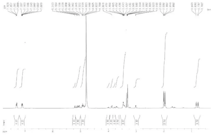
1612 (aromatic), 1512 (aromatic), 1755 (carbonyl), 1072 (C-O), 1042 (C-O); 1H and 13C NMR spectral data, see Table 1; ESI-MS m/z positive mode: 1079 [M + Na]+, 1095 [M + K]+; HRMS: 1079.3578 [M + Na]+ (calc. for [C48H64O26 + Na]+: 1079.3584).
Supplementary Information
Supplementary data including IR, LC-DAD, 1 H and 13C NMR and HRMS spectra are available free of charge
at http://jbcs.sbq.org.br as PDF file.
Acknowledgments
The authors wish to thank CAPES for a fellowship (B. B. Cota) and FIOCRUZ for financial support.
References
1. Giulietti, A. M.; Harley, R. M.; Queiroz, L.P.; Wanderley, M. G. L; Van Den Berg, C.; Conserv. Biol. 2005, 19, 632. 2. Tagushi, H.; Yosioka, I.; Yamasaki, K.; Kim, I. H.; Chem.
Pharm. Bull. 1981, 29, 55.
3. Noda, N.; Kobayashi, Y.; Miyahara, K.; Fukahori, S.; Phytochemistry 1995, 39, 1247.
4. Lin, J. H.; Liu, Y. C.; Hau, J. P.; Wen, K. C.; Phytochemistry 1996, 42, 549.
5. Kizu, H.; Kaneko, E. I.; Tomimori, T.; Chem. Pharm. Bull. 1999, 47, 1618.
6. Sheng-Yang, H.; Shi, J. G.; Mo, S. Y.; Wang, S.J.; Yang, Y. C.; J. Asian Nat. Prod. Res. 2004, 6, 49.
7. Williams, C. A.; De Brito, A. L.; Toscano, Harborne, J. B.; Eagles, J.; Waterman, P. G.; Phytochemistry 1994, 37, 1045. 8. Reis, M. G.; Singer, R. B.; Gonçalves, R.; Marsaioli, A. J.;
Nat. Prod. Comm. 2006, 1, 757.
9. Pabst, G. F. J.; Dungs, F.; Orchidaceae Brasiliensis. Brücke Verlag: Hildesheim, 1975.
10. NAPRALERT (Natural Producst Alert) Database; http://www. napralert.org/, accessed in January 2008.
11. Johnson, M. K.; Alexander, K. E.; Lindquist, N.; Loo, G.; Comp. Biochem. Physiol. C: Pharmacol. Toxicol. 1999, 122, 211. 12. Wilson, D. M.; Fenical, W.; Hay, M.; Lindquist, N.; Bolser, R.;
Phytochemistry 1999, 50, 1333.
13. Gitterman, A.; Parry, R. J.; Dufresne, R.F.; Sternbach, D. D.; Cabelli, M. D.; J. Am. Chem. Soc. 1980, 102, 2074.
14. Aasen, A.; Behr, D.; Leander, K.; Acta Chem. Scand., B 1975, 29, 1002.
15. Wagner, H. M.; Bladt, S.; Zgainski, E. M.; Plant Drug Analysis, 1st ed., Springer: Berlin, 1984.
16. Güvenalp, Z.; Demirezer, L.O.; Turk. J. Chem. 2005, 29, 163. 17. Yeskaliyeva, B.; Mesaik, M. A.; Abbaskhan, A.; Kulsoom, A.;
Burasheva, G. S. H.; Abilov, Z. H. A.; Choudhary, M. I.; Atta-Ur-Rahman; Phytochemistry 2006, 67, 2392.
20. Pagani, F.; Bull. Chim. Farm. 1976, 115, 407.
21. Appolonia, C.; Marston, A.; Hostettmann, K.; J. Nat. Prod. 1986, 49, 725.
22. Huang, S. Y.; Li, G. Q.; Shi, J. G.; Mo, S. Y.; J. Asian Nat. Prod. Res. 2004, 6, 49.
23. Morikawa, T.; Xie, H.; Matsuda, H.; Yoshikawa, M. J. Nat. Prod. 2006, 69, 881.
24. Zhang, D.; Shi, J.; Wang, Y.; Zhang, D.; Gao, M.; Yang, Y.; Huang, S.; PCT/CN2003/001155. 2004 (WO/2004/058244).
25. Zhang, D.; Liu, G.; Shi, J. G.; Zhang, J. J.; Basic Clin. Pharmacol. Toxicol. 2006, 98, 55.
26. Mabry, T. J.; Markham, K. R.; Thomas, M. B.; The Systematic Identification of Flavonoids, Springer Verlag: New York, 1970.
27. Stockholm University. Structural analysis of carbohydrates. Kontaktuppgifter: Stockholm. Available at: http://www.casper. organ.su.se/sop/, accessed in February 2008.
S
u
p
p
le
m
e
n
ta
ry
In
fo
rm
a
ti
o
n
J. Braz. Chem. Soc., Vol. 19, No. 6, S1-S8, 2008. Printed in Brazil - ©2008 Sociedade Brasileira de Química 0103 - 5053 $6.00+0.00
*e-mail: [email protected]
Chemical Constituents of
Habenaria petalodes
Lindl. (Orchidaceae)
Betania B. Cota,a Alviclér Magalhães,b Adriano M. C. Pimenta,c Ezequias P. Siqueira,a
Tânia M. A. Alvesa and Carlos L. Zani*,a
a Laboratório de Química de Produtos Naturais, CPqRR-FIOCRUZ, Av. Augusto de Lima, 1715,
30190-002 Belo Horizonte-MG, Brazil
b Instituto Tecnológico de Fármacos, FIOCRUZ, Rua Sizenando Nabuco, 100, 21041-250 Rio de Janeiro-RJ, Brazil
c Departamento de Bioquímica e Imunologia, ICB – UFMG. Av. Antônio Carlos, 6.627,
31270-901 Belo Horizonte-MG, Brazil
Figure S1. Infrared spectrum of 4 (KBr, cm-1).
Cota et al. S3 Vol. 19, No. 6, 2008
Figure S3.1H NMR spectrum of 4 (CD
3OD, ppm).
Figure S4. 1H NMR spectrum of 4 (CD
Figure S5.1H NMR spectrum of 4 (CD
3OD, ppm).
Figure S6.1H NMR spectrum of 4 (CD
Cota et al. S5 Vol. 19, No. 6, 2008
Figure S7. 1H NMR spectrum of 4 (CD
3OD, ppm).
Figure S8. 1H-1H COSY NMR spectrum of 4 (CD
Figure S9.13C NMR spectrum of 4 (CD
3OD, ppm).
Figure S10. 13C NMR spectrum of 4 (CD
Cota et al. S7 Vol. 19, No. 6, 2008
Figure S11.1H-13C HMBC NMR spectrum of 4 (CD
3OD, ppm).
Figure S12. 1H-13C HMBC NMR spectrum of 4 (CD
Figure S13. 1H-13C HMBC NMR spectrum of 4 (CD