Agreement between dual x-ray absorptiometers
using pencil beam and fan beam: indicators of
bone health and whole-body plus appendicular
tissue composition in adult athletes
Óscar Tavares1 João P Duarte 2,3 Daniela C. Costa 2 Paulo Sousa-e-Silva 2 Diogo Martinho 2,4 Leonardo G. O. Lus 2,5
Pedro Duarte-Mendes 6
João Valente-dos-Santos 2,7,8 Jorge Conde 9 José M. Casanova 10 Edilson S. Cyrino 11 Manuel J. Coelho-e-Silva 2
1. Department of Medical Imaging and Radiation Therapy, School of Health and Technology, Polytechnic Institute of Coimbra, Coimbra, Portugal;
2. CIDAF - UID/DTP/04213/2016, Faculty of Sport Sciences and Physical Education, University of Coimbra, Coimbra, Portugal; 3. Portuguese Foundation for Science and Technology - SFRH/BD/101083/2014, Lisbon, Portugal; 4. Portuguese Foundation for Science and Technology - SFRH/BD/121441/2016, Lisbon, Portugal; 5. LACAPS, Federal University of Alagoas (Ufal), Arapiraca, Brasil; 6. SHERU - Sport, Health & Exercise Research Unit – Polytechnic Institute of Castelo Branco, Portugal; 7. Faculty of Physical Education and Sport, Lusófona University of Humanities and Technologies, Lisbon, Portugal; 8. Portuguese Foundation for Science and Technology - SFRH/BPD/100470/2014, Lisbon, Portugal; 9. Instituto Politécnico de Coimbra, Coimbra, Portugal; 10. Faculty of Medicine, University of Coimbra, Coimbra, Portugal; 11. Metabolism, Nutrition and Exercise Laboratory, Physical Education and Sport Center, Londrina State University.
http://dx.doi.org/10.1590/1806-9282.64.04.330
SUMMARY
OBJECTIVE: The current study was aimed to examine intra-individual variation on indicators of bone health in addition to whole-body plus appendicular tissue measurements using two concurrent assessments based on pencil beam and fan beam dual energy X-ray absorptiometry (DXA) systems in adult athletes from several sports.
METHOD: Thirty-two male participants (27.6±10.1 years) were measured on anthropometry including multifrequency bioelectric im-pedance and air-displacement plethysmography. Bone mineral content (BMC), bone area, fat and lean soft tissue were derived using pencil beam (Lunar DPX-MD+) and fan beam (Lunar iDXA) absorptiometry. Bone mineral density (BMD) was obtained for the femoral neck, trochanter and triangle of ward. Finally, the right thigh was defined as a region of interest (ROI). Analyses comprised intra-class correlation (ICC), Effect size (d) from mean differences of repeated measurements, coefficient of variation (CV)
RESULTS: ICC were >0.900 for all measurements. Intra-individual differences were large for BMC (d=1,312; CV=2,7%), bone area (d=1,761; CV=2,7%), fat tissue (d=1,612; CV=11%) and all indicators of appendicular lean soft tissue (d=1,237-1687; CV=2,0-4,1%). A very large difference (d=4,014; CV=8.4%) was diagnosed for lean soft tissue of the ROI.
CONCLUSION: Although differences among concurrent instruments for BMC and bone area, the effect size of mean differences was negligible for BMD. Fat and lean soft tissue derived from DXA should be interpreted as reference values (not criterion) due to equip-ment-related variation, more apparently in the ROI values.
KEYWORDS: Absorptiometry, photon. Bone density. Body composition. Athletes.
DATE OF SUBMISSION: 31-Aug-2017 DATE OF ACCEPTANCE: 09-Sep-2017
CORRESPONDING AUTHOR: Manuel Coelho-e-Silva Estádio Universitário de Coimbra
Pavilhão III – Coimbra – Portugal – 3040-156
E-mail: mjcesilva@hotmail.com
INTRODUCTION
Assessment of body composition corresponds to a branch of human biology1, 2 and traditionally, body
composition models comprise two compartments,3
dividing body mass into fat and fat free mass. Tech-nological advancements have allowed the appear-ance of nondestructive methods of evaluating body composition, being, in general, considered in vivo methodologies. Dual energy X-ray absorptiometry (DXA) assessment is based on a model of body com-position evaluation, allowing quantification of fat tissue, lean soft tissue and bone mineral content (BMC), both for the whole body and for standardized segments (head; trunk, subdivided into ribs, pelvis and spine; upper limbs and lower limbs), and it is also possible to obtain the aforementioned compo-nents in regions of interest, termed ROI. From the clinical point of view, there is considerable interest in assessment of the proximal femoral area, often the target of prosthesis, by surgery, and addition-ally the lumbar spine (especiaddition-ally L1-L4), a segment with high informative value regarding the general state of the skeletal structure.
DXA technology has gained popularity over oth-er technologies for its speed, low cost and reduced radiation exposure.4, 5 Previously, the available
tech-nology would use iodine-125 as a source of radiation, the methodology being single-photon absorptiometry
(SPA), aimed to estimate the mineral content of long bones distal portion, such as radius and ulna, being described as valid.6 The SPA technology was used
to obtain one of the most popular equations to de-termine the percentage of fat mass from the triceps and subscapular fat folds.7 DXA technology
mea-sures the attenuation of X-rays emitted at frequen-cies of two different energies, with iodine-125 being replaced by gadolinium-153, which emits radiation at 44 and 100 KeV. There are currently several DXA devices and manufacturers Hologic and Lunar are the most mentioned ones.8-10 DXA has been used
mainly in the determination of BMC and, conse-quently, bone mineral density (BMD), by combining BMC and bone area. In fact, this is a criticism of the method, since the BMD should consider BMC by bone volume and not so much by area, assuming that there are inter-individual variations of those observed in relation to thickness measurement. Each of the manufacturers has developed models for reduction of radiological exposure, scanning time and precision gains [either by improving
detec-tors resolution or by the technology of X-ray emis-sion tubes, highlighting procedures “pencil beam” (PB) and “fan beam” (FB)]. DXA, like the generality of the technologies, has assumptions, such as the heating of tissues during the procedures of applica-tion of the method, and values of water and carbon dioxide are lost, making it necessary to transform the BMC quantified to obtain the bone mineral it-self.5 Additionally, it is assumed that fat and lean
mass have attenuation constants and the exposure of said tissues to low and high voltages allows deter-mining the proportions per unit area, assuming that the thickness in the antero-posterior plane does not affect the estimates.
Most of the studies focus on the comparisons between equipment from different manufacturers (Hologic vs. Lunar) or between equipment of the same manufacturer adopting different technolo-gies for X-ray emission, i.e., PB as opposed to FB11
or even among methodologies: DXA vs. comput-ed tomography scan12-14 or by the methodology of
corporal potassium measurement.15 As a general
rule, studies point to an error associated with DXA technology ranging from 1% to 3%.16, 17 The interest
of Sports Sciences for body composition is due, in large part, to the search for indices of metabolic ef-ficiency. Recently, studies have emerged to express the oxygen uptakes per unit of fat-free body mass18
,
since the fat corresponds to a nonrelevant metabol-ic tissue, or even considering oxygen consumption in the running pattern expressed per liter of lower limb volumetry (or even mL.L-1.kg-1). The
exprsion per appendicular mass of the lower limb is es-pecially relevant when the movement pattern does not oblige the athlete under evaluation to support the whole body mass and this happens in the cy-cle ergometer to obtain maximum and average me-chanical power19 or in the dynamometer isokinetic
technique to evaluate the moments of maximum strength (peak torques) of the muscles responsible for knee extension and flexion actions.20 However,
the search for informative and noninvasive proto-cols on limbs volume and body mass has justified a line of study21-23 devoted to more parsimonious
methodologies in terms of costs and morosity (an-thropometry) based on geometric models.24 The
com-peting equipment to estimate the determination accuracy of fat tissue, lean soft tissue and BMC are not abundant for the whole body and particular seg-ments. In fact, the possible absence of agreement among equipments shall partially compromise pro-posals of calibration of new equations.21-23
The present study aims to examine agreement among indicators resulting from the application of competing DXA from manufacturer Lunar, namely taking into account Lunar DPX-MD+ (PB technolo-gy) and Lunar iDXA (FB technology), having been carried out with healthy young adults from multi-ple sports.
METHODS
Procedures and sample
The present study is cross-sectional in nature and adopts procedures recommended for human re-search25
, having been approved by a Research Ethics
Committee (REC) (CE/FCDEF-UC/00102014). The research design comprises several models of body composition assessment and, in the case of DXA, two pieces of equipment from manufacturer Lunar (DPX-MD+ and iDXA). All measurements were carried out on the same day and by qualified technicians in duly certified laboratory units. Sample subjects were in-formed about the study objectives and the nature of the procedures, having signed an Informed Consent Form (ICF). The final sample corresponds to 32 mod-erately active adult males (27.6 ± 10.1 years) practic-ing various sport modalities.
Anthropometry
Height and sitting height were measured to the nearest 0.1 cm (stadiometer Harpenden, model 98,603, Holtain Ltd., Crosswell, GB, and Harpenden Sitting Height Table). Lower limbs length was calcu-lated by the difference from previous measurements. Body mass was obtained by a SECA (model 770, Ha-nover, MD, USA) scale with a reduction of 0.1 kg. All measurements were performed by the same evaluator.
Bioelectric impedance
Participants adopted a standing position, follow-ing the manufacturer’s instructions. After removfollow-ing shoes, socks and other clothes, total body water was evaluated by a multifrequency bioelectrical imped-ance analyzer, 1, 5, 50, 250, 500, 1,000 kHz (InBody 770 scanner: In-body Bldg, Seul, Korea).
Plethysmography of displaced air
Information on body volume was obtained through displaced air plethysmography (BODPOD composition system, model BODPOD 2006, Life Measurement, Inc, Concord, CA, USA). Participants were evaluated twice consecutively following the manufacturer’s in-structions. Body density was calculated by dividing body mass (kg) by body volume (L).
Dual energy x-ray absorptiometry (DXA)
The two DXA instruments used were: Lunar DPX-MD+ (Software: enCORE version 4,00,145, GE Lunar Corporation, 726 Heartland Trail, Madison, WI 53717-1915 USA) and Lunar iDXA (enCORE ver-sion 13,60,033, GE Medical Systems Lunar3030, Ohmeda Drive, Madison, WI 53718 USA). For each of the equipments, a full body scanner was carried out. Additionally, area near the femur [information on the BMD of the femur neck, Ward triangle, tro-chanter and body of the femur (or shaft)] was car-ried out for each equipment. Subsequently, in the processing phase, an ROI (right thigh) was defined, as described in previous studies.22, 23 For the whole
body and each and every segment, the information includes BMC and bone area to subsequently deter-mine the BMD and the component of the fat tissue and lean soft tissue. Calibration was performed on the same day, before the first one, using the model
(“phantom”) and the procedures recommended by
the manufacturer.
For the Lunar DPX-MD+ equipment, technical documentation specifies that it is a device with X-ray emission by PB technology, having a potential of 76 kV, with an accuracy of < 1% for BMD (whole body) and also 1% for body composition without specifying whether it applies to both tissues (fat tissue and lean soft tissue). In turn, one of the in-novations announced in the iDXA technical leaflet concerns the FB issue. Although iDXA has an FB
is different from previous equipment by having a high resolution detector (CZT-HD).
Statistical analysis
Analyses comprised descriptive statistics (range, mean, standard error of the mean, confidence in-tervals of the mean and standard deviation) for the entire sample (n = 32), as well as the verification of normality assumptions. Subsequently, intra-indi-vidual differences (time 1 and time 2) have been de-termined, parallel to the calculation of the technical error of measurement (TEM).26 Then, and based on
TEM, the coefficient of variation was determined (CV %: expressed as a percentage of the combined mean of repeated measurements). Also based on as-sociative statistics, it was possible to determine the intraclass correlation coefficient (ICC) and its 95% confidence interval. The differences of the means of the repeated measurements were evaluated based on the effect size(Cohen’s d),which have been qual-itatively interpreted as follows:27 < 0.2 (trivial);
0.2-0.6 (small); 0.2-0.6-1.2 (moderate); 1.2-2.0 (large); 2.0-4.0 (very large); > 2.0-4.0 (extremely large). Statistical procedures were carried out using resources from software IBM SPSS v. 23 for Mac OS software (SPSS Inc., IBM Company, NY, USA).
RESULTS
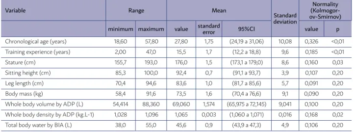
Table 1 summarizes the characteristics of the sample. BMC, bone area used in determination of BMD, whole body lean soft tissue and whole body
TABLE 1 DESCRIPTIVE STATISTICS FOR THE TOTAL SAMPLE AND TEST FOR NORMALITY ON ILLUSTRATIVE VARI-ABLES (N=32)
Variable Range Mean
Standard deviation
Normality (Kolmogor-ov-Smirnov)
minimum maximum value standard error 95%CI value p
Chronological age (years) 18,60 57,80 27,80 1,75 (24,19 a 31,06) 10,08 0,326 <0,01 Training experience (years) 2,00 47,0 15,5 1,7 (12,2 a 18,8) 9,6 0,185 <0,01
Stature (cm) 155,7 193,0 176,0 1,5 (173,1 a 179,0) 8,6 0,160 0,03
Sitting height (cm) 85,3 100,0 92,4 0,7 (91,1 a 93,7) 3,9 0,107 0,20
Leg length (cm) 70,4 94,6 83,6 1,0 (81,7 a 85,6) 5,7 0,091 0,20
Body mass (kg) 58,4 91,6 73,5 1,6 (70,4 a 76,6) 9,1 0,090 0,20
Whole body volume by ADP (L) 54,414 88,360 69,060 1,574 (65,975 a 72,145) 9,041 0,100 0,20 Whole body density by ADP (kg.L-1) 1,028 1,096 1,065 0,003 (1,060 a 1,071) 0,016 0,168 0,02 Total body water by BIA (L) 38,0 55,0 45,6 0,9 (43,9 a 47,3) 4,9 0,106 0,20
ADP (air displacement plethysmography), BIA (body impedance), 95%CI (95% confidence intervals).
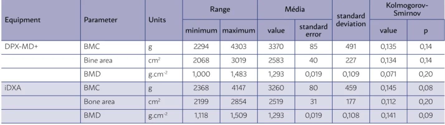
fat tissue, are presented in Table 2. Also the out-puts related to BMD in the proximal femoral area are presented in Table 2 [femur neck, triangle of ward, trochanter and shaft]. Table 2 includes data from Lunar DPX-MD+ and Lunar iDXA. Violation of assumptions of normal distribution was uniquely noted for fat tissue with regard to the whole body.
Comparison of results by two competing pieces of equipment (Table 3) suggests a substantial intra-individual difference for whole body BMC (mean of intraindividual difference = 110 g, magnitude of the wide effect: d = 1,312) and also for the bone area used in calculating BMD (mean intraindividual difference = 65 cm2, effect: d = 1.761). However, for the BMD
intraindividual differences were negligible (–0,001 g.cm-2) and the magnitude effect was trivial. In
addi-tion, magnitude of the intraindividual difference was large for the fat tissue, with the average being 11.87 kg for equipment Lunar DPX-MD+ and 13.56 kg for equipment Lunar iDXA, corresponding to a mean of intraindividual differences of 1.70 kg (d = 1.612, mag-nitude of the wide effect). Differences for the lean tis-sue were trivial, that is, 0.04 kg.
TABLE 2 DESCRIPTIVE STATISTICS AND TEST FOR NORMALITY ON OUTPUTS DERIVED FROM EACH OF THE TWO DUAL ENERGY X-RAY ABSORPTIOMETRY EQUIPMENTS USED IN THE PRESENT STUDY (N=32).
Equipment Parameter Units
Range Média
standard deviation
Kolmogorov- Smirnov
minimum maximum value standard error value p
DPX-MD+ BMC g 2294 4303 3370 85 491 0,135 0,14
Bine area cm2 2068 3019 2583 40 227 0,134 0,14
BMD g.cm-2 1,000 1,483 1,293 0,019 0,109 0,071 0,20
iDXA BMC g 2368 4147 3260 80 459 0,145 0,08
Bone area cm2 2199 2854 2519 31 177 0,112 0,20
BMD g.cm-2 1,118 1,509 1,293 0,019 0,108 0,141 0,09
DPX-MD+ Lean soft tissue kg 48,483 66,415 57,508 0,881 5,062 0,104 0,20 Fat tissue kg 4,488 28,222 11,865 1,100 6,321 0,171 0,02 iDXA Lean soft tissue kg 47,391 66,874 57,466 0,939 5,395 0,101 0,20 Fat tissue kg 6,749 27,216 13,564 0,960 5,516 0,17 0,02
DPX-MD+ Femural neck g.cm-2 0,847 1,615 1,218 0,032 0,186 0,092 0,20
Traingle of Ward g.cm-2 0,636 1,734 1,145 0,038 0,220 0,081 0,20
Trochanter g.cm-2 0,807 1,322 1,057 0,036 0,147 0,094 0,20
Shaft g.cm-2 1,119 2,073 1,469 2,073 0,220 0,220 0,09
iDXA Femural neck g.cm-2 0,843 1,624 1,219 0,031 0,175 0,114 0,20
Traingle of Ward g.cm-2 0,613 1,704 1,125 0,039 0,225 0,079 0,20
Femural neck g.cm-2 0,802 1,331 1,041 0,027 0,155 0,104 0,20
Shaft g.cm-2 1,110 2,069 1,444 0,037 0,215 0,141 0,09
DPX-MD+ LST: trunk kg 21,923 31,503 26,101 0,472 2,709 0,109 0,20
LST: upper limbs kg 5,094 8,536 6,986 0,169 0,974 0,092 0,20 LST: lower limbs kg 16,526 23,875 20,406 0,346 1,985 0,113 0,20 LST: right thigh kg 3,370 6,348 4,976 0,117 0,672 0,142 0,09
iDXA LST: trunk kg 22,567 31,302 26,883 0,441 2,533 0,118 0,20
LST: upper limbs kg 5,164 8,940 7,211 0,181 1,038 0,109 0,20 LST: lower limbs kg 15,809 23,719 19,886 0,377 2,164 0,126 0,20 LST: right thigh kg 4,321 7,203 5,494 0,125 0,717 0,112 0,20
For lean soft tissue indicators, differences were observed for all segments and ROI: d = 1.687 (large differences) for the trunk; d = 1.237 (large effect size) for upper limbs; d = 1.402 (also large effect) for the lower limbs. For the ROI, the lean soft tissue showed a vary large variation between equipments (d = 4.014). With regard to the ICCs, for all measures above, ICC > 0.900 was obtained. The CV % fluctuated be-tween 2.3% and 2.7% for the measures used in
cal-culation of BMD. For tissue, CV % is only 1.5% for the lean soft tissue component and 11.2% for the fat tissue component. For the variables of the proximal femoral area, TEM was always less than 5% of the combined mean [i.e., CV % equal to 2.4% for the fem-oral neck, 2.5% for the Ward triangle, 2.0% for the trochanter and 4.5% for the shaft], the data quali-ty being corroborated by ICC coefficients always higher than 0.950. For lean soft tissue, CV % = 2.1
and ICC = 0.972 for the trunk, CV % = 4.2 and ICC = 0.967 for the upper limbs; CV % = 3.2 and ICC = 0.966 for the lower limbs were observed. For the ROI, the lean soft tissue showed a higher variation between equipment (CV % = 8.43, ICC = 0.931).
DISCUSSION
In the present study, agreement among indica-tors resulting from the application of competing equipments used in DXA, one being a PB technology
(Lunar DPX-MD+) and another, FB (Lunar iDXA) was
examined. Regarding BMC, the bone area for deter-mining BMD, BMD, fat tissue and lean soft tissue, healthy adults and sportsmen of various sports were assessed. Negligible differences were found for BMD, despite a trend for Lunar DPX-MD+ to produce higher values for BMC and also for the bone area. In a study of women between the ages of 21 and 80,11 there was
a trend for the FB mode to underestimate (by compar-ison to the PB mode) the bone area used to calculate BMD. This study, previously mentioned, was carried out with the equipment Hologic QDR-2000, that has the possibility of adopting the two modes mentioned above (FB and PB).5 However, other studies28, 29 shoed
that among FB technology equipment, the subject’s thickness constitutes a source of discrepancy. The
Lunar manufacturer’s first equipment had a beam angle of about 30 degrees, considering wide-angle
FB,30 having been replaced by narrow-angle FB
equip-ment (in the Lunar Prodigy equipment the angle is 4.5 degrees), and considered several overlapping scans, which takes place in the Lunar iDXA(equally a nar-row-angle FB, with the added advantage of being equipped with a CZT-HD high resolution detector). In the present study, a high ICC was always obtained between the aforementioned narrow-angle FB (iDXA) equipment and the PB (Lunar DPX-MD+) equipment. For measurements of the proximal femoral area, which are widely used in clinical settings, BMD pre-sented differences between equipment which fluc-tuated between trivial and moderate, although CV % and ICC confirmed a certain idea of data quality, especially for the femur neck, trochanter and Ward triangle, revealing shaft as a more problematic pa-rameter. The literature confirms this trend for vari-ation in the discrepancy between FB and PB modes, namely in a study of 63 women11 which made it
pos-sible to conclude that there was an overestimation of +1.5% by FB in the lumbar spine, in parallel with an underestimation of –0.7% in the femoral neck and –1.8% in the trochanter. In the latter region, a correla-tion of +0.36 was found among the residuals of the two modes (FB/PB) and the body mass of the women
TABLE 3 COMPARISONS BETWEEN EQUIMENTS (DPXMD+ MINUS IDXA)
Dependent
variable Units
95%LC mean differencesIntra-individual Effect size TEM ICC
DPX-MD+ iDXA value 95%CI d qualitative value %CV value (IC 95%)
BMC g (3202;3537) (3103; 3416) 110 (86 a 134) 1,312 (larga) 91 2,7% 0,995 (0,990 a 0,998) Bone area cm2 (2506; 2661) (2458; 2579) 65 (39 a 91) 1,761 (larga) 69 2,7% 0,966 (0,931 a 0,983)
BMD g.cm-2 (1,255; 1,330) (1,256; 1,330) -0,001 (–0,016 a 0,014) 0,000 (trivial) 0,029 2,2% 0,961 (0,922 a 0,981)
Lean soft tissue kg (55,781; 59,235) (55,625; 59,307) 0,04 (–0,395 a 0,478) 0,043 (trivial) 0,85 1,4% 0,986 (0,972 a 0,993) Fat tissue kg (9,709; 14,022) (11,682; 15,446) -1,70 (–2,084 a –1,314) 1,612 (larga) 1,41 11,1% 0,992 (0,983 a 0,996)
Femural neck g.cm-2 (1,154; 1,281) (1,159; 1,279) 0,001 (-0,016 a 0,014) 0,031 (trivial) 0,030 2,4% 0,986 (0,971 a 0,933)
Traingle of Ward g.cm-2 (1,070; 1,221) (1,048; 1,202) 0,020 (0,008 a 0,033) 0,512 (pequena) 0,028 2,4% 0,994 (0,988 a 0,977)
Trochanter g.cm-2 (1,007; 1,107) (0,989; 1,094) 0,015 (0,006 a 0,024) 0,495 (pequena) 0,021 1,9% 0,993 (0,986 a 0,977)
Shaft g.cm-2 (1,394; 1,544) (1,370; 1,517) 0,025 (-0,007 a 0,058) 0,656 (moderada) 0,066 4,5% 0,954 (0,908 a 0,977)
LST: trunk g (25,177; 27,026) (26,019; 27,747) -0,78 (-1,090 a -0,474) 1,687 (larga) 0,81 2,0% 0,972 (0,943 a 0,986) LST: upper limbs g (6,654; 7,318) (6,857; 7,565) -0,23 (-0,352 a -0,098) 1,237 (larga) 0,29 4,1% 0,967 (0,934 a 0,984) LST: lower limbs g (19,728; 21,083) (19,148; 20,624) 0,52 (0,254 a 0,785) 1,402 (larga) 0,63 3,1% 0,966 (0,932 a 0,983) LST: right thigh g (4,747; 5,205) (5,250; 5,739) -0,52 (-0,644 a -0,393) 4,014 (muito larga) 0,44 8,4% 0,931 (0,860 a 0,966)
(Software: enCORE GE Healthcare 2011 version 13,60) and Lunar iDXA (ME+210160 Software: enCORE GE Healthcare 2012 version 15,00), intra-individual mean differences and respective 95% confidence intervals (n=32) and technical error of measurement (TEM), coefficient of variation (%CV) and intra-class correlation coefficient (ICC).
BONE MINEARAL DENSITY
LEAN SOFT TISSUE
evaluated. Also, in another study with 40 postmeno-pausal women, accuracy of repeated BMD measure-ments was 1.1-1.6% for the lumbar spine and 2.2-2.5% for the femoral neck, with intraindividual variation being highest for obese women.31
For fat tissue, it is possible that variations in en-ergy and data processing associated with each of the equipments contribute to a substantive intraindivid-ual difference and caution is recommended for the acceptance of fat mass data, expressed in kg, from the DXA technology. In the present study, this vari-able showed the highest value for CV % (11.2%) and the magnitudes of intraindividual differences were large. Regarding lean soft tissue, the data between DXA-MD+ and iDXA appeared to be in agreement, except for ROI, where it is considered an additional source of error, that is, the error introduced by the ob-server, also involving data processing and not just ac-quisition. Use of DXA for determination of trunk fat, combined with measurements and thickness of sub-cutaneous fat folds, was tested as a protocol to obtain an intra-abdominal fat quantification using computed tomography14
, having explained 91% of the
interindi-vidual variance, although with a CV % of 14.8%. Appendicular composition has also been of in-terest in several studies using DXA technology. For example, a study of 41 male rugby players (16.3 to 20.7 years old) calibrated the geometric models by anthropometry to determine the lower limb volume-try, based on data obtained by DXA (Hologic, Explor-er W, Waltham, Massachusetts, USA, software QDR version 12.4) to obtain data on fat mass and fat-free mass of the lower limb.21 Thus, it was possible to
determine correlation coefficients of 0.81 and 0.90 between the anthropometric method and the DXA reference. More recently, the same researchers22 has
calibrated the geometric models based on two coni-cal structures (only for the thigh) in 168 school-age children using the DXA equipment (Hologic
Explor-er W, Waltham, Massachusetts, USA, software QDR version 12.4). In this last study, the appendicular thigh volume corresponded to an ROI defined be-tween the transverse planes that pass bebe-tween two anthropometric references: ischium and suprapatel-lar. Finally, another study23 has been carried out with
42 adolescent volleyball players (14.0-17.9 years) aim-ing at anthropometric calibration of thigh volumes obtained by anthropometry and DXA (Lunar DPX NT/Pro/MD+/Duo/Bravo). However, intra-observer reliability for the same equipment has not been de-termined, particularly with regard to ROI, which re-quires more expertise from the observer.
CONCLUSIONS
In general, the various parameters revealed good reproducibility and allowed to confirm a certain idea of the quality of the indicators resulting from the application of competing DXA equipment (Figure 1). Negligible differences were found for BMD, despite a trend for equipment Lunar DPX-MD+ to produce higher numbers for BMC and also for the area. It is recommended, however, that measurements of whole body fat tissue and in the case of lean soft tis-sue in the thigh, when obtained by DXA, be not taken as a criterion, but rather as a reference. Such an un-derstanding has implications for the interpretation of intraindividual discrepancies that would comprise measurement error in each of the competing vari-ables and not only in the predictive variable.
CONFLICT OF INTEREST
The authors declare having no conflict of interest. The study was partially funded by the Portu-guese Foundation for Science and Technology: uid/ dtp/04213/2016, SFRH/BD/101083/2014, SFRH/ BD/121441/2016, SFRH/BPD/100470/2014.
RESUMO
OBJETIVO: O presente estudo examinou a concordância entre os indicadores de saúde óssea e composição tecidual resultantes da
apli-cação de equipamentos concorrentes de absorciometria de raios X de dupla energia (DXA).
REFERENCES
1. Heymsfield SB, Pietrobelli A, Wang Z, Saris WH. The end of body composition methodology research? Curr Opin Clin Nutr Metab Care. 2008;8(6):591-4.
2. Silva AM, Minderico CS, Teixeira PJ, Pietrobelli A, Sardinha LB. Body fat measurement in adolescent athletes: multicompartment molecular mod-el comparison. Eur J Clin Nutr. 2006;60(8):955-64.
3. Wells JC, Fewtrell MS. Measuring body composition. Arch Dis Child. 2006;91(7):612-7.
4. Stewart AD, Hannan WJ. Prediction of fat and fat-free mass in male ath-letes using dual X-ray absorptiometry as the reference method. J Sports Sci. 2000;18(4):263-74.
5. Lohman TG, Chen Z. Dual energy X-ray-absorptiometry. In: Heymsfield SB, Lohman TG, Wang Z, Going SB, eds. Human body composition. Champaign: Human Kinetics; 2005. p.63-78.
6. Cameron JR, Sorensen J. Measurement of bone mineral in vivo: an im-proved method. Science. 1963;142(3589):230-2.
7. Slaughter MH, Lohman TG, Boileau RA, Horswill CA, Stillman RJ, Van Loan MD, et al. Skinfold equations for estimation of body fatness in chil-dren and youth. Hum Biol. 1988;60(5):709-23.
8. Kohrt WM. Body composition by DXA: tried and true? Med Sci Sports Exerc. 1995;27(10):1349-53.
9. Sardinha LB, Lohman TG, Teixeira PJ, Guedes DP, Going SB. Comparison of air displacement plethysmography with dual-energy X-ray absorptiom-etry and 3 fields methods for estimating body composition in middle-aged men. Am J Clin Nutr. 1988;68(4):786-93.
10. Foster BJ, Platt RW, Zemel BS. Development and validation of a predictive equation for lean body mass in children and adolescents. Ann Hum Biol. 2012;39(3):171-82.
11. Ruetsche AG, Lippuner K, Jaeger P, Casez JP. Differences between dual-X ray absorptiometry using pencil beam and fan beam modes and their de-terminants in vito and in vitro. J Clin Densitom. 2000;3(2):157-66. 12. Svendsen OL, Harboo J, Hassager C, Christiansen C. Accuracy of
mea-surements of total body soft tissue composition by dual-energy X-ray absorptiometry in vivo. Am J Clin Nutr. 1993;57(5):605-8.
13. Svendsen OL, Harboo J, Hassager C, Christiansen C. Accuracy of mea-surements of total-body soft-tissue composition by dual energy X-ray absorptiometry in vivo. Basic Life Sci. 1993;60:381-3.
14. Svendsen OL, Hassager C, Bergmann I, Christiansen C. Measurement of abdominal and intra-individual fat in postmenopausal women by dual en-ergy X-ray absorptiometry and anthropometry: comparison with comput-erized tomography. Int J Obes Relat Metab Disord. 1993;17(1):45-51. 15. Heymsfield SB, Smith R, Aulet M, Bensen B, Lichtman S, Wang J, et al.
Appendicular skeletal muscle mass: measurement by dual-photon ab-sorptiometry. Am J Clin Nutr. 1990;52(2):214-8.
16. Modlesky CM, Lewis RD, Yetman KA, Rose B, Rosskopf LB, Snow TK, et al. Comparison of body composition and bone mineral mea-surements from two DXA instruments in young men. Am J Clin Nutr. 1996;64(5):669-76.
17. Korth WM. Preliminary evidence that DEXA provides an accurate assess-ment of body composition. J Appl Physiology (1985). 1988;84(1):372-7. 18. Valente-dos-Santos J, Sherar L, Coelho-e-Silva MJ, Pereira JR, Vaz V,
Cupido-dos-Santos A, et al. Allometric scaling of peak oxygen uptake in male roller hockey players under 17 years old. Appl Physiol Nutr Metab. 2013;38(4):390-5.
19. Carvalho HM, Coelho-e-Silva MJ, Figueiredo AJ, Gonçalves CE, Phi-lippaerts RM, Castagna C, et al. Predictors of maximal short-term power outputs in basketball players 14-16 years. Eur J Appl Physiol. 2011;111(5):789-96.
20. Carvalho HM, Coelho-e-Silva MJ, Valente-dos-Santos J, Gonçalves RS, Philippaerts R, Malina RM. Scaling lower-limb isokinetic strength for bi-ological maturation and body size in adolescent basketball players. Eur J Appl Physiol. 2012;112(8):2881-9.
21. Carvalho HM, Coelho-e-Silva MJ, Franco S, Figueiredo AJ, Tavares OM, Ferry B, et al. Agreement between anthropometric and dual-energy X-ray absorptiometry assessments of lower-limb volumes and compo-sition estimates in youth-club rugby athletes. Appl Physiol Nutr Metab. 2012;37(3):463-71.
22. Coelho-e-Silva MJ, Malina RM, Simões F, Valente-dos-Santos J, Martins RA, Vaz Ronque ER, et al. Determination of thigh volume in youth with anthropometry and DXA: agreement between estimates. Eur J Sport Sci. 2013;13(5):527-33.
23. Tavares ÓM, Valente-dos-Santos J, Duarte JP, Póvoas SC, Gobbo LA, Fer-nandes RA, et al. Concurrent agreement an anthropometric model to pre-dict thigh volume and dual-energy X-Ray absorptiometry assessment in female volleyball players aged 14-18 years. BMC Pediatr. 2016;16(1):190. 24. Jones PR, Pearson J. Anthropometric determination of leg fat and
muscle plus bone volumes in young male and female adults. J Physiol. 1969;204(2):63P-66P.
25. Harriss DJ, Atkinson G. Ethical standards in sport and exercise science re-search: 2016 update. Int J Sports Med. 2015;36(14):1121-4.
26. Lohman T, Roche A, Martorell R. Anthropometric standardization refer-ence manual. Champaign: Human Kinetics; 1988.
27. Hopkins WG, Marshall SW, Batterham AM, Hanin J. Progressive statistics for studies in sports medicine and exercise science. Med Sci Sports Exerc. 2009;41(1):3-13.
28. Tothill P, Hannan WJ. Comparisons between hologic QDR 1000W, QDR 4500A,and lunar expert dual-energy X-ray absorptiometry scanners used for measuring total body bone and soft tissue. Ann N Y Acad Sci. 2000;904:63-71.
29. Genton LD, Hans D, Kyle UG, Pichard C. Dual-energy X-ray absorptiom-etry and body composition: differences between devices and comparison with reference methods. Nutrition. 2002;18(1):66-70.
30. Griffiths MR, Noakes KA, Pocock NA. Correcting the magnification error of the beam densitometers. J Bone Miner Res. 1997;12(1):119-23. 31. Patel R, Blake GM, Rymer J, Fogelman I. Long-term precision of DXA
scanning assessed over seven years in forty postmenopausal women. Os-teoporos Int. 2000;11(1):68-75.
RESULTADOS: Foram obtidos CCI>0,900 para todas as medidas, com diferenças intraindividuais largas apenas para CMO (d = 1,312; CV = 2,7%), área de tecido ósseo (d = 1,761; CV = 2,7%), tecido gordo total (d = 1,612; CV = 11%) e tecido magro em todos os segmentos (d = 1,237-1,687; CV = 2,0-4,1%). A massa magra da ROI apresentou uma variação intraindividual muito larga (d = 4,014; CV = 8,4%).
CONCLUSÃO:Foram encontradas diferenças negligenciáveis para a DMO de corpo todo. As medidas de massa gorda e massa magra obtidas por DXA não devem ser tidas como critério, mas antes como referência, muito especialmente quando se delimita uma ROI.