UNIVERSIDADE DA BEIRA INTERIOR
Ciências da Saúde
Antioxidant properties of white tea in
(pre)diabetic rats
Ana Carolina Lourenço Silveira
Dissertação para obtenção do Grau de Mestre em
Ciências Biomédicas
(2º ciclo de estudos)
Orientadora: Professora Doutora Branca M. Silva
Co-Orientador: Doutor Luís Pedro Rato
Co-Orientador: Doutor Marco Alves
ii O conteúdo do presente trabalho é da exclusiva responsabilidade do autor:
iii
Acknowledgements
First of all, I would like to thank my supervisor, Professor Branca Silva for accepting me to be part of this project and for all the support, knowledge, enthusiasm and critical revision of the text.
To my co-supervisor Luís Pedro Rato for all the laboratory help, support, availability, patience, good advice, knowledge and critical review of the text. I have no words to express my thankfulness.
I would also like to thank my co-supervisor, Professor Marco Alves for his suggestions during this year and critical review of the text and for the financial support (PTDC/BIM-MET/4712/2014).
This work is also supported by Professor Pedro F. Oliveira (PTDC/QUI-BIQ/121446/2010 and PEst-C/SAU/UI0709/2011), to whom I also want to thank.
Special thanks to my colleague, Antónia Diniz for all the support and words of encouragement throughout this year, in fact I could not have had better company this year.
I thank my laboratory colleagues especially Sandra, Vanessa, Jane, Catarina, Margarida and Joana for the relaxed and joyful work environment they have always provided.
To my family and friends, thank you.
To my parents, to my brother and to Tiago, a special thanks for all the strength, affection and patience throughout this academic journey. Thank you for believing in me when I do not believe in myself, I hope to make you proud.
iv
Resumo
A Diabetes Mellitus (DM) é uma das doenças metabólicas mais prevalentes no mundo ocidental e resulta de uma falha na secreção e/ou ação da insulina. Esta hormona, produzida nas células β pancreáticas permite que as células captem glucose para suprir as suas necessidades energéticas. Quando a insulina está ausente ou quando a sua função não é normal as células não absorvem glucose que permanece na corrente sanguínea causando hiperglicémia. A DM está associada à formação excessiva de radicais livres e, quando as defesas antioxidantes endógenas não estão presentes em quantidade suficiente para neutraliza-los causam danos celulares. A DM está ainda associada ao envelhecimento e com a progressão da idade há uma tendência para surgirem várias complicações em diferentes orgãos. Uma das complicações que ocorre mais tardiamente é a insuficiência respiratória que resulta das alterações fisiológicas e estruturais do tecido pulmonar e que diminuem as trocas gasosas alveolares. A pré-diabetes (PrDM) é um estado em que alguns, mas não todos os critérios para a DM são verificados sendo que representa um elevado risco para o desenvolvimento da DM. A sua prevalência tem vindo a aumentar significativamente nas últimas décadas. É por isso essencial encontrar alternativas terapêuticas no combate aos efeitos negativos desta patologia e à sua progressão para DM.
O chá branco é rico em polifenóis e metilxantinas, motivo pelo qual apresenta um elevado potencial antidiabético e antioxidante. Este estudo pretende avaliar, pela primeira vez, os efeitos da PrDM no pulmão e verificar se o consumo regular de chá branco contribui para a melhoria da capacidade antioxidante total do tecido pulmonar de ratos PrDM. Para isso usou-se um modelo de rato Wistar com PrDM induzida com uma dousou-se baixa de estreptozotocina em que num dos grupos o consumo de água foi substituído por chá branco. Após dois meses recolheu-se o tecido pulmonar e avaliou-se o potencial antioxidante, as atividades de várias enzimas envolvidas nas defesas antioxidantes endógenas e alguns parâmetros oxidativos, como a oxidação de proteínas (nitração e carbonilação), a peroxidação lipídica e os danos no ácido desoxirribonucleico. Neste estudo, verificou-se que a PrDM diminuiu a atividade da supéroxido dismutase e da glutationa peroxidase. Além disso, aumentou os níveis de nitração proteica e de peroxidação lipídica e diminuiu os níveis de histonas H2A deste tecido. Ou seja, com a diminuição da atividade das enzimas antioxidantes do pulmão ocorreu um aumento nos danos oxidativos das suas células. Os nossos resultados demonstram que o consumo regular de chá branco repôs a capacidade antioxidante total do tecido pulmonar, diminuiu os níveis de nitração de proteínas e de peroxidação lipídica e aumentou os níveis de histonas H2A deste tecido. Estes resultados mostram, pela primeira vez, que o consumo regular de chá branco melhora a capacidade antioxidante total do tecido pulmonar de ratos PrDM, sugerindo que
v pode ser uma alternativa terapêutica natural e económica para fazer face aos efeitos deletérios da PrDM no pulmão e evitar a progressão para DM.
Palavras-chave
Pré-diabetes, Chá branco, Camellia sinensis (L.), Stress oxidativo, Função Pulmonar, Antioxidantes
vi
Resumo Alargado
A Diabetes Mellitus (DM) é uma doença metabólica que resulta de uma falha na secreção e/ou ação da insulina e que culmina com o aumento dos níveis de glucose na corrente sanguínea levando a um estado de hiperglicémia caraterístico da doença. O número de pessoas mundialmente afetadas por este problema tem vindo a aumentar atingindo em 2017 cerca de 425 milhões de pessoas. Pode ser classificada em dois tipos principais, a DM tipo 1 (T1DM) e a DM tipo 2 (T2DM). A T1DM carateriza-se pela destruição das células β pancreáticas e tem como consequência a necessidade de administração de insulina exógena para garantir a sobrevivência. A T2DM costuma ocorrer em idades mais avançadas e carateriza-se pela diminuição da secreção de insulina e/ou resistência à mesma. Por outro lado, além destes dois tipos principais de DM existe ainda uma fase prodomal da doença designada por pré-diabetes (PrDM) em que os indivíduos apresentam níveis de glucose acima dos normais, mas abaixo dos valores da DM. Além disso, são intolerantes à glucose e/ou resistentes à insulina, sendo que este é um estado de elevado risco para o desenvolvimento da DM. Tal como se verifica na DM a prevalência também tem aumentado e estima-se que atinja 471 milhões de pessoas em 2035.
De forma geral, a DM pode tornar-se uma doença perigosa pois está associada ao aparecimento de várias complicações como problemas cardiovasculares, retinopatia, nefropatia, doenças neurodegenerativas e diminuição da função pulmonar. Sabe-se que em 2012 cerca de 1,5 milhões de pessoas com DM morreram, estando a principal causa de morte associada às complicações. Um dos danos mais comuns causados pela DM é ao nível pulmonar, no entanto, como o aparecimento é mais tardio tem sido dada uma menor relevância. Com o passar dos anos o pulmão começa a ser a afetado e surge maior predisposição a infeções, pneumonias, doença pulmonar obstrutiva crónica, asma, fibrose pulmonar e dificuldades respiratórias, principalmente durante o sono. Verifica-se que a estrutura e fisiologia dos pulmões sofrem alterações que resultam numa diminuição das trocas gasosas alveolares. Uma caraterística da DM é o aumento da produção de radicais livres que gera um estado de stress oxidativo (OS). Estes radicais livres são produzidos através da oxidação da glucose, peroxidação lipídica da lipoproteína de baixa densidade, formação de produtos finais de glicação avançada, via poliol, via proteína quinase C e cadeia transportadora de eletrões. Estes podem ser neutralizados por defesas antioxidantes endógenas como a supéroxido dismutase (SOD), glutationa peroxidase (GPx), glutationa redutase (GR) e catalase (CAT) que os convertem em produtos menos tóxicos para o organismo. No entanto, quando estas defesas estão enfraquecidas estes radicais ficam livres no organismo e provocam danos em proteínas, lípidos e no ácido desoxirribonucleico (DNA).
vii A terapia medicamentosa usada atualmente na DM tem efeitos secundários para o organismo e que podem ser evitados caso se encontrem outras terapias alternativas eficazes. Um produto natural que já mostrou as suas valências é o chá branco. Estudos anteriores realizados pelo nosso grupo de investigação demonstraram pela primeira vez a sua ação antioxidante e protetora no coração, cérebro e testículo de ratos PrDM.
O chá branco é composto em grande parte por polifenóis, proteínas, polissacarídeos, minerais, aminoácidos e ácidos orgânicos, ligninas e metilxantinas (teofilina, cafeína e teobrominina). Em comparação com outros chás, por exemplo o chá verde, o chá branco tem maiores concentrações de polifenóis, nomeadamente catequinas (como epigalhocatequina, epicatequina galhato e epigalhocatequina-3-galhato), de cafeína, ácido gálhico e teobromina. Além do seu poder antioxidante, é também conhecido pelo seu potencial antidiabético, cardioprotetor e anti-carcinogénico. Na sequência dos estudos anteriores, neste trabalho pretende-se avaliar os efeitos da PrDM no tecido pulmonar e perceber se o consumo regular de chá branco pode contribuir para diminuir essas lesões provocadas pelo ambiente pró-oxidante induzido pela PrDM. Para o efeito usou-se um modelo de rato Wistar com PrDM induzida por uma dose baixa de estreptozotocina (40 mg/kg). Os animais foram divididos em três grupos, sendo o primeiro constituído por seis animais saudáveis, o segundo por seis animais com PrDM e que consumiram água e o terceiro por seis animais com PrDM mas que consumiram chá branco (em vez de água) durante dois meses. Após esse período, recolheu-se o tecido pulmonar e avaliou-se a sua capacidade antioxidante total, as suas defesas antioxidantes (SOD, GPx, GR e CAT) e os danos provocados nas suas proteínas, lípidos e DNA. De forma geral, verificou-se que na PrDM tanto as atividades da SOD como a da GPx se encontravam diminuídas. Em relação à atividade da CAT e da GR não se verificaram alterações nem no grupo de animais PrDM que consumiu água, nem no que consumiu chá branco. Analisou-se também o potencial antioxidante do tecido pulmonar através do poder redutor do ferro. No pulmão de ratos PrDM a capacidade antioxidante encontrou-se ligeiramente diminuída, sendo que o consumo regular de chá branco aumentou esta capacidade. Como o chá branco mostrou ter um efeito antioxidante benéfico verificou-se se poderia evitar as lesões provocadas pelo aumento do OS causado pela PrDM. De facto, verificou-se que o consumo de chá branco restaurou a atividade da SOD e da GPx e os níveis de nitração proteica, peroxidação lipídica e histonas H2A no tecido pulmonar.
Assim, os resultados desta dissertação demonstram, pela primeira vez, que o consumo de chá branco contribuiu para melhorar a capacidade antioxidante do tecido pulmonar e que evita danos principalmente ao nível da nitração proteica e da peroxidação lipídica e repõe os níveis de histonas H2A presentes no DNA em tecido pulmonar de ratos PrDM. Portanto, o consumo
viii regular de chá branco pode vir a ser uma terapia alternativa (ou complementar) aos fármacos convencionais, já que além de ser um produto natural é também muito mais económico.
ix
Abstract
Diabetes Mellitus (DM) is one of the most prevalent metabolic diseases in the western world and results from failure of insulin secretion and/or action. This hormone, produced in pancreatic β cells allows cells to capture glucose to meet their energy needs. When insulin is absent or when its function is not normal the cells do not absorb glucose that remains in the bloodstream causing hyperglycaemia. DM is associated with excessive formation of free radicals and when endogenous antioxidant defenses are not present in sufficient amounts to neutralize them cause cell damage. DM is associated with excessive free radical formation and when endogenous antioxidant defenses are not present in sufficient amounts to neutralize them, they cause cell damage. DM is still associated with aging and with age progression there is a tendency for several complications to arise in different organs. One of the complications that occurs later is respiratory failure resulting from physiological and structural changes in lung tissue and decreasing alveolar gas exchange. Prediabetes (PrDM) is a condition in which some, but not all, of the criteria for DM are verified and represent a high risk for the development of DM and whose prevalence has been increasing significantly in recent decades. It is therefore essential to find therapeutic alternatives in the fight against the negative effects of this pathology and its progression to DM.
White tea is rich in polyphenols and methylxanthines, which is why it has a high antidiabetic and antioxidant potential. This study intends to evaluate for the first time the effects of PrDM in the lung and to verify if the regular consumption of white tea contributes to the improvement of the antioxidant capacity of the lung tissue of PrDM rats. A Wistar rat model with PrDM induced with a low dose of streptozotocin was used in which one of the groups consumption of water was replaced by white tea. After two months the lung tissue was collected and the antioxidant potential, the activities of seceral enzymes involved in the endogenous antioxidant defenses and some oxidative parameters, such as protein oxidation (nitration and carbonylation), lipid peroxidation and damage in the deoxyribonucleic acid. In this study, PrDM was found to decrease the activity of the superoxide dismutase and glutathione peroxidase. In addition, it increased levels of protein nitration and lipid peroxidation and decreased histone H2A levels of this tissue. That is, with the decrease in the activity of antioxidant enzymes of the lung occurred an increase in the oxidative damage of its cells. Our results demonstrate that regular consumption of white tea restored the total antioxidant capacity of lung tissue, decreased levels of protein nitration and lipid peroxidation, and increased levels of histone H2A from this tissue. These results show for the first time that regular consumption of white tea improves the antioxidant capacity of the lung tissue of PrDM rats, suggesting that it may be a natural and economical alternative to treat the deleterious effects of PrDM in the lung and avoid progression to DM.
x
Keywords
Prediabetes, White tea, Camellia sinensis (L.), Oxidative stress, Pulmonary Function, Antioxidants
xi
Table of Contents
Introduction ... 1
1. Diabetes Mellitus – brief overview ... 2
1.1. Prediabetes ... 5
1.2. Diabetes and reactive oxygen species ... 5
1.3. Biomarkers of oxidative stress ... 9
1.3.1 Protein oxidation ... 9
1.3.2 Lipid Peroxidation ... 10
1.3.3 H2A and P-H2A Histones ... 11
2. Diabetes Mellitus and Lung ... 12
3. White Tea ... 14
3.1. White tea composition ... 16
3.2. Antioxidant and antidiabetic potential ... 19
Aim of the study ... 23
Material and Methods ... 25
1. Chemicals ... 26
2. White tea infusion ... 26
3. Animal model and experimental design ... 26
4. Insulin and glucose tolerance test ... 27
5. Enzymatic assays ... 27
xii
5.2. Gluthatione Peroxidase Activity ... 27
5.3. Glutathione Reductase Activity ... 28
5.4. Catalase Activity... 28
6. Ferric reducing antioxidant power assay ... 29
7. Analysis of protein nitration, lipid peroxidation group, H2A histones and P-H2A ... 29
8. Analysis of carbonyl groups ... 29
9. Statistical analysis ... 30
Results ... 31
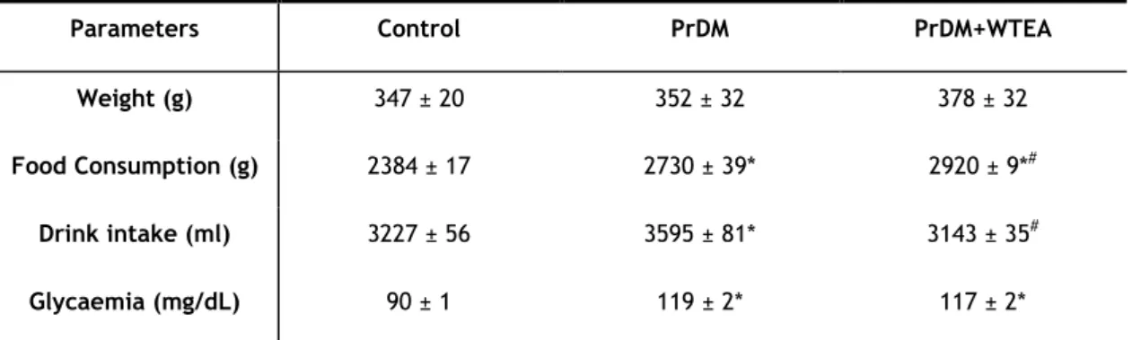
1. The animal model developed prediabetes ... 32
2. White tea consumption by prediabetic rats improves the antioxidant defenses of lung tissue ... 34
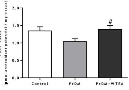
3. White tea consumption by prediabetic rats improves the total antioxidant capacity of the lung tissue ... 36
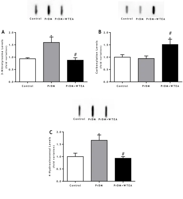
4. White tea consumption by prediabetic rats restores protein nitration and lipid peroxidation in the lung tissue ... 37
5. Consumption of white tea by prediabetic rats restores the levels of H2A histones of the lung tissue ... 39
Discussion ... 40
Conclusions ... 46
xiii
List of Figures
Figure 1 - Schematic summary of diabetes types, symptoms and complications... 4 Figure 2 - Schematic representation of the generation of free radicals through the activation of the polyol pathway, the PKC pathway, the AGEs and the electron transport chain ... 7 Figure 3 - Representation of the action of antioxidant enzymes SOD, GPx, GR, CAT in order to
neutralize free radicals and eliminate H2O2 formed ... 9
Figure 4 - Representation of the main production steps of different types of tea ... 15 Figure 5 - Schematic representation of the chemical structures of A) EC; B) EGC; C) ECG; D) EGCG present in WTEA ... 17 Figure 6 - Schematic representation of the chemical structures of methylxanthines present in WTEA, A) caffeine; B) theobromine; C) theophylline. ... 18 Figure 7 - Effect of the consumption of WTEA in the activities of antioxidants defenses A) SOD; B) GPx; C) GR; D) CAT of the control, PrDM and PrDM+WTEA groups ... 35 Figure 8 - Effect of WTEA consumption in the antioxidant potential of lung tissue. The antioxidant potential is expressed through the FRAP value (μmol of antioxidant potential/mg tissue).. ... 36 Figure 9 - Effect of WTEA consumption on oxidative damages in lung tissue A) protein nitration levels; B) carbonylation levels C) lipid peroxidation levels in the control group, PrDM rats and PrDM+WTEA. ... 38 Figure 10 - Effect of WTEA uptake by PrDM rats on A) DNA histone H2A and B) DNA histone P-H2A levels in control group rats, PrDM rats and PrDM rats consuming WTEA ... 39
xiv
List of Tables
Table 1: Characterization of some features of the animal model over 60 days of treatment. 32 Table 2: Glucose tolerance and Insulin resistance in rats from the experimental groups. ... 33
xv
Abbreviations
3-NT 3-Nitrotyrosine
4-HNE 4-hydroxynonenal
4-HPNE 4-hydroperoxy-2E-nonenal
AGEs Advanced glycation end products
AUC Area under the curve
BTEA Black tea
CAT Catalase
COX-2 Cyclooxygenase-2
DAG Diacylglycerol
DNA Deoxyribonucleic acid
EC Epicatechin
ECG Epicatechin gallate
EDTA Ethylenediaminetetraacetic acid
EGC Epigallocatechin
EGCG Epigallocatechin-3-gallate
ERK Extracellular signal-regulated kinases
ETC Electron transport chain
FADH2 Flavin adenine dinucleotide
FRAP Ferric reducing antioxidant power
GPx Glutathione peroxidase
GR Glutathione reductase
GSH Reduced glutathione
GTEA Green Tea
H2O2 Hydrogen peroxide
iNOS Nitric oxide synthase
LDL Low density lipoprotein
MAPK Mitogen-activated protein kinases
NADH Nicotinamide and adenine dinucleotide
NADPH Nicotinamide adenine dinucleotide phosphate
NF-kB Nuclear transcription factor kappa-light-chain-enhancer of activated B cells
NO Nitric oxide
OS Oxidative stress
PBS Phosphate buffered saline
PI3K Phosphatidylinositol 3 kinase
xvi
PrDM Prediabetes
PVDF Polyvinylidenedifluoride
ROS Reactive oxygen species
SOD Superoxide dismutase
STZ Streptozotocin
T1DM Diabetes Mellitus type 1
T2DM Diabetes Mellitus type 2
TBARS Thiobarbituric acid reactive substances
t-Bu-OOH tert-Butyl hydroperoxide
TPTZ 2,4,6-Tripyridyl-S-Triazine
Tyr 4-Hydroxyphenylalanine
WTEA White tea
1
2
1. Diabetes Mellitus – brief overview
Diabetes mellitus (DM) is one of the most prevalent metabolic diseases in developed countries (Shaw et al. 2010). Overall, the prevalence of DM doubled, as the number of adults suffering from DM reached 425 million in 2017 (Federation 2017), compared with 171 million at the
beginning of the 21th century. According to recent projections, this number will continue to
increase, and it is expected that by 2045 about 629 million adults will suffer from DM (Federation 2017). Alarmingly, there is also a strikingly increasing trend of DM initiation among children and adolescents (Chen et al. 2011). It generally results from a failure in the secretion and/or action of insulin, a hormone produced by pancreatic β cells that allows glucose to be uptaken by cells in order to guarantee energy required for metabolism (Canivell et al. 2014). When insulin is decreased or when its function is compromised, the cells do not absorb glucose, which remains in the bloodstream causing hyperglycaemia (Asmat et al. 2016). In healthy subjects, glycaemia is regulated, and fasting plasma glucose levels are maintained between 4.0 and 5.5 mmol/L (72-99 mg/dl) (Ferrannini et al. 2011). When DM develops, blood glucose levels are no longer controlled by the body and these values are changed. Classically DM has been classified as type 1DM (T1DM) and type 2DM (T2DM) (Figure 1).
T1DM is characterized by an autoimmune response of T lymphocytes to insulin-producing β cells (Yoon et al. 2005). The loss of pancreatic β cells occurs progressively and results in marked decrease or even absence of insulin production. As a consequence, these individuals depend on the exogenous administration of insulin to survive and, therefore, this disease is also known as insulin-dependent DM (Figure 1). It affects 5-10% of the diabetic community and the first symptoms of T1DM appear as a rule in the first years of life and are therefore known as juvenile DM. T1DM when uncontrolled can lead to ketoacidosis, which can be lethal (Canivell et al. 2014, Asmat et al. 2016). As with DM in general, also the T1DM has been increasing over the years. It is estimated that by 2020 the prevalence of T1DM in individuals under 5 years of age will increase to double (Canivell et al. 2014). Studies indicate that the causes of this self-destruction are genetic, but it is also believed that environmental factors are progressively contributing to the increase in these numbers (Canivell et al. 2014, Asmat et al. 2016).
In turn, T2DM represents almost 95% of diabetics and is known as non-insulin-dependent DM. The tendency is to appear in advanced ages and, in the initial state, a decrease in insulin sensitivity begins (Canivell et al. 2014). Subsequently those affected with this disease have insulin resistance and, to compensate for this condition, β-pancreatic cells increase the rate of insulin secretion, leading to hyperinsulinemia. However, with the progression of T2DM,
β-3 pancreatic cells lose the ability to release adequate amounts of insulin in an attempt to compensate for hormone resistance and individuals develop a more or less severe insulin deficiency (Figure 1). The risk of developing T2DM has a genetic predisposition, but the main risk factors are increased age, obesity and lack of physical exercise (Canivell et al. 2014, Asmat et al. 2016).
DM is characterized by significant metabolic changes, associated with the development of various complications such as cardiovascular problems, foot ulcers, sexual dysfunction, retinopathy, nephropathy, neurodegenerative diseases (Nishikawa et al. 2007, Canivell et al. 2014, Asmat et al. 2016) and impairment of lung function (Nandhini et al. 2012) (Figure 1). However, depending on the degree of severity, polyuria, polydipsia (Canivell et al. 2014), polyphagia (Canivell et al. 2014, Asmat et al. 2016), weight loss, blurred vision (Canivell et al. 2014), difficulty in healing and increased vulnerability to infections (Canivell et al. 2014) also occur (Figure 1). The studies aimed at finding a cure for DM have been increasing over the years, but the most common treatment continues to be dietary and lifestyle modifications and drug therapy with hypoglycemic agents, such as sulfonylureas, α-glycosidase inhibitors and thiazolidenediones. However, when it is not possible to control blood glucose levels in this way it is necessary to administer exogenous insulin, which is verified in T1DM. Nonetheless, a healthy diet is essential to control the pathogenesis of DM, as it may help delay the complications associated with DM or even prevent the development of DM (Bastaki 2005). When it is no longer possible to avoid it, the treatment goes through changes in lifestyle and use of drug therapy such as biguanides, thiazolidinediones, α-Glucosidase Inhibitors, GLP-1 analogies. Metformin is one of the most commonly used drugs and its use is not recent. However, all of this medication is associated with side effects that can be avoided if alternative therapies are found (Bansal 2015).
4
5
1.1. Prediabetes
The difficulty in diagnosing DM led to the creation of an intermediary state called prediabetes (PrDM). PrDM is less severe than DM once patients have glycaemic values above the values of healthy people, but below the values of DM and therefore cannot be considered healthy individuals nor with DM (Figure 1). According to the American Diabetes Association it is characterized by altered fasting glycemia with values ranging from 5.6-6.9 mmol/L (100-125 mg/dl). Individuals also exhibit decreased glucose tolerance and/or have insulin resistance (Association 2018). It is also associated with obesity, hypertension, dyslipidemia and decreased glucose metabolism (Kasturi et al. 2008). The prevalence of this disease among young people has been increasing, potentiating the risk for the development of T2DM. When β-pancreatic cells fail to compensate for insulin resistance, the transition from the state of PrDM to T2DM arises (Engelgau et al. 2000). Worldwide, the prevalence of this disease has been increasing and is estimated to reach more than 470 million people by 2030 (Tabák et al. 2012). Annually, about 5-10% of individuals with PrDM become diabetic and lifestyle can contribute to increase these values (Nathan et al. 2007). PrDM may also lead to complications such as nephropathies, neuropathies, retinopathy, macrovascular diseases (Tabák et al. 2012) and decreased lung function (Li et al. 2013).
1.2. Diabetes and reactive oxygen species
Free radicals are very unstable and reactive molecules of short half-life, that have one or more unpaired electrons (Asmat et al. 2016).Their presence in the body in small quantities is essential to life because the radicals activate cell signalling pathways, such as the mitogen-activated protein kinases (MAPK) pathway, the extracellular signal-regulated kinases (ERK) pathway that alters gene expression (Cho et al. 2003). In addition, some radicals are produced by neurons and will act as neurotransmitters (Fang et al. 2002). Also, macrophages and neutrophils are responsible for generating these species that act as mediators in immunity, for example in the respiratory burst in order to eliminate antigens (Fang et al. 2002, Freitas et al. 2010). Some reactive oxygen species (ROS) are involved in gene transcription. The ROS also intervene in leukocyte adhesion, in angiogenesis (Fang et al. 2002), contribute to vasoregulation by preventing thrombosis, are involved in fibroblast proliferation and increase the expression of antioxidant enzymes (Tiwari et al. 2013). However, when present in excess the defense system is not able to neutralize the ROS and these when yielding the unpaired electron originate oxidation of the cellular components leading to the increase of oxidative stress (OS) (Pandey et al. 2010, Halliwell et al. 2015, Asmat et al. 2016). OS has long been defined as a disturbance in the balance of antioxidants and pro-oxidants (Asmat et al. 2016). It is known that increased glucose levels can stimulate
6 the production of free radicals and, therefore, OS is strongly associated with DM (Baynes 1991), together with a decrease in antioxidant defenses (McLennan et al. 1991, Saxena et al. 1993), one of the main responsible for the progression and for the appearance of complications of this disease (Maritim et al. 2003). This association between DM and ROS is no longer recent and has been discussed since the 1990s (Baynes 1991), being related to the damage of cellular proteins, membrane lipids and nucleic acids (Maritim et al. 2003). Langerhans islands are very damaged by ROS since these islands have very low levels of antioxidant defenses (Tiwari et al. 2013).
The main source of free radicals is the oxidation of glucose. In the enediol form the glucose is oxidized giving rise to an enediol anion radical. This can be converted into reactive ketoaldehydes and superoxide anion radicals. When antioxidant enzymes are not present in sufficient amounts to neutralize it, superoxide anion radical are very reactive and capable of causing cellular damage (Maritim et al. 2003). In addition, superoxide anion radicals can also react with nitric oxide (NO) and give rise to reactive peroxynitrite radicals (Hogg et al. 1993). Hyperglycaemia is also a source of radical production because it promotes lipid peroxidation of low density lipoprotein (LDL), contributing to the formation of free radicals (Kawamura et al. 1994). Also, the interaction of glucose with proteins leads to the formation of a Schiff base and consequently the formation of the Amadori product that results in advanced glycation end products (AGEs) (Hori et al. 1996). AGEs can lead to the formation of radicals in two different ways. One of them is through its receptors that will inactivate enzymes and alter their structure and function (Figure 2) (McCarthy et al. 2001). The other form is when the intracellular OS increases leading to activation of the Ras/MAPK pathway which in turn activates the nuclear transcription factor kappa-light-chain-enhancer of activated B cells
(NF-kB) which regulates several genes. Thus, NF-kB increases the production of NO which is
considered to cause damage in pancreatic β cells (Mohamed et al. 1999). The polyol pathway converts glucose into sorbitol through the aldose reductase dependent nicotinamide adenine dinucleotide phosphate (NADPH). When glucose levels are within healthy levels, aldose reductase has low affinity for glucose. However, under hyperglycaemic condition, the activity of this enzyme increases and more sorbitol is produced. As the conversion of glucose to sorbitol increases, consequently the levels of NADPH required for this conversion decrease. Thus, there is less NADPH to reduce glutathione reductase (GR) so that it can regenerate
glutathione peroxidase (GPx). Without GPx the hydrogen peroxide (H2O2) generated by the
free radicals is not converted to H2O. Thus, it indirectly contributes to the formation of ROS
(Figure 2) (Srivastava et al. 2005, Rains et al. 2011). The protein kinase C (PKC) pathway is another mechanism involved in the production of OS in DM. Generally, PKC isoforms are activated by diacylglycerol (DAG), a second lipid messenger. When glycemia increases, there is also an increase in dihydroxyacetone phosphate, a glycolytic intermediate that is reduced
7 to glycerol 3-phosphate, contributing to the increase in DAG synthesis. This increase leads to the activation of PKC (Rains et al. 2011). It is known that PKC plays an important role in the activation of NADPH oxidase, a major source of ROS in non-phagocytic cells. Increased activation of PKC caused by hyperglycemia also increases the production of free radicals and thus the OS (Figure 2) (Soetikno 2012). In addition to all these mechanisms, the formation of ROS is also due to the overproduction of superoxide radicals in the mitochondria. Under normal conditions, nicotinamide and adenine dinucleotide (NADH) and pyruvate are generated, in which NADH donates electrons and pyruvate donates equivalent reductants that enter the tricarboxylic acid (TCA) cycle to produce NADH and flavin adenine dinucleotide
(FADH2) (Rains et al. 2011). In the electron transport chain (ETC), NADH and FADH2 provide
the electrons that feed the chain and contribute to the production of adenosine triphosphate. When glycaemia increases the number of substrates entering the TCA cycle also increases and thus the number of reducing equivalents that donate electrons to the ETC is also increased. Hyperglycaemia increases the proton gradient, which results in overproduction of electron donors through the TCA cycle, which in turn causes a marked increase in superoxide production (Figure 2) (Du et al. 2001).
Figure 2 - Schematic representation of the generation of free radicals through the activation of the polyol pathway, the PKC pathway, the AGEs and the electron transport chain (Araki et al. 2010).
8 In order to eliminate the harmful effects of free radicals the organism has mechanisms to neutralize these radicals and thus keep the cells protected from their effects. The antioxidant defenses such as superoxide dismutase (SOD), GPx, GR and catalase (CAT) have the ability to neutralize these radicals and convert the formed products (Pham-Huy et al. 2008). SOD is the first line of defense against cellular damage caused by ROS (Tiwari et al.
2013). It catalyzes the dismutation of the superoxide anion in H2O2 and O2, compounds less
toxic to the organism (Faraci et al. 2004, Wang et al. 2012), reducing the possibility of superoxide anion interacting with NO and forming a reactive peroxynitride. Subsequently, in
the presence of other enzymes H2O2 will be converted to O2 and H2O (Davari et al. 2013).
When overexpressed or when antioxidants identical to SOD are administered, DM is prevented (Wang et al. 2011). GPx and GR are two enzymes that can be found in the cytoplasm,
mitochondria, and nuclei of cells. GPx has the function of metabolizing H2O2 into two H2O
molecules using reduced glutathione (GSH) as a hydrogen donor. When donated a hydrogen, GSH is transformed into oxidized glutathione (GSSG) and GSH is renewed by GR using NADPH, obtained through glucose-phosphate dehydrogenase, as a cofactor (Maritim et al. 2003). In turn, CAT is an antioxidant enzyme present in almost all living organisms that metabolizes
H2O2 by converting it into H2Oand O2. When CAT appear to be decreased, pancreatic β cells,
which contain a large amount of mitochondria, undergo OS producing ROS in excess, leading to the dysfunction of these cells and aggravating DM (Jamieson 1986). Although SOD is the only one to act directly in the free radicals, GPx, GR and CAT are equally important because
they convert H2O2 present in the organism (Figure 3), that in an excessive concentration can
cause significant damages to proteins, deoxyribonucleic acid (DNA), ribonucleic acid and lipids (Takemoto et al. 2009).
9
Figure 3 - Representation of the action of antioxidant enzymes SOD, GPx, GR, CAT in order to neutralize
free radicals and eliminate H2O2 formed (Finosh et al. 2013).
1.3. Biomarkers of oxidative stress
1.3.1 Protein oxidation
Proteins are involved in many physiological functions, including cell signalling and transport through cells but they are ROS targets and both structure and function can be affected. There are many side chain targets for the oxidation of proteins, including cysteine, methionine, and tyrosine.
4-Hydroxyphenylalanine (Tyr) is a non-essential amino acid present in most proteins in nature. Tyr is moderately hydrophilic, which is explained by its aromatic hydrophobic benzene ring with a hydroxyl group (Bartesaghi et al. 2007, Ryberg et al. 2007). As a result, Tyr is exposed to the surface of proteins allowing modifications. Tyr nitration in proteins is associated with OS resulting in the formation of 3-nitrotyrosine (3-NT) (Tsikas 2012, Ahsan 2013). 3-NT is a covalent modification of the protein and is used as an OS biomarker. In
10 healthy individuals the levels of 3-NT are residual and may be around 31±6 pmol/ml in human plasma (Kamlsakl et al. 1996). However, in various pathological conditions 3-NT levels change to values higher than those considered normal for healthy individuals. Measurements of 3-NT
in biological samples are generally performed by methodologies such as
immunohistochemistry, high performance liquid chromatography, gas chromatography, immunochemical detection and slot-blot analysis (Teixeira et al. 2016).
Carbonyl groups are markers of protein oxidation (Suzuki et al. 1999). The carbonyl groups are chemically stable, which is useful both for their detection and for storage. The increased carbonylated protein content was reported in different cells and plasma of diabetic patients (Suzuki et al. 1999, Pandey et al. 2010). The carbonyl content can be measured by using several techniques, such as slot blot, Western blot, enzyme-linked immunosorbent assay and spectrophotometrically (Dalle-Donne et al. 2003).
1.3.2 Lipid Peroxidation
Lipids are the main targets of ROS, with hydroperoxides being the most toxic in cells, not only directly but also through the degradation of highly toxic hydroxyl radicals. The hydroperoxides can also react with the transition metals and produce highly reactive aldehydes, such as 4-hydroxynonenal (4-HNE) (Guo et al. 2012). 4-HNE may have an enzymatic or non-enzymatic origin. The enzymatic origin results from the transformation of polyunsaturated fatty acids by 15-lipoxygenases. The major precursors of 4-HNE are 13-hydroperoxyoctadecadienoic acid which is produced by the oxidation of linoleic acid through 15-lipoxygenases-1 and 15-hydroperoxy eicosatetraenoic acid produced by the oxidation of arachidonic acid by 15-lipoxygenases-2. On the other hand, it can be formed by the non-enzymatic process in five different mechanisms. I) It may be formed by reduction of a hydroperoxide in a lipid alkoxy radical by transition metal ions and followed by β-scission; II) The peroxyl radical cyclizes to form a dioxetane which is oxygenated in peroxy-dioxetane which is then cleaved to form the 4-hydroperoxy-2E-nonenal (4-HPNE) precursor of 4-HNE III) The hydroperoxide radical can be oxygenated in dioxetane and undergo fragmentation which results in the formation of 4-HPNE which is converted to 4-HNE; IV) The hydroperoxide reacts with the reduced form of a transition metal producing alkoxyl radicals which, after reaction
with O2, hydrogen abstraction and fragmentation, produce 4-HNE; V) The alkoxyl radical
undergoes cyclization, oxygenation, transition metal oxidation, hydrolysis and rearrangement yielding 4-HNE (Ayala et al. 2014). In DM there are reports of significant changes in both lipid metabolism and structure (Fowler 2008). The increase in lipid peroxidation is also an indication of decline in antioxidant defense mechanisms (Saddala et al. 2013) and can be measured by slot-blot by quantifying 4-HNE (Jesus et al. 2016) and by the thiobarbituric acid reactive substances (TBARS) method through quantification of malondialdehyde that is
11 formed when chains of polyunsaturated fatty acids are attacked by hydroxyl radicals (Alves et al. 2015).
1.3.3 H2A and P-H2A Histones
Histones have an important role in the regulation of genes, being affected by the presence of ROS that damage them (Sohal et al. 1996). H2A is one of the five histone types that organize DNA into the chromatin. Chromatin consists of the nucleosome of eight histones, two from each of the four core types (H2A, H2B, H3, H4). Histone H2A is required to disrupt the cell cycle and to repair DNA damage after double stranded DNA breaks. DNA damage results in the rapid phosphorylation of H2A in Serine-139 by kinases similar to phosphatidylinositol 3 kinase (PI3K) resulting in formation of P-H2A (Yuan et al. 2010). In addition to repairing DNA damage, H2A is required for apoptosis and in response to atopic signals is phosphorylated by several kinases including ataxia-telangiectasia mutated kinase, ataxia telangiectasia and Rad3-related protein and DNA-dependent protein kinase. This rapid response is required to recruit proteins that respond to DNA damage, including Mediator of DNA damage checkpoint protein 1, Nijmegen breakage syndrome 1, DNA repair protein RAD50, Double-strand break repair protein MRE11A and breast cancer 1 (Yuan et al. 2010). However, H2A can also be phosphorylated in Tyr142. When this occurs, the proteins required for DNA repair are not recruited which promotes the binding of pro-apoptotic factors (Cook et al. 2009). Studies in human spermatozoa prove that OS induces the phosphorylation of H2A (Gao et al. 2013). Although phosphorylation of H2A is poorly studied in DM, a study by Dong et al. (2015) found under in vitro conditions that OS-induced DM caused phosphorylation of H2A in embryonic cells. Thus, phosphorylation of H2A occurs in an attempt to recruit proteins that respond to DNA damage induced by OS. The quantification of histones H2A and P-H2A can be done by western blot (Dahabieh et al. 2017), confocal laser scanning microscopy (Mazzucchelli et al. 2017) and slot blot (Ramos et al. 2007).
12
2. Diabetes Mellitus and Lung
The main cause of death in patients with DM are complications associated with the disease
and induced by glycotoxicity (Zheng et al. 2017). As the lung has ventilatory reserves, which
is not the case of other organs, the symptoms and complications appear later (Eren et al.
2014). As a consequence
,
until a few years ago, the medical community disregarded lungdamage caused by diabetic complications (Zheng et al. 2017). Pulmonary disease associated with DM includes a higher predisposition to infections, pneumonia, onset of chronic obstructive pulmonary disease, asthma, pulmonary fibrosis, tuberculosis, and sleep apnea (Ehrlich et al. 2010, Kent et al. 2014, Hsiao et al. 2015). Pulmonary function decreases over the years in patients with DM. In recent years, this subject has been considered relevant and, contrary to the disregard given previously, the number of studies indicating physiological and structural abnormalities in the lungs of patients with DM has been increasing which has influenced some authors to make revisions on the subject (Eren et al. 2014). McCloud et al. (2004) found that after three weeks upon induction of DM by alloxan (65 mg/kg), male New Zealand white rabbits presented morphological abnormalities in the lungs, such as capillary dilatation, severe parenchymal haemorrhage and agglomeration of erythrocytes. One of the main causes may be microangiopathy induced by uncontrolled hyperglycaemia leading to lung injury (Williams et al. 1984). Behind the studies performed in the animal models, autopsies in diabetic individuals also showed morphological abnormalities, such as thickening of the alveolar epithelium, centrilobular emphysema and pulmonary microangiopathy. In fact, microangiopathy and alveolar thickening are mainly responsible for the respiratory insufficiency associated with DM. However, hyperglycaemia is known to be associated with reduced lung function, respiratory muscle weakness and decreased gas exchange at the alveolar level (Vracko et al. 1979). Hyperglycaemia is characterized by increased blood glucose levels and is associated with increased protein catabolism. It is known that the latter leads to a decrease in muscle contractility and may lead to muscle weakness. Thus, if the activity of the respiratory muscles decreases, there will be a decrease in gas exchange and, consequently, a decrease in lung function (El-Azeem et al. 2013). Hyperglycaemia also affects the lungs through non-enzymatic glycosylation of proteins, such as collagen and elastin. As these two proteins promote lung expansion, when they are modified the lung expelling capacity is also affected and as a consequence the volume of inspiration (Marvisia et al. 2001). Studies by Nandhini et al. (2012) in diabetic individuals show significant reductions in forced vital capacity, forced expiratory volume in one second and forced expiratory flow by 25-75% indicating weakness of the respiratory muscles. In addition, the authors observed a reduction of the forced inspiratory flow by 25-75% that suggests the mechanical and neuromuscular pulmonary properties were decreased. In this way, it was observed that the efficiency of the ventilatory pump is affected in this disease, as well as the respiratory
13 muscles that show weakness. Davis et al. (2000) found that the diabetic lung had a reduced air flow compared to a healthy lung. This may be explained since DM is associated with high levels of inflammatory mediators that in conjunction with microangiopathy, cause changes in the lung matrix proteins. For instance, the metabolism of collagen is also altered by the glycosylation of proteins. This glycosylation leads to irreversible crosslinking of collagen making it less susceptible to proteolysis than native collagen (Ofuwe et al. 1988). This causes the modified collagen to accumulate and there is less of the native collagen resulting in significantly lower lung volumes (El-Azeem et al. 2013). In addition to these alterations, OS, non-enzymatic protein glycosylation, polyol pathway, NF-kB signaling pathway and PKC pathway contribute to lung injury (Zheng et al. 2017). Protein glycosylation may lead to
increased OS by the release of O2 and H2O2 and the activation of phosphogenic via the
receptor of AGEs (Yan et al. 1994). In addition, phagocytic cells generate large amounts of NO and ROS. Furthermore, peroxynitrite is formed when NO reacts with superoxide, thus
increasing peroxynitrite formation increases NO which prompts protein nitration (Hensley et
al. 2000). Thus, an exacerbation of the protein nitration (Hensley et al. 2000) and lipid peroxidation together with the reduction of antioxidant defenses will contribute to increased ROS-induced lung damages (Gumieniczek et al. 2002). On the other hand, the polyol pathway also contributes to lung damage because as aforementioned when the glucose concentration in the cell increases, aldose reductase is activated to reduce glucose to sorbitol (Luo et al. 2016). Then, sorbitol can induce cellular osmotic pressure, leading to cell death (Wu et al. 2016). On the other hand, as the polyol pathway also consumes NADPH and produces NADH, redox cellular imbalance can occur and trigger OS, leading to changes in the integrity and
function of the cellular membrane of the lung (Zheng et al. 2017). The NF-kB signalling
pathway is involved in the regulation of gene expression in inflammatory responses. This pathway is responsible for regulating the expression of tumor necrosis factor alpha, interleukin 1 beta, interleukin 6 (Sun et al. 2009), NO synthase (iNOS) and cyclooxygenase-2 (COX-2) (Yagi et al. 2006). NF-κB levels were found to be increased in diabetic animals as well as levels of iNOS and COX-2 (Zhang et al. 2015). These evidences demonstrate that the diabetic lungs are subject to OS mediated by the activation of the NF-kB pathway (Zheng et al. 2017). Finally, PKC also contributes to lung damage. The activation of PKC by the state of hyperglycaemia can activate the NADPH oxidase, which generates superoxide anion, that is, more ROS which contribute to the increase of the OS (Bey et al. 2004). Thus, through all the explained mechanisms, OS plays a key role in the development of respiratory dysfunction
associated to DM (Eren et al. 2014).The study of the effects of DM on lung tissue is recent
14
3. White Tea
Tea (Camellia sinensis (L.)) is used by the Chinese since the year 3000 B.C as a medicinal beverage because of its therapeutic potential. It is considered one of the most consumed beverages worldwide, following water (Graham 1992), not only due to the health benefits, but also because of its low cost (Baptista et al. 1998). Tea is produced from an infusion of leaves of C. sinensis (Lopez et al. 2011), cultivated in more than 30 countries (Graham 1992).
C. sinensis is available in two varieties: C. sinensis var. sinensis and C. sinensis var. assamica
(de Mejia et al. 2009). The first, originating in China, develops in a cold climate and appears as a small-leafed plant. The second, originating in India, is developed in countries with a semitropical climate and is characterized by being a tree that unlike the previous one presents large leaves. C. sinensis var. sinensis originates most of the leaves used for the production of green tea (GTEA) and white tea (WTEA) while C. sinensis var. assamica originates the leaves used for the production of black tea (BTEA). The leaves for production of WTEA are very young and the buds are covered with silver hairs, both harvested seasonally in early spring (Rusak et al. 2008). These buds, when protected from light, show little chlorophyll formation that gives them a white appearance (Alcázar et al. 2007). In order to avoid oxidation, soon after harvest the leaves and buds are vaporized and dried (Rusak et al. 2008).
The leaves from Camellia sinensis (L.) may originate different types of tea, depending on the processing and collection (de Mejia et al. 2009): unfermented, partially fermented and completely fermented. When it is unfermented it originates the GTEA and the WTEA, when it is partially fermented it gives the oolong tea and when it is totally fermented it gives origin to the BTEA (Dias et al. 2013) (Figure 4). At collection, the leaves undergo a process called "fermentation" that occurs through exposure to air (McKay et al. 2002). This process is catalyzed by endogenous enzymes such as polyphenol oxidase (McKay et al. 2002) and enzymatic oxidation of leaves leads to the formation of theaflavins and thearubigins, two of those responsible for color, hence BTEA has a darker shade (Palmer 1984). Thearubigins are characterized by having high molecular weights and having a red color that is responsible for the coloring of the teas, such as BTEA. In other words, the BTEA has a darker coloration because it is more oxidized than the others, and therefore has a greater amount of this polyphenol. In contrast, GTEA and WTEA are lighter in color. Besides thearubigins also the theaflavins are responsible for coloring the tea. They are characterized by the structure of the benzotropolone ring and the red-orange color, thus contributing to the aroma of tea (Khan et al. 2007).
15
Figure 4 - Representation of the main production steps of different types of tea (Dias et al. 2013).
It is estimated that in 2016 world consumption of tea will have been approximately 5.5 million tons (FAO 2018). According to the International Tea Committee (2006), the BTEA is the most consumed in India (694.5 thousand tons in 2005) and in turn, the GTEA is most consumed in China (484.9 thousand tons in 2005). According to the Food and Agriculture Organization of the United Nations (2018), in 2016 BTEA was the most produced and most exported followed by GTEA. In turn, the WTEA is less known by the population and is thought to be the least consumed (Alcázar et al. 2007) and also the least studied (Almajano et al. 2008).
In general, the several types of teas present bioactive compounds that confer health benefits (Maron et al. 2003, Afifa Khatun et al. 2017, Charehsaz et al. 2017, Schimidt et al. 2017, Thitimuta et al. 2017, Fan et al. 2018, Fujiki et al. 2018). WTEA in particular has a potential antidiabetic (Islam 2011), antioxidant (Alves et al. 2015, Nunes et al. 2015, Oliveira et al. 2015, Dias et al. 2016), cardioprotective (Alves et al. 2015), anti-carcinogenic (Hajiaghaalipour et al. 2015), neuroprotective (Nunes et al. 2015) and anti-obesity effect (Sohle et al. 2009). These effects of WTEA are related to its chemical composition, which is largely composed by polyphenols and methylxanthines.
16
3.1. White tea composition
In general, tea is composed of polyphenols, proteins, polysaccharides, minerals, amino acids and organic acids, lignins and methylxanthines (theophylline, caffeine and theobromine) (Seeram et al. 2006).
The phenolic compounds of tea are the constituents that have attracted the most attention, particularly flavonoids (Seeram et al. 2006). There is a wide variety of flavonoids that are grouped into six major groups, flavanoids, flavones, flavonoids, isoflavones, flavanones and anthocyanidins (for review see (Dias et al. 2017)). One of the main classes of polyphenols present in tea is the catechins group that belongs to the family of flavanols and is characterized by its strong antioxidant power. The main catechins that are present in WTEA are epicatechin (EC), epicatechin gallate (ECG), epigallocatechin (EGC) and epigallocatechin-3-gallate (EGCG) (Figure 5) (de Mejia et al. 2009). The EC has on carbon 3 of C-ring a hydroxyl group and on carbons 3’ and 4’ of B-ring an ortho-dihydroxy group. In the ECG, on the carbon 3 of C-ring, a portion of esterified gallate is present, whereas the EGC has on the carbon 3’, 4’ and 5’ of B-ring a trihydroxyl group. EGCG is the most abundant catechin in both WTEA and GTEA (Dias et al. 2013, Nunes et al. 2015) and has a trihydroxyl group on carbons 3’, 4’ and 5’ on B-ring and a gallate esterified on C-ring carbon 3 (Graham 1992). The beneficial effects of these teas are generally associated with the presence of this catechin (Song et al. 2012). According to Saha et al. (2017) a tea prepared from 500 mg of leaves in 10 ml of water contains about 29% of polyphenols being 13% of catechins. In addition, it contained 3.9 g/100 g of caffeine and 1 g/100 g of total flavonoids. According to Belentine et al. (1997), WTEA shows higher concentrations of catechins and lower concentrations of theaflavins and thearubigins, and the catechin content in tea depends on how the leaves are processed before drying and on the geographical location during growth. The concentration of flavonoids also depends on how the tea is prepared, such as the amount of leaves used, the fermentation time and the temperature at which it is prepared (McKay et al. 2002). Thus, it is known that hot tea is the one with the highest concentration of flavonoids (541-692 μg/ml) (Hakim et al. 2000) and the smallest are in the instant preparations (90-100 μg/ml) and in tea containing ice (Arts et al. 2000). Other bioactive compounds also found in tea are gallic, p-coumaric, caffeic and amino acid acids (Seeram et al. 2006).
17
Figure 5 - Schematic representation of the chemical structures of A) EC; B) EGC; C) ECG; D) EGCG present in WTEA (Adapted from (Dias et al. 2013)).
The most abundant free amino acid in teas is L-theanine which accounts for 50% of the total free amino acids present and is responsible for the aroma (Dufresne et al. 2000). L-theanine constituted between 1-3% of the dry weight of the tea however, this percentage may vary according to the location of the crop and time of collection (Vuong et al. 2011). It is considered an antioxidant and has antidiabetic effect (Adhikary et al. 2017).
The methylxanthines present in tea are heterocyclic compounds that belong to the purine family (Figure 6) (Monteiro et al. 2016). In the WTEA, theophylline, theobromine and caffeine are present, the latter representing the second largest WTEA compound (Dias et al. 2014). Caffeine is the most studied methylxanthine and its effects have already been observed in
18 several diseases. The therapeutic action of the various methylxanthines has already been observed in respiratory diseases such as asthma and chronic obstructive pulmonary disease. Theophylline is a potent bronchodilator used in asthma disease that diffuses better in bronchial tissue than other methylxanthines (Monteiro et al. 2018). Theobromine (0.1% in methylcellulose) has also been shown to be effective in eliminating cough induced by citric acid in Dunkin Hartley pigs (Usmani et al. 2005). Caffeine, in turn, improves ventilation and respiratory muscle function. It is an efficient eliminator of hydroxyl radicals and is also considered an ally in T2DM (Monteiro et al. 2018).
Figure 6 - Schematic representation of the chemical structures of methylxanthines present in WTEA, A) caffeine; B) theobromine; C) theophylline (Adapted from (Monteiro et al. 2016)).
Other constituents, although not the main ones, that also play an important role are chlorophylls, lipids, carotenoids and volatile compounds (Balentine et al. 1997). These are others responsible for developing the aroma of tea (Dufresne et al. 2000).
19 Dias et al. (2014) studied the major constituents of WTEA and found that they were consisted mainly of EGCG (82 g/kg of tea extract), caffeine (71 g/kg of tea extract), sucrose (60 g/kg of tea extract), EGC 46 g/kg tea extract), L-theanine (19 g/kg of tea extract), glucose (6 g/kg of tea extract) and EC (5 g/kg of tea extract). In addition, it compared the composition of this tea with that of the GTEA and found that WTEA had higher concentrations of EGC, EGCG, caffeine, alanine and sucrose (Dias et al. 2014).
3.2. Antioxidant and antidiabetic potential
The search for a natural therapy used as complement to classical medical drugs, or even as replacement, has aroused great interest not only because it is affordable but also because it presents fewer side effects (Xiao et al. 2014). The WTEA shows a strong antioxidant potential mainly due to its composition (Almajano et al. 2008). Antioxidants scavenge free radicals (McLennan et al. 1991, Saxena et al. 1993) together with endogenous antioxidant defenses counteracting the adverse effects of ROS (Rietveld et al. 2003). When these mechanisms fail, an imbalance is generated and the excessive production of ROS causes damage to proteins, lipids and DNA (Sohal et al. 1996).
The antioxidant potential of WTEA was verified in a study by Almajano et al. (2011) in which
striatal cells were subjected to OS through exposure to H2O2 (250 μM) for 24 hours. In the
cells treated with WTEA (25 μg/ml, 100 μg/ml or 200 μg/ml), the oxidative damage decreased due to the high antioxidant potential of this tea conferred by its main constituents. In addition to this study, Yen et al. (2013) verified the protective effect of WTEA on Clone 9
cells subjected to H2O2 (0.2 mM) induced OS. It was observed that the cells treated with
WTEA (0.2 mg/ml) presented higher percentage of cell viability and higher activity of CAT and GPx associating these improvements with the antioxidant capacity of WTEA.
The effect of WTEA was also studied in animals. In a study by Ruiz et al. (2018) in which Sprague-Dawley rats were injected with Adriamycin (10 mg/kg body weight), an oxidising agent, the WTEA consumption (both 0.15 mg/kg and 0.45 mg/kg) protected the liver and brain of the injuries caused by adriamycin. This evidence suggests that WTEA acts as an antioxidant when OS is increased. Koutelidakis et al. (2009) also evidenced the antioxidant capacity of GTEA and WTEA in heart, lung, spleen, liver, kidney and brain of male mice. After diary administration by gavage of 0.1 ml of GTEA or WTEA during 5 consecutive days it was observed an increased antioxidant capacity in the heart tissue in mice that ingested GTEA and WTEA when compared to those animal who ingested water. In mice receiving GTEA, these results were also found in the lung, showing how GTEA and WTEA may bring benefits to the antioxidant capacity of these tissues.
20 When the antioxidant capacity is decreased, the proteins are more prone to the damage caused by free radicals. In order to verify whether WTEA could counteract these damages, Espinosa et al. (2012) performed a study in Sprague-Dawley rats in which rats injected with adriamycin (10 mg/kg body weight) ingested for 12 months different doses (0.15 mg/kg and 0.45 mg/kg) of WTEA. The consumption of 0.15 mg/kg WTEA restored the levels of the protein carbonyl groups in the liver and heart, however to observe the same effects in the brain, it was required a higher dose (0.45 mg/kg). Also Kumar et al. (2012) verified the protective effects of GTEA and WTEA in an animal model where OS was induced by administration of benzo(a)pyrene (125 mg/kg body weight). The authors used male Balb/c mice and treated them for 35 days with GTEA and WTEA (2%). The authors observed that in untreated animals the antioxidant defenses were decreased in both lung and liver tissues. On the contrary, it was observed that GTEA and WTEA restored the levels of antioxidant capacity to those observed in the control group. Similarly, the consumption of GTEA and WTEA also decreased DNA damages.
Previous work from our group using a PrDM animal model induced by intraperitoneal injection of 40 mg/kg streptozotocin (STZ) has also showed the benefits of WTEA consumption. Alves et
al. (2015) found that the regular consumption of WTEA altered the glycolytic metabolism of
cardiac tissue and subsequently its oxidative profile. It was observed that the total antioxidant capacity decreased in the PrDM group and, the regular consumption of WTEA significantly increased this capacity. The lipid damages assessed by TBARS increased in the animals with PrDM compared to the control group, being restored by the consumption of WTEA. In turn, levels of protein carbonyl groups increased significantly in the PrDM group compared to the control group. However, there were no significant changes with the consumption of WTEA.
In addition to the effects observed in cardiac tissue, the daily consumption of WTEA also changed the oxidative parameters of the testicular tissue since Oliveira et al. (2015) observed a decrease in the testicular protein carbonylation in PrDM animal treated with WTEA. WTEA consumption was also effective in increasing the antioxidant capacity that was decreased in the testis of PrDM rats. In addition, lipid damage decreased, which, through the analysis of TBARS levels, was revealed to be increased in PrDM rats.
The benefits of tea consumption were also observed in the cerebral cortex where the regular consumption of WTEA increased the antioxidant capacity (Nunes et al. 2015). Interestingly, the expression of CAT, an antioxidant enzyme considered to be one of the most important antioxidant defenses in this tissue, was diminished in PrDM rats and WTEA intake reestablished this expression confirming its antioxidant power (Nunes et al. 2015). Also, lipid peroxidation and protein carbonyl levels increased in PrDM rats compared to the control
21 group, decreasing with the consumption of WTEA (Nunes et al. 2015). A study by Al-Shiekh et
al. (2014) in STZ-induced DM rats (55 mg/kg body weight, dissolved in 0.1M sodium citrate
buffer) analyzed the concentration of GSH and the activity of SOD, GPx and CAT. The authors found that all these enzymes were decreased in the liver of DM animals when compared to the control group composed of healthy animals. Thus, with the consumption of WTEA (2%) for 4 weeks, it was found that the expression of GSH and the activity of SOD, GPx and CAT increased significantly to values similar to the animals of the control group.
Studies on the effect of WTEA are still scarce and therefore its antioxidant potential in humans has not yet been studied. However, there are already studies on the effect of GTEA and caffeine (one of the main WTEA compounds) in humans. Erba et al. (2005) verified the effect of 2 cups of GTEA (approximately 250 mg of catechins) for 42 days on the lipid profile of human plasma. The authors observed that the consumption of GTEA increased the total antioxidant activity in the plasma protecting the cells from oxidative damage. Iso et al. (2006) found that both the consumption of GTEA (6 or more cups) and caffeine (416 mg/d) decreased the risk for the development of T2DM in Japanese adults. The authors of this article relate this decrease to the antioxidant potential of each of these products.
In addition to antioxidant properties, WTEA also has an antidiabetic potential. In vitro studies have demonstrated the antidiabetic potential of WTEA. Anderson et al. (2002) found under in
vitro conditions that catechins, including EGCG, and theaflavins prevented hyperglycaemia
since these polyphenols increased insulin sensitivity, probably by protecting pancreatic β-cells from damages. This has been observed by Han et al. (2003) which exposed RINm5F pancreatic β-cell at 0-200 µg/ml doses of EGCG during 24 hours. Also in this in vitro study it was found that EGCG reduced β-cell damage.
Over the years animal studies have emerged reinforcing the antidiabetic potential of this tea. A study in male albino Wistar diabetic rats induced by STZ (60 mg/kg) and given daily EGCG (25 mg/kg dissolved in water) for 8 weeks was found to reduce serum glucose levels which were increased in animals with DM. The levels of total cholesterol, triglycerides and LDL in DM rats were increased, and the consumption of EGCG contributed to the decrease of these
values (Roghani et al. 2010). Also the Islam study (2011),in STZ-induced male Sprague-Dawley
rats (65 mg/kg), consumption of WTEA (0.5% dissolved in water) for 4 weeks reduced hyperlipidemia, decreased insulin resistance and increased insulin sensitivity, evidencing the antidiabetic potential of WTEA. In a study by Nunes et al. (2015) rats administrated with 40 mg/kg STZ developed PrDM characteristics, namely glucose intolerance and insulin resistance. In the PrDM group that consumed WTEA (1 g/100 ml water) an improvement of insulin resistance was found although glycaemia did not decrease. The authors suggest that the main
22 responsible for those alterations could be the polyphenols present in WTEA since they are characterized by a strong antioxidant action.
Studies on the antidiabetic effect of WTEA have also begun to appear in humans. MacKenzie
et al. (2007) evidenced that the ingestion of 375 mg of tea capsules for 3 months in
individuals with T2DM ameliorated the hyperglycemic levels. These data were also corroborated by Venables et al. (2008) who observed in patients with T2DM that the ingestion of a tea capsule (equivalent to 3.5 cups) reduced glucose intolerance and increased insulin sensitivity and, thus, may reduce the risk of T2DM. Taken together, these studies suggest that both potentials are related since generally the antidiabetic potential of WTEA is related to its antioxidant properties. In addition, they show that the consumption of WTEA can be a cheap and natural alternative to reduce the effects caused by DM. After all, the antioxidant and antidiabetic effects depends on the amount consumed and its bioavailability (Holst et al. 2008).
23
24 DM is one of the most prevalent metabolic diseases in developed countries representing a high risk for the population. PrDM, an initially less serious condition than DM, has been increasing, especially among young people, being a risk factor for the development of T2DM.
The lung is one of the organs affected by this disease becoming more susceptible to infections, pneumonia, asthma and chronic obstructive pulmonary disease because it undergoes structural and physiological changes that result in the reduction of gas exchanges. The WTEA is known for its antioxidant and antidiabetic power, however its effect on the lung tissue of PrDM rats has not yet been studied.
Thus, the objective of this study was to verify the effects of PrDM on the lung and evaluate the effects of the regular consumption of WTEA on the activity of endogenous antioxidant enzymes (SOD, GPx, GR, CAT), total antioxidant capacity and damages caused in proteins, lipids and histones H2A of the lung tissue of PrDM rats.
25