Biomimetic Synthetic
Self-Assembled Hydrogels for Cell
Transplantation
Daniela Barros
1,2, Isabel Freitas Amaral
1,2,3and Ana
Paula Pêgo
1,2,3,41 INEB – Instituto de Engenharia Biomédica, Universidade do Porto, Porto, Portugal
2 i3S - Instituto de Investigação e Inovação em Saúde, Universidade do Porto, Porto, Portugal 3 FEUP – Faculdade de Engenharia, Universidade do Porto, Porto, Portugal
4 ICBAS – Instituto de Ciências Biomédicas Abel Salazar, Universidade do Porto, Porto, Portugal
Originally published in Current Topics in Medicinal Chemistry, Volume 15, Issue 13, 2015 DOI
10.2174/1568026615666150330111057
“The published manuscript is available at EurekaSelect via
http://www.eurekaselect.com/openurl/content.php?genre=article&doi=10.2174/1568026615666150 330111057.”
ABSTRACT
The development of three-dimensional matrices capable of recapitulating the main features of native extracellular matrix and contribute for the establishment of a favorable microenvironment for cell behavior and fate is expected to circumvent some of the main limitations of cell-based therapies. In this context, self-assembly has emerged as a promising strategy to engineer cell-compatible hydrogels. A wide number of synthetically-derived biopolymers, such as proteins, peptides and DNA/RNA, with intrinsic ability to self-assemble into well-defined nanofibrous structures, are being explored. The resulting hydrogels, in addition to closely resembling the architecture of native cellular microenvironments, present a versatile and dynamic behavior that allows them to be designed to undergo sol-to-gel transition in response to exogenous stimulus. This review presents an overview on the state-of-the-art of the different strategies being explored for the development of injectable synthetic self-assembled hydrogels for cell transplantation and/or recruitment of endogenous cells, with an emphasis on their biological performance, both in vitro and in vivo. Systems based on peptides are the most widely explored and have already generated promising results in pre-clinical in vivo studies involving different repair/regenerative scenarios, including cartilage, bone, nerve and heart. On the other hand, systems based on DNA and hybrid hydrogels are now emerging for application in the biomedical field with high potential. Finally, the main challenges hampering the translation of these systems to the clinic and the issues that need to be addressed for these to progress from bench-to-bedside are discussed.
As our understanding of cell biology and ability to control cell behavior, both in vitro and in vivo, improves, cell-based therapies are growing in interest within the field of regenerative medicine [1]. Their effectiveness relies on the ability of transplanted cells to mediate a therapeutic effect that potentiates the repair/regeneration processes [2]. Nevertheless, before the effective translation of these therapies into the clinic, a number of issues still have to be solved, such as low cell survival, lack of control of cell fate and poor cell engraftment following transplantation [3, 4].
Within biological systems, cells reside in the extracellular matrix (ECM), a complex and highly dynamic microenvironment that provides structural, mechanical, and biochemical support to cells, ultimately determining their behavior, function and fate. In this sense, and to improve the efficacy of cell transplantation therapies, biomaterial-based matrices mimicking the ECM are being designed to be used as vehicles for cell delivery. The goal is to create a permissive niche for the transplanted cells, promoting cell survival, proliferation, differentiation, and integration within the host tissue. One of the major challenges one faces when designing a cell-compatible matrix is the recapitulation of the instructive cellular niches of natural tissues and the control of the bidirectional interaction between cells and the surrounding microenvironment.
The design of such matrices must take into account a number of biological and physical criteria, as follows:
(i) Biocompatibility. The developed matrix should be able of perform its function without causing any toxic or undesirable effects.
(ii) Mechanical properties. The matrix mechanical properties should closely match those of the target tissue, since these will impact cell behavior and, consequently, determine the therapeutic efficacy of the transplanted cells.
(iii) Permeability. The matrix should allow the continuous exchange of gases, ions, nutrients and metabolites with the surrounding microenvironment, which is vital for the survival and proliferation of transplanted/encapsulated cells.
(iv) Degradation. Envisaging an application in the context of tissue repair/regeneration a degradable matrix is preferred in the majority of cases. It is paramount that the rate and mechanism of matrix degradation are adjusted to the process of tissue remodeling.
In the last few years, the incorporation of biochemical and biophysical ECM cues – cell adhesion, cell-triggered proteolytic degradation, mechanical stimulation and ECM/growth factor binding - into matrices of both synthetic and natural origin was explored to better mimic the native cellular microenvironment, with different degrees of development and success (consult references [5-7] for a comprehensive review on engineered matrices as ECM mimics).
Hydrogels as attractive vehicles for cell transplantation
Hydrogels are reticulated structures comprising cross-linked hydrophilic polymers with high water content (over 90% of water) [8]. A number of unique features make hydrogels highly promising matrices for cell transplantation and even for endogenous cell recruitment in the context of tissue
repair/regeneration. These include a highly hydrated nature, good permeability and elasticity, resembling the nature of soft tissue microenvironments [5]. These properties will favor cell-cell and cell-matrix interactions, ultimately influencing cell behavior and fate. Moreover, the structural and mechanical properties of hydrogels can be easily tuned to mimic the tissue-specific ECM microenvironment, by varying the polymer molecular weight and/or its concentration, or by adjusting the cross-linking degree of the network [1]. This is crucial for the development of cell-compatible hydrogels, as cells also respond to biophysical properties of the microenvironment not specifically related to its molecular composition, but to the overall structural and mechanical characteristics of the matrix.
Cells are particularly sensitive to mechanical stimuli, and small changes in matrix stiffness or rigidity can alter cell behavior in many aspects, such as morphology, migration, proliferation, differentiation and, in the case of stem cells, lineage specification and commitment [9, 10]. For instance, mesenchymal stem cells (MSCs) cultured on variably compliant poly(acrylamide) gels were found to undergo neurogenic differentiation on gels with elastic moduli (E) mimicking brain tissue ( 1 kPa), while on gels mimicking skeletal muscle (E = 11 kPa) and collagenous bone (E = 34 kPa) these cells differentiated along the myogenic and osteogenic lineages, respectively [10]. The mechanism by which cells convert mechanical signals into biochemical responses is termed mechanotransduction, a dynamic process orchestrated by the ECM, mechanosensitive molecules and cellular components (e.g. integrins, focal adhesions, stretch-activated ion channels, cadherins, cytoskeleton filaments, nuclei) [11]. Particularly important for matrix elasticity sensing are focal adhesions, multiprotein complexes formed upon integrin binding to ECM adhesion ligands, which link the ECM to the intracellular cytoskeleton. Focal adhesions act as mechanosensitive receptors, through which alterations in the conformation of ECM molecules caused by mechanical stimuli (e.g. tension/loading) are transmitted to cytoskeleton proteins, leading to cytoskeletal (re)organization, contraction, and activation of mechanotransduction signaling pathways affecting cell morphology, migration, and gene expression profile.
Hydrogels, rather than implanted, can be designed to be injected and create a three-dimensional (3D) network in situ. This is highly advantageous when envisaging clinical translation as it (i) allows the homogenous encapsulation of cells and bioactive molecules; (ii) enables the use of minimally invasive procedures for implantation; and (iii) ensures the conformational filling of the injured site, which usually has an irregular shape, leaving no gaps between the matrix and the degenerated/damaged tissue. In situ forming hydrogels will thus favor a more close contact and, consequently, a more efficient integration of the matrix within the host tissue. Injectable hydrogels could be further designed to have thixotropic properties (becoming less viscous when subjected to an applied stress). This is achieved through the control of different variables (e.g. temperature, pH and ionic strength) that influence directly the in situ gelling process (for a more extensive review on strategies explored to design in situ forming hydrogels see [12, 13]). When the cross-linking occurs in the presence of cells, either mediated by (bio)chemical (e.g. enzymatically, Michael-type addition or photopolymerization) or physical (e.g. ionic interactions or self-assembly) processes [13], care must be taken so that the impact on cell viability is limited and minute.
These properties have made hydrogels very attractive in the design of either acellular systems, specifically designed to allow the infiltration of endogenous cells, or cellular grafts to promote tissue remodeling and regeneration [14].
Naturally-derived polymers (e.g. collagen, fibrin or alginate) have been widely explored in the design of hydrogels due to their inherent biocompatibility and ability to mimic several features of native ECM. Nevertheless, their use is very often hindered by batch-to-batch variability, risk of immunogenicity and limited range of mechanical properties [15, 16]. Therefore, recent strategies have focused on the use of hydrogels derived from either purely synthetic polymers (e.g. poly(ethylene glycol) (PEG) and poly(vinyl alcohol)) or synthetically-derived biopolymers (e.g. proteins, peptides and DNA/RNA). Their synthetic nature, as well as their well-defined and versatile structure allows them to be engineered, through the incorporation of bioactive sequences (e.g. cell adhesive and protease sensitive), originating bioactive and bioresponsive hydrogels [16, 17]. In this context, synthetically-derived biopolymers are being more extensively explored, due to the several advantageous features of these materials, when compared with purely synthetic polymers. In contrast with the simpler and stochastic structure of purely synthetic polymers, synthetically-derived biopolymers can be engineered to attain a complex and reproducible structure. Moreover, by being synthesized using solid-phase synthesis or recombinant technology (eventually combined with genetic engineering) [18], they present a monodisperse distribution of the molecular weights. Finally, their inherent ability to be cleaved by enzymes allows a more tight control over the kinetics of hydrogel degradation and, as a result, over the remodeling process.
Self-assembled hydrogels
An important feature to take into account when engineering matrices to serve as ECM mimics is the architecture of native ECM. Attaining 3D networks structurally mimicking the nanofibrous structure of the ECM is highly desirable. This is expected to favor the creation of an instructive microenvironment for cell anchorage and cell migration, ultimately leading to the implant, colonization and integration within the host tissue. In this sense, “top-down” approaches, such as electrospinning and phase separation, have been explored in the design of scaffolds for such purposes. However, with these techniques one has a limited control over the scaffold structure at the nanoscale level, as well as over the reproducibility of the process. In the last few years, molecular self-assembly has emerged as the most promising and attractive strategy in this context [19-21]. This is a natural and spontaneous “bottom-up” approach driving the association and organization of single molecules into supramolecular structures. Self-assembly results from the establishment of multiple non-covalent intermolecular interactions (e.g. hydrogen bonding, electrostatic interactions, Van der Waals forces and aromatic π-stacking) [22, 23]. A wide number of biopolymers existing in nature (e.g. proteins, peptides, polysaccharides and DNA/RNA) present intrinsic ability to self-assemble into highly-ordered structural elements. As a result, their building blocks are being explored to develop synthetic self-assembled networks [18], leading to hydrogels with a nanofibrillar structure, closely resembling the architecture of native ECM. Moreover, the weak and transient nature of established interactions allows the design of hydrogels that undergo a sol-to-gel transition in response to exogenous stimulus (e.g. pH, temperature and ionic strength), which provides matrices with a “smart” and dynamic behavior [19, 24]. In the following sections, we present the state-of-the-art of synthetic self-assembled hydrogels, focusing on their application as matrices for cell transplantation in the context of regenerative medicine.
Engineered protein-based hydrogels
A number of naturally-occurring proteins (e.g. elastin, collagen and silk) have remarkable structural and biological properties that prompted their use in the design of self-assembled hydrogels [25]. In
particular, the ones derived from constituents of the mammalian ECM proteins, like collagen, fibronectin or laminin, additionally present biological active peptide domains recognized by cell adhesion receptors and cell-secreted proteolytic enzymes. These features have made protein-based hydrogels highly advantageous in the context of cell therapies [26]. However, the use of proteins derived from natural sources is very often hampered by low scalability, reproducibility, stability and price. In this sense, in the last few years, engineered proteins have arisen as a promising alternative for the design of self-assembled hydrogels. The synthetic nature of these proteins allows the combination in one structure of key attributes of native ECM, including cell adhesion, growth factor and ECM binding domains, as well as sequences that confer to the final protein elasticity and proteolytic degradation. But while maintaining the main attractive biological features of naturally derived proteins, precise control over the primary amino acid sequence and molecular weight can be achieved, allowing the fine-tuning of the gelation process and of the hydrogel structural and mechanical properties.
As the number of engineered proteins that can be used for the development of self-assembled hydrogels [26, 27] is too large to be effectively covered by this review, only the engineered protein-based hydrogels that have shown the most promising results, both in vitro and in vivo, were addressed. These include hydrogels based on elastin- and silk-like polypeptides, as well as hydrogels based on de novo engineered proteins, such as leucine zipper coiled-coil-based polypeptides and amphiphilic block co-polypeptides.
Elastin-like polypeptides
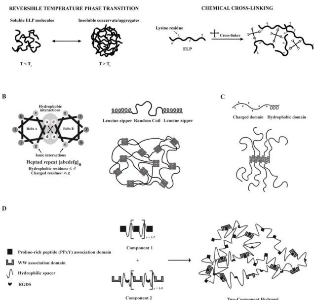
Elastin is an ECM structural protein, essentially composed of β-sheet structures, that mediates important biological functions, such as cell-cell and ECM-cell interactions, providing tissues with unique physical characteristics in terms of tensile strength and elasticity [25]. A broad number of synthetic polypeptides have been designed inspired by this natural protein, among which elastin-like polypeptides (ELPs). These are genetically engineered sequences containing the pentapeptide repeat - (VPGXG)n - in which X can be any amino acid except proline [28]. ELPs are
thermally-responsive and thus can undergo a reversible inverse phase transition in response to temperature changes (Fig. 1A) [25, 29]. At temperatures below a specific transition temperature (Tt), ELP
molecules exist in a disordered and bulky conformation, while above the Tt they self-associate
through the establishment of hydrophobic interactions, forming an insoluble and highly viscous coacervate (Fig. 1A). The Tt of an ELP is highly dependent on its composition, molecular weight and
concentration, and thus can be easily tuned by changing any of these parameters [30, 31]. The thermal responsiveness of ELPs provides a natural trigger for self-assemble that can be exploited for the development of injectable defect-filling hydrogels [32, 33] (for a more extensive review on ELPs stimulus responsive behavior see [34, 35]). These molecules have been shown to be biocompatible, biodegradable and non-immunogenic, which makes them highly attractive for tissue engineering purposes [26].
The self-assembly of ELP-based hydrogels can also be achieved by chemical cross-linking (Fig. 1A) [36]. For this purpose, ELPs are engineered to include lysine residues, enabling primary amine reactive cross-linking [37]. The mechanical properties of the formed hydrogels, as well as the rate of the gelation process, can be easily tuned by varying the number of lysine residues and the pH of the cross-linking reaction [38]. Chemical cross-linking was found not to affect cell viability, as shown by studies using mouse fibroblasts [37, 39].
ELP-based hydrogels can be engineered to incorporate specific bioactive sequences, such as cell adhesive and/or proteolytic sensitive domains, to impart them with bioactivity. As an example, a chemically cross-linked biomimetic matrix comprising ELP structural domains modified with the RGD cell adhesion motif and a sequence sensitive to urokinase plasminogen activator (uPA) was designed for purposes of neural regeneration [40]. Apart from being able to support PC-12 neuronal-like cell line adhesion, proliferation and differentiation, the resulting hydrogel showed highly tunable degradation rates, depending on the degree of exposure to uPA [40]. Also, a chemically cross-linked ELP hydrogel designed with an RGD cell-adhesion motif have shown to be able of promote neurite outgrowth of encapsulated dorsal root ganglia (DRG) cells [41].
To improve the mechanical and biological properties of ELPs, a different class of genetically engineered materials - silk-elastin-like polypeptides (SELPs), was developed. These are composed by repeating units of silk (GAGAGS) and elastin (GVGXP) peptide sequences [25, 42-44]. Silk-like blocks provide chemical and thermal stability, due to their ability to form hydrogen-bonded β-sheets, while elastin-like blocks introduce flexibility and aqueous solubility to SELPs, by reducing their degree of crystallization [25]. The sequence and length of silk- and elastin-like blocks, as well as their ratio strongly influence the extent of the gelation process, as well as the biological and physicochemical properties of the developed hydrogels [42, 45].
Leucine zipper coiled-coil-based polypeptides
Engineered protein-based hydrogels assembled through the leucine zipper coiled-coil domains have been widely explored in the context of tissue engineering. Leucine-zipper coiled-coil domain is an α-helical structural motif with a heptad repeat sequence (abcdefg)n, comprising hydrophobic (a and d),
charged (e and g) and polar (b, c and f) residues that form an amphiphilic α-helical structure, through the establishment of hydrophobic and ionic interactions (Fig. 1B). The design rules guiding the formation of a coiled-coil structure were extensively reviewed by Mason and Arndt [46]. The formed structure reversibly self-assembles into coiled-coils, whose physical association will favor the formation of an engineered hydrogel network [47]. Based on the advantageous features of this self-assembly module, a number of genetically engineered triblock copolymers were synthesized. These are composed by a random coil water-soluble segment (Ala-Gly-rich sequence), flanked by two short leucine zipper end motifs (each containing six heptad repeats) that drive the formation of the network through a reversible gelation process triggered by changes in pH and temperature (Fig. 1B) [47-49]. The formed physically cross-linked hydrogels (Fig. 1B) display shear-thinning properties and rapid recovery after injection, allowing encapsulated cells to survive [49]. Moreover, the degradation rate of the developed hydrogels can be easily controlled by engineering the amino acid composition of the leucine zipper coiled-coil [50]. Due to their modular structure these hydrogels can be endowed with bioactivity through the incorporation, within the central domain of the triblock polypeptide, of cell-adhesion ligands and proteolytic-sensitive sequences. Indeed, when modified with the RGD-cell adhesion ligand, the developed matrices have shown to be able to support human foreskin fibroblasts and human umbilical vein endothelial cells (HUVECs) attachment, spreading and proliferation, as well as focal adhesion formation [51-53].
Amphiphilic block co-polypeptides
A different approach that is being increasingly used in the development of protein-based self-assembled hydrogels explores amphiphilic block co-polypeptides composed by a repeating sequence
of charged amino acids (Lysine (K) or Glutamate (E)), usually ranging from 80 to 380 residues in length, and another of hydrophobic residues (Leucine (L) or Valine (V)), comprising 10 to 40 residues in length (Fig. 1C) [54]. Such block co-polypeptides usually present sequences like KxLy, KxVy or ExLy.
Self-assembling occurs by a process not found in nature and that is dependent on physical interactions between hydrophobic α-helical domains (Fig. 1C) [54]. These domains associate into “twisted fibrillar tapes with helices packed perpendicular to the fibril axes” [54]. This arrangement contributes to the formation of a hydrogel with increased stability at high temperatures and with ability to rapidly recover after stress, as a result of the weak and reversible nature of the interactions mediating the self-assembly process [54, 55].
A number of self-assembled hydrogels based on di- [55, 56], tri- [57] and pentablock [58] copolymers, differing in the length and/or composition of the hydrophobic domain, have been proposed. The length of the hydrophobic domain significantly impacts the stability of the formed hydrogels, with shorter segments resulting in weaker hydrogels.
Due to the similarity between the stiffness of some of these amphiphilic block co-polypeptides and the brain tissue (~200 Pa), their influence on cellular behavior has been further assessed in this system [56].
Mixing-induced two-component hydrogel
The concept of protein-protein interaction between specific association domains was explored by Heilshorn´s group for the design of mixing-induced two-component hydrogels [59, 60]. The first component of such system comprises proline-rich peptides (PPxY) linked by hydrophilic spacers (Fig. 1D). The second is composed by engineered repeats of WW domain linked together by hydrophilic spacers containing the cell adhesion motif RGDS (Fig. 1D). The inclusion of such domain imparts flexibility and biofunctionality to component 2. Upon mixing, under physiological conditions, the two components are physically cross-linked leading to the formation of a self-assembled hydrogel. The strength of the resultant material can be tuned by changing the number of repeats per component and by adjusting the strength of protein-protein interactions mediating the self-assemble process [60].
As in this system, self-assembly occurs without the need of an external trigger like temperature, pH and ionic strength, physiological conditions are kept unaltered during the cross-linking process, which is highly advantageous when envisaging the use of such systems for cell transplantation [59]. Indeed, mixing-induced two-component hydrogels have shown to be able to support the growth and differentiation of PC12 neuronal-like cells, HUVECs, and primary murine neural stem cells (NSCs) [59], constituting highly promising injectable materials for cell transplantation. Moreover, these hydrogels present shear-thinning properties, resulting from the weak nature of the established interactions [59].
the strength of this interaction varies according to the specific WW domain used. WW domains are linked by hydrophilic spacers containing the RGDS cell-adhesion motif. Adapted from [59].
Recently, hydrogels formed based on peptide self-assembly constitute one of the most actively investigated class of biomaterials in the context of tissue engineering. Peptide-based molecules, unlike other biopolymers (e.g. proteins and DNA/RNA), can be obtained with high purity, reproducibility and good yields, crucial features when envisaging a clinical application. Depending on the nature of the molecular building blocks (natural and non-natural amino acids), peptide sequences with acidic, basic, polar, non-polar or aromatic features can be developed [18, 63]. These features can influence, per se, different biological functions, including cell adhesion, proliferation and differentiation [18], thus circumventing the need of using more complex proteins. Nevertheless, the bioactivity and biofunctionality of the resulting systems can be further improved through the incorporation of specific cell adhesion domains (e.g. fibronectin- or collagen-derived RGDS [64-66], laminin-derived IKVAV [67], bone-marrow homing peptides (BMHP) -1 and -2 [66] and the osteogenic growth peptide ALK [68]), or proteolytic-sensitive sequences (e.g. matrix metalloproteinase (MMP)-2 [69] and -13 [70] specific cleavage sites)).
The mechanisms behind the self-assembly processes mediating the formation of peptide-based hydrogels was thoroughly reviewed by Zhang and co-workers [71, 72]. These hydrogels are usually engineered to self-assemble according to the design rules derived from natural proteins, by adopting basic secondary structural motifs, such as β-sheets, β-hairpins or α-helices. However, a number of new approaches based on electrostatic, amphiphilic or aromatic interactions are being widely explored for the development of new self-assembled hydrogels.
In the following sections, we present the state-of-the-art of the two main categories of peptide-based self-assembled hydrogels.
Self-assembling peptides
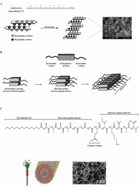
Self-assembling peptides based on naturally-derived amino acids have been proposed as promising structural elements for the development of novel hydrogels for tissue engineering applications [71]. Inspired by the yeast protein zuotin [73], Zhang’s group synthesized a number of ionic self-complementary peptides, including EAK16 [74, 75], RADA16 [76, 77], FEK16 [78] and KLD-12 [79]. These are composed by alternating charged hydrophilic and hydrophobic natural amino acids and have an intrinsic ability to self-assemble into highly ordered β-sheet nanofibrous scaffolds (Fig. 2A). The self-assembly process can be further accelerated by changes in pH or in salt solution concentration [74-76]. Ultimately, the non-covalent nature of the established interactions, in combination with the dependence on external stimulus (e.g. pH, temperature and ionic strength), allows the self-assembly process to be reversible and, as a result, the development of “smart” hydrogels [77]. The stimulus-responsiveness of the developed hydrogels allows a more tight control over the self-assembling process, which can be of interest for obtaining an in situ forming gel for cells and/or biomolecules delivery. These ionic self-complementary peptides have been successfully applied in a number of tissue engineering applications, namely as 3D matrices for in vitro cell culture [76, 79-82] and as vehicles for cell transplantation [80] and/or endogenous cell recruitment [83]. Peptides based on RADA16 self-assembling motif are the most frequently employed due to the ease with which it is synthesized and modified for specific applications. RADA16-based hydrogels are able of effectively support the differentiation of adult rat liver progenitor cells into functional hepatocyte-like spheroid clusters [81], PC-12 neuronal-hepatocyte-like cell viability, neurite outgrowth and synapse formation [80], and hippocampal neuronal cell culture [82].
To afford peptide-based self-assembled hydrogels with additional biological activity, a number of specific bioactive sequences were incorporated. For instance, RADA16-based hydrogels incorporating cell adhesion motifs from collagen VI (RGDS), as well as BMHP- 1 and -2, have been developed and their in vitro biological performance evaluated using mouse adult NSCs [66]. As compared with non-functionalized matrices, the bioactive hydrogels showed improved ability to support NSC survival, proliferation and differentiation. [66]. Similar hydrogels comprising bioactive sequences known to enhance osteoblast behavior, such as the osteogenic growth peptide ALK, osteopontin cell-adhesion motif (DGR) and 2-unit RGD motifs (PGR), were also designed, and shown to support mouse pre-osteoblastic MC3T3-E1 cells adhesion, proliferation and migration, as well as osteogenic differentiation [68].
β-sheet self-assembly was also explored in the development of triblock peptides, comprising an amphiphilic triblock peptide motif (Fig. 2B). The central B block comprises alternating hydrophilic (glutamine) and hydrophobic (leucine) residues ((QL)n=1-6 repeat), whose interactions lead to the
formation of peptide dimmers [84]. These in turn, form β-sheet hydrogen bonds with each other, which keep them aligned perpendicularly to the peptide backbone axis, allowing the formation of highly-ordered nanofibrous matrices (Fig. 2B). On the other hand, the A block is composed by two to four positively charged lysine residues which, due to electrostatic repulsions, will affect the conditions required for peptide self-assembly at physiological conditions. Therefore, a balance of repulsive and attractive forces must be attained to control the self-assembly of triblock peptides into well-defined nanostructures and the properties of the resultant hydrogels [84]. The modular nature of amphiphilic triblock peptides can be harnessed to favor a more tunable control over hydrogel mechanical properties. For instance, the careful selection of the amino acid residues composing A and B blocks significantly influences the gelation conditions, the length and diameter of the formed nanofibers, and the viscoelastic properties of the resultant hydrogel [85]. To improve the bioactivity of developed hydrogels, an MMP-2 specific cleavage site and an RGD cell adhesion motif were incorporated in the central B block and in the A block, respectively [69]. The combination of these two bioactive motifs resulted in increased viability, spreading and migration of MSCs from human exfoliated deciduous teeth within the hydrogel when compared with the non-functionalized ones. An alternative design based on β-sheet motifs was developed, in which an amphiphilic β-hairpin secondary structure was used as self-assembling motif [8, 86]. Here, peptide sequences fold in response to different external stimulus, including pH [87], temperature [88] and ionic strength [89]. The formed hydrogels present a controllable shear-thin recovery kinetic, due to the weak and transient nature of the interactions mediating the self-assemble process [90]. This allows the encapsulation and subsequent delivery of cells to the injured sites, by minimally invasive procedures. β-hairpin-based hydrogels were shown to be cytocompatible, being able to mediate the adhesion, migration and proliferation of NIH3T3 murine fibroblasts (seeded on their surface), even in the absence of serum proteins or cell-adhesive motifs [91]. Through the incorporation of sequences sensitive to MMP-13, the degradation of such hydrogels can be efficiently controlled [70].
Self-assembling peptide amphiphiles
Peptide amphiphiles (PAs) are composed by a peptide sequence with a covalently linked chemical tail (e.g. short hydrophobic sequences or aromatic moieties) with intrinsic ability to self-assemble. These molecules can combine, thus, the structural features of amphiphilic molecules with the chemical functionality/bioactivity of small peptide sequences [92-94]. In the last decade, pioneering
work of Stupp and co-workers resulted in the proposal of a broad range of PAs comprising a hydrophilic sequence covalently linked to an alkyl chain [94]. These molecules are typically composed by four distinct domains (Fig. 2C): (1) short hydrophobic tail responsible to drive the self-assembly process; (2) β-sheet peptide domain, which, through the formation of hydrogen bonds, promotes the cohesion of the formed nanofibers; (3) charged residues to confer water solubility; and (4) bioactive domain to mediate a specific biological response. Their self-assembly into long and stable nanofibers (Fig. 2C) is driven by the hydrophobic tail and occurs under specific pH, temperature or ionic strength conditions [95]. The resulting structures further undergo a sol-to-gel transition under physiological conditions in the presence of multivalent ions, which allows injection and in situ gel formation [64]. As result of the self-assembly process, the peptide sequence will be orientated towards the aqueous environment. This, in combination with the versatility and modular nature of PAs, allows the easy incorporation of bioactive sequences at the extremity of the peptide domain (Fig. 2C) and the development of bioactive hydrogels. As an example, PAs functionalized with the IKVAV epitope from laminin, revealed higher ability to promote NSC adhesion, migration and differentiation into a neuronal phenotype, as well as neurite outgrowth, when compared to laminin-coated surfaces [67]. These promising outcomes were associated to the high density of IKVAV epitopes present on the nanofibers surface [67]. Therefore, both the epitope density and their presentation on the nanofiber surface were found to contribute to such a pronounced biological response. Also in the context of nerve regeneration, a hydrogel based on a PA engineered with a heparin sulfate mimetic and an IKVAV epitope was developed [96]. The two bioactive moieties have demonstrated a cooperative effect in the promotion of PC12 neuronal-like cells neurite outgrowth, even when in presence of inhibitory components, such as chondrotin sulfate proteoglycans [96]. For application in bone regenerative therapies, a hydrogel was engineered with BMP-receptor binding peptides, termed osteopromotive domains [97]. The developed hydrogels showed ability to promote cell survival and osteoblastic differentiation of human bone marrow stromal cells (BMSCs) [97].
Aromatic PAs comprising short (e.g. di- or tri-) peptide sequences capped, at the N-terminal, with a synthetic aromatic moiety have been also explored for the development of self-assembled hydrogels. These PAs have the intrinsic ability to self-assemble and drive the formation of highly-ordered amphiphilic hydrogels, by a combination of hydrophobic and π-π interactions [98]. A number of PAs comprising the fluoren-9-ylmethyloxycarbonyl (Fmoc) aromatic stacking ligand were developed [99-101]. The simplicity of the aromatic moieties (only 2-3 amino acids in length) allows the easy modification of the peptide sequence and a more tunable control over the structure and mechanical properties of the resultant hydrogels [99-101]. The structure of the self-assembled PAs, resembles in several aspects the native ECM, namely in their hydrated nature, nanofibrous architecture and rigidity (G’~10 kPa) [100-102]. To endow these hydrogels with bioactivity, a modular design strategy combining an Fmoc-RGD peptide with the Fmoc-diphenylalanine (Fmoc-FF) sequence was explored [65]. The developed hydrogels showed ability to support the adhesion, migration and proliferation of human adult dermal fibroblasts, in an integrin-dependent manner. By changing the ratio in which the two components are mixed, a more tunable control over hydrogel mechanical and biological properties may be achieved. Alternatively, some authors explored the use of naphthalene (Nap), rather than the Fmoc moiety, as aromatic capping group to synthesize the Nap-FFGEY peptide. The tyrosine residue (Y) present in the resultant aromatic PA could be easily phosphorylated by the action of a kinase, leading to the formation of a molecule with increased hydrophilicity, not capable of self-assemble and form a hydrogel. Interestingly, when injected in a mouse model it was observed that the phosphorylated PA was converted to a gel by the action of naturally occurring phosphatase
enzymes [103, 104]. Therefore, injectable hydrogels based on these PAs can be developed by taking advantage of this enzymatic switch.
DNA-based hydrogels
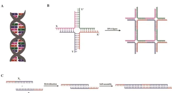
Despite the high costs associated at present with DNA synthesis, this molecule has several advantageous features that make it highly attractive for the development of self-assembled hydrogels [107]. These include its hydrophilic nature, inherent biocompatibility and ability to form stable and flexible secondary structures through specific Watson-Crick base-pairing (Fig. 3A) [107]. Moreover, DNA can be easily tailored either by varying the number and type of nucleic bases or through the incorporation of bioactive motifs, while retaining the ability to self-assemble into highly-ordered nanostructures [18, 107, 108]. Ultimately, one of the most important and unique properties of these molecules is the wide range of molecular biology tools available for their synthesis and modification [109]. In fact, no other molecule simultaneously meets all the aforementioned properties, making DNA a highly attractive and versatile material for the development of self-assembled hydrogels. The unique features, as well as the inherent ability of DNA to self-assemble allow it to be used as structural component of nanoscale materials, thus behaving per se as a biopolymer.
To design complex and stable nanoscale structures based on DNA, Lu and co-workers, taking advantage of the Watson-Crick base-pair rules aforementioned (Fig. 3A), engineered branched DNA molecules with complementary sticky-ends. The self-assembly and the formation of a cross-linked hydrogel network occurs under physiological conditions and in the presence of a DNA ligase (Fig. 3B) [110]. Therefore, the developed matrices can be used to encapsulate live mammalian cells, without any deleterious effect on their viability [110]. To promote a more tunable control over the self-assembly process and over the structure of the resultant hydrogel, the adjustment of the concentration and type of branched DNA monomers [110], as well as the development of stimuli-responsive (e.g. pH [111], temperature [112] or enzymatic activity [112]) hydrogels, can be exploited. Indeed, a number of DNA-based hydrogels with responsive properties that self-assemble rapidly without the need of any chemical cross-linker were developed [111, 112]. The properties and applications of such hydrogels were comprehensively reviewed by Tan and co-workers [108]. More recently, DNA hydrogels formed by self-assembly of linear DNA molecules comprising sticky ends have been developed (Fig. 3C) [113]. These hydrogels are thermoresponsive, allowing a more tight control over the self-assembling process, which can be of interest to obtain an in situ forming gel for cell and biomolecule delivery. To the best of our knowledge, the effect of such hydrogels on cell behavior has not been assessed yet.
Despite the recent advances on the use of DNA as structural component for the development of self-assembled hydrogels, the weak nature of the designed matrices [114], as well as their poor stability against extra- and intra-cellular degradation, mainly mediated by nucleases [115], still constitutes some of the main obstacles to the application of DNA-based hydrogels in the biomedical field. Therefore, in the last few years a number of chemical modifications to native DNA have been explored in an attempt to favor the formation of more stable and resistant hydrogels [115]. Also, to develop mechanically robust hydrogels, DNA are being explored as cross-linking agent, rather than biopolymer, to mediate the assembly of synthetic polymers, forming DNA-hybrid hydrogels [114, 116], which will be addressed in the following section.
Hybrid hydrogels
Hybrid hydrogels combine the advantageous properties of biological (macro)molecules with the tunable and reproducible structural and mechanical features of synthetic polymers [117, 118]. The combination of these two distinct classes of molecules, either through covalent or non-covalent interactions, can result in hydrogels with a higher level of structural organization and improved properties, when compared with their individual components. The biological components are usually molecules adopting basic conformational secondary structures (β-sheets or α-helices), such as peptides, proteins, or DNA/RNA. Their use allows the tunable control over the structural organization and properties of developed hydrogels [117, 118]. The synthetic segments contribute, normally, for the improvement of the mechanical performance of the network. Despite the wide variety of synthetic polymers that can be used in the design of hybrid hydrogels, PEG and poly(N-(2-hydroxypropyl)methacrylamide) (pHPMA) are the ones usually selected, due to their hydrophilicity, biocompatibility and non-immunogenicity. Moreover, the incorporation of these polymers can contribute to increase the stability of the final network in time by delaying its (enzymatic) degradation, which may be highly advantageous when envisaging a clinical translation.
The concept of using biopolymers to guide the self-assembly of synthetic polymeric matrices has been increasingly explored. Indeed, a number of hybrid block copolymers containing biologically-inspired proteins/peptides adopting β-sheet or coiled-coil structures, have been engineered for the development of self-assembled hybrid hydrogels (for a more extensive review see [118]). Despite their conjugation with a synthetic polymer, these coiled-coil and β-sheet forming biopolymers retain their ability to self-assemble and thus can be used to induce the formation of a highly-ordered nanofibrous network. One of the first and well-known examples of hybrid hydrogels formed based on this concept, explores a hybrid diblock copolymer comprising β-sheet forming peptide sequences, derived from the natural occurring β-amyloid protein, conjugated with a PEG polymer [119, 120]. Since then, a number of other β-sheet-forming proteins/peptides have been explored for the design of self-assembled hybrid hydrogels. As an example, block copolymers comprising ELPs ((VPGVG)4
and (VPAVG)4) conjugated with a PEG backbone were used. Here, the self-assembly is driven by a
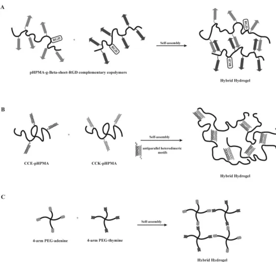
reversible temperature transition, culminating in the formation of an insoluble and supramolecular thermoresponsive coacervate [121, 122]. Further, hybrid hydrogels in which the synthetic polymer self-assembly is guided by the chemical cross-linking of elastin domains modified with lysine residues were also developed [123, 124]. The resulting hydrogels present elastomeric and shear-thinning properties, allowing these to be injected and form a matrix in situ [123, 124]. Moreover, these hydrogels enable a tunable control over the density of elastin cell adhesion domains, as well as of matrix stiffness, which is highly desirable to modulate cell behavior and when studying cell-matrix interactions. Indeed, ELP-PEG hybrid hydrogels have shown to be able of support fibroblasts growth, migration and proliferation [123, 124]. β-sheet forming peptides were also explored as component of hybrid graft copolymers. A pHPMA polymer grafted with complementary β-sheet peptides were developed and endowed with bioactivity through the coupling of RGD cell-adhesion domains to each complementary hybrid graft copolymer (Fig. 4A). These hydrogels have shown ability to support MC3T3-E1 cell viability and proliferation.
The self-assembly properties of coiled-coil forming peptides were also explored to create hybrid hydrogels. Diblock copolymers of PEG and coiled-coil forming peptides that self-assemble into a two-stranded α-helical coiled-coil conformation in aqueous solution, illustrate such strategy [125, 126]. It has been shown that the conjugation of coiled-coil domains with a synthetic polymer favors their
thermal stability, resulting from the formation of a PEG hydrophilic shell around the coiled-coil structure [125, 126]. Further, genetically engineered coiled-coil forming peptides (CC1 and CC2), non-covalently grafted to a pHPMA synthetic backbone have been explored for the development of hybrid graft copolymers [127]. Here, the physical cross-linking and the consequent hybrid hydrogel formation were favored by a temperature-induced conformational change of coiled-coil peptides [127]. The temperature-responsiveness of these hydrogels allows the development of “smart” materials and a more tunable control over the material properties. Another design involving the pHPMA hydrophilic polymer and peptide grafts forming homodimers and higher aggregates were also described [128, 129]. In this case, a minimum of four heptad repeats was found to be needed to induce self-assembly and the formation of homodimers [128, 129]. The number and concentration of the copolymer, as well as temperature proved to have a crucial role on the control of the gelation process [129]. Still, this design is very often limited by the possibility of dimer formation between grafted peptides attached to the same macromolecule. To avoid this and to guarantee the formation of a well-ordered 3D structure, a modified approach was proposed. In this new design, a pHPMA synthetic backbone was grafted with a pair of oppositely charged coiled-coil forming peptides (CCE and CCK) (Fig. 4B) [130, 131]. The oppositely charged peptide grafts self-assemble into antiparallel heterodimeric motifs and induce the formation of a physically cross-linked hydrogel [130, 131]. Here, a reversible self-assembly occurs when the graft copolymers, CCE-P and CCK-P, are mixed equimolarly at neutral pH.
A different approach for the design of hybrid hydrogels consists in using star-shaped macromolecules, such as 4-arm PEG, in combination with biological molecules, responsible for mediating the self-assembly process. As an example, four-arm star-shaped PEG polymers functionalized with either thymine or adenine nucleotides were used to create injectable hydrogels, assembled by base pairing between the two complementary nucleotides (Fig. 4C) [56, 116]. These hydrogels were explored for soft tissue engineering applications, namely for the encapsulation of adipose-derived stem cells. The gelation time (and therefore cell entrapment time) was shown to be dependent on the type of nucleotides used, and the developed hydrogels found to support cell viability and growth [116].
In vivo biological performance of synthetic self-assembled
hydrogels
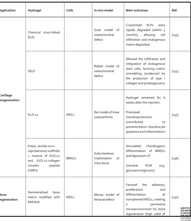
The self-assembled hydrogels discussed so far in this review were designed envisaging their application as 3D scaffolds for cell transplantation. Proof-of principle of the suitability of these hydrogels in terms of ability to create a permissive microenvironment for cell growth and differentiation, in vitro, has been highlighted. However, several of these systems have already been evaluated in vivo. In this section, an overview of the in vivo biological performance of several of these hydrogels, when explored for cell delivery, is presented in Table 1, namely that of the most recent and promising bioactive self-assembled hydrogels designed for cell transplantation and/or endogenous cell recruitment, in the context of cartilage, bone, nerve and cardiac regeneration.
Translation of synthetic self-assembled hydrogels into the clinic:
progress and future challenges
As discussed in the previous section, the potential of several synthetic self-assembled hydrogels for use in cell transplantation and/or endogenous cell recruitment has been already demonstrated in a
number of pre-clinical in vivo studies involving different repair/regenerative scenarios (Table 1). Although the promising results have fueled the interest of the research community and manufacturers into the use of these materials, just now these begin to enter clinical testing. To the best of our knowledge, only one product based on self-assembled hydrogels is presently on the market - NuCoreTM Injectable Nucleus developed by Spine Wave (Shelton, CT) [156]. This system
consists of a recombinant protein copolymer comprising blocks derived from silk and elastin structural proteins and it was already evaluated in a pilot clinical trial to assess product safety for potential application in the repair and regeneration of invertebral disc [157]. The trial did not involve the transplantation of cells. No complications or adverse effects to the patient have been reported. Moreover, the system seems to slow the degenerative process over time. Despite the promising results obtained in this pilot study, further investigation on product safety and efficacy need to be performed prior its clinical use.
While trying to bring synthetic self-assembled hydrogels from bench-to-bedside, a number of requisites need to be fulfilled and few key issues must be taken into consideration. A wide number of self-assembled systems (proteins, peptides, DNA, synthetic polymers or hybrid structures) have been proposed for the design of cell-compatible hydrogels. Therefore, one of the most important issues to be addressed upon the development of these products and that most possibly significantly contributes to delay their translation, is the selection of the most suitable system for each particular application. The chosen building blocks should be biocompatible, biodegradable and able to mimic the cellular microenvironment of the surrounding tissues, as discussed in previous sections. The characterization of the proposed systems in terms of degradation rate and degradation by-products, as well as of biofunctionality (including inflammatory response) should be carried out in a number of in vitro and in vivo set-ups, before considering clinical translation. Scalability is also an important issue to take into account, since some building blocks, such as proteins and DNA, are difficult to obtain in large scale and in good yields, what has put a strain in costs. Nevertheless, the efforts made in the last few years towards the efficient chemical synthesis of such systems will possibly contribute to enhance their feasibility for biomedical applications. Also, production-related issues, such as purification and sterilization will be paramount to define if these will reach the market.
A challenge while developing injectable in situ forming hydrogels is to gain control over the 3D architecture of the hydrogel fibrillar network. The building blocks of synthetic self-assembled hydrogels typically assemble into randomly oriented networks, and the nanofibrillar structures formed in several peptide hydrogels are also randomly oriented. To better mimic the natural anisotropic structure and functional features of the ECM, control over fibrillar orientation both at the nano- and the micro-scale should be achieved. This is particularly important in hydrogels envisaged for application in highly organized tissues (e.g. nerve and bone), in which the structural and mechanical properties of the ECM fulfill important biomechanical properties and regulate cell behavior. For instance, longitudinally-oriented aligned structures have been shown to induce directional axonal re-growth and alignment of transplanted cells, by providing contact guidance, which can be of most importance in therapies for the treatment of spinal cord and peripheral nerve injuries [158]. Attempts to develop in situ forming hydrogels of aligned self-assembled PA nanofibers were made. This was achieved by exposing isotropic PA solutions to elevated temperatures, under which plaque-like structures are formed, which break apart into aligned bundles of multiple PA nanofibers upon cooling [159]. Upon pipetting the resultant liquid crystalline solution into CaCl2
-containing physiological buffer, string-like gels -containing aligned nanofibers are formed. When modified with IKVAV or RGDS epitopes, aligned PA hydrogels containing neurons or neural
progenitor cells (added to the CaCl2 solution prior hydrogel formation), were able to guide neurite
extension from neurons along the direction of the nanofibers in vitro, and to promote the growth of oriented processes from transplanted neural progenitor cells within the spinal cord, in vivo [160]. Control over the architecture of in situ forming hydrogels is therefore worthy to explore, and likely to be most rewarding.
The complexity and highly dynamic nature of biological systems constitutes another major challenge in the design and application of cell-compatible hydrogels. Hydrogels should be engineered not only to serve as vehicles for cell and/or biomolecule delivery, but also to guide cell behavior and fate. Therefore, hydrogels able to change their biological and/or physical properties along the different stages of cell engraftment and tissue regeneration are much awaited. In this context, the exploration of molecular self-assembly constitutes a highly promising strategy, once by being mediated by multiple weak and transitory interactions the supramolecular hydrogel structure can be easily reconfigured.
Another advantage of these synthetic hydrogels is that they can be combined with a number of bioactive molecules, including ECM proteins, growth factors or drugs that can additionally or synergistically modulate cell behavior and fate. Unsolved is the spatial and temporal control over the availability of these cues. These would allow the mirroring of those observed in the native ECM in the context of repair/regenerative processes in which gradients of biochemical and physical cues are key players of the process.
In conclusion, the development of biomimetic synthetic self-assembled hydrogels is rapidly progressing, supported in part by technology advances in the field of recombinant proteins, solid-phase synthesis and polymer chemistry. In particular, with hybrid hydrogels one can combine the advantageous properties of biological (macro)molecules with the tunable and reproducible structural and mechanical features of synthetic polymers, conferring superior properties to the resulting systems. However, when envisaging the use of such hydrogels for cellular therapies, mimicking the complexity and functionality of the natural ECM without compromising important features for application in a clinical setting, like injectability, still remains a major challenge.
Acknowledgments
The authors would like to acknowledge the FEDER funds through the Programa Operacional Factores de Competitividade – COMPETE and the Portuguese funds through FCT – Fundação para a Ciência e a Tecnologia (HMSP-ICT/0020/2010, PTDC/SAU-BMA/118869/2010 and PEst/SAU/LA0002/2013) that supported this work. D Barros is supported by FCT (SFRH/BI/52379/2013) and I.F. Amaral by QREN through program ON.2, in the framework of "Project on Biomedical Engineering for Regenerative Therapies and Cancer” (NORTE-07-0124-FEDER-000005).
References
[1] M. Guvendiren, J.A. Burdick, Engineering synthetic hydrogel microenvironments to instruct stem cells, Curr. Opin. Biotechnol. 24(5) (2013) 841-6.
[2] C. Wang, R.R. Varshney, D.A. Wang, Therapeutic cell delivery and fate control in hydrogels and hydrogel hybrids, Adv. Drug Deliv. Rev. 62(7-8) (2010) 699-710.
[3] O. Lindvall, Z. Kokaia, Stem cells for the treatment of neurological disorders, Nature 441(7097) (2006) 1094-6.
[4] M.P. Lutolf, P.M. Gilbert, H.M. Blau, Designing materials to direct stem-cell fate, Nature 462(7272) (2009) 433-41.
[5] M.W. Tibbitt, K.S. Anseth, Hydrogels as Extracellular Matrix Mimics for 3D Cell Culture, Biotechnol. Bioeng. 103(4) (2009) 655-663.
[6] H. Geckil, F. Xu, X. Zhang, S. Moon, U. Demirci, Engineering hydrogels as extracellular matrix mimics, Nanomedicine 5(3) (2010) 469-84.
[7] K.B. Fonseca, P.L. Granja, C.C. Barrias, Engineering proteolytically-degradable artificial extracellular matrices, Prog. Polym. Sci. 39(12) (2014) 2010-2029.
[8] P.M. Kharkar, K.L. Kiick, A.M. Kloxin, Designing degradable hydrogels for orthogonal control of cell microenvironments, Chem. Soc. Rev. 42(17) (2013) 7335-72.
[9] C.M. Lo, H.B. Wang, M. Dembo, Y.L. Wang, Cell movement is guided by the rigidity of the substrate, Biophys. J. 79(1) (2000) 144-52.
[10] A.J. Engler, S. Sen, H.L. Sweeney, D.E. Discher, Matrix elasticity directs stem cell lineage specification, Cell 126(4) (2006) 677-89.
[11] D.E. Ingber, Cellular mechanotransduction: putting all the pieces together again, FASEB J. 20(7) (2006) 811-27.
[12] R. Jin, In-situ Forming Biomimetic Hydrogels for Tissue Regeneration, in: C. Lin (Ed.), Biomedicine, InTech2012.
[13] J.-A. Yang, J. Yeom, B.W. Hwang, A.S. Hoffman, S.K. Hahn, In situ-forming injectable hydrogels for regenerative medicine, Prog. Polym. Sci. 39(12) (2014) 1973-1986.
[14] F. Wang, Z.Q. Li, M. Khan, K. Tamama, P. Kuppusamy, W.R. Wagner, C.K. Sen, J.J. Guan, Injectable, rapid gelling and highly flexible hydrogel composites as growth factor and cell carriers, Acta Biomater. 6(6) (2010) 1978-1991.
[15] G.A. Saracino, D. Cigognini, D. Silva, A. Caprini, F. Gelain, Nanomaterials design and tests for neural tissue engineering, Chem. Soc. Rev. 42(1) (2013) 225-62.
[16] J. Zhu, R.E. Marchant, Design properties of hydrogel tissue-engineering scaffolds, Expert Rev. Med. Devices 8(5) (2011) 607-26.
[17] E.S. Place, J.H. George, C.K. Williams, M.M. Stevens, Synthetic polymer scaffolds for tissue engineering, Chem. Soc. Rev. 38(4) (2009) 1139-51.
[18] N. Stephanopoulos, J.H. Ortony, S.I. Stupp, Self-Assembly for the Synthesis of Functional Biomaterials, Acta Mater. 61(3) (2013) 912-930.
[19] T. Lu, Y. Li, T. Chen, Techniques for fabrication and construction of three-dimensional scaffolds for tissue engineering, Int. J. Nanomedicine 8 (2013) 337-50.
[20] H. Cao, T. Liu, S.Y. Chew, The application of nanofibrous scaffolds in neural tissue engineering, Adv. Drug Deliv. Rev. 61(12) (2009) 1055-64.
[21] M. Nune, P. Kumaraswamy, U.M. Krishnan, S. Sethuraman, Self-assembling peptide nanofibrous scaffolds for tissue engineering: novel approaches and strategies for effective functional regeneration, Curr. Protein Pept. Sci. 14(1) (2013) 70-84.
[22] G.M. Whitesides, B. Grzybowski, Self-assembly at all scales, Science 295(5564) (2002) 2418-2421.
[23] S.G. Zhang, Fabrication of novel biomaterials through molecular self-assembly, Nature Biotechnol. 21(10) (2003) 1171-1178.
[24] R.J. Mart, R.D. Osborne, M.M. Stevens, R.V. Ulijn, Peptide-based stimuli-responsive biomaterials, Soft Matter 2(10) (2006) 822-835.
[25] R. Silva, B. Fabry, A.R. Boccaccini, Fibrous protein-based hydrogels for cell encapsulation, Biomaterials 35(25) (2014) 6727-38.
[26] A.M. Jonker, D.W.P.M. Lowik, J.C.M. van Hest, Peptide- and Protein-Based Hydrogels, Chem. Mater. 24(5) (2012) 759-773.
[27] S. Banta, I.R. Wheeldon, M. Blenner, Protein engineering in the development of functional hydrogels, Annu. Rev. Biomed. Eng. 12 (2010) 167-86.
[28] T. Kowalczyk, K. Hnatuszko-Konka, A. Gerszberg, A.K. Kononowicz, Elastin-like polypeptides as a promising family of genetically-engineered protein based polymers, World J. Microb. Biot. 30(8) (2014) 2141-2152.
[29] W.F. Daamen, J.H. Veerkamp, J.C.M. van Hest, T.H. van Kuppevelt, Elastin as a biomaterial for tissue engineering, Biomaterials 28(30) (2007) 4378-4398.
[30] D.W. Urry, Physical chemistry of biological free energy transduction as demonstrated by elastic protein-based polymers, J. Phys. Chem. B 101(51) (1997) 11007-11028.
[31] D.E. Meyer, A. Chilkoti, Quantification of the effects of chain length and concentration on the thermal behavior of elastin-like polypeptides, Biomacromolecules 5(3) (2004) 846-51.
[32] H. Betre, S.R. Ong, F. Guilak, A. Chilkoti, B. Fermor, L.A. Setton, Chondrocytic differentiation of human adipose-derived adult stem cells in elastin-like polypeptide, Biomaterials 27(1) (2006) 91-9. [33] H. Betre, L.A. Setton, D.E. Meyer, A. Chilkoti, Characterization of a genetically engineered elastin-like polypeptide for cartilaginous tissue repair, Biomacromolecules 3(5) (2002) 910-6. [34] S.R. MacEwan, A. Chilkoti, Elastin-like polypeptides: biomedical applications of tunable biopolymers, Biopolymers 94(1) (2010) 60-77.
[35] A. Chilkoti, T. Christensen, J.A. MacKay, Stimulus responsive elastin biopolymers: Applications in medicine and biotechnology, Curr. Opin. Chem. Biol. 10(6) (2006) 652-7.
[36] J. Lee, C.W. Macosko, D.W. Urry, Elastomeric polypentapeptides cross-linked into matrixes and fibers, Biomacromolecules 2(1) (2001) 170-179.
[37] D.W. Lim, D.L. Nettles, L.A. Setton, A. Chilkoti, Rapid cross-linking of elastin-like polypeptides with (hydroxymethyl)phosphines in aqueous solution, Biomacromolecules 8(5) (2007) 1463-70. [38] K. Trabbic-Carlson, L.A. Setton, A. Chilkoti, Swelling and mechanical behaviors of chemically cross-linked hydrogels of elastin-like polypeptides, Biomacromolecules 4(3) (2003) 572-80.
[39] D.W. Lim, D.L. Nettles, L.A. Setton, A. Chilkoti, In situ cross-linking of elastin-like polypeptide block copolymers for tissue repair, Biomacromolecules 9(1) (2008) 222-30.
[40] K.S. Straley, S.C. Heilshorn, Independent tuning of multiple biomaterial properties using protein engineering, Soft Matter 5(1) (2009) 114-124.
[41] K.J. Lampe, A.L. Antaris, S.C. Heilshorn, Design of three-dimensional engineered protein hydrogels for tailored control of neurite growth, Acta Biomater. 9(3) (2013) 5590-9.
[42] Z. Megeed, J. Cappello, H. Ghandehari, Genetically engineered silk-elastinlike protein polymers for controlled drug delivery, Adv. Drug Deliv. Rev. 54(8) (2002) 1075-91.
[43] A. Nagarsekar, J. Crissman, M. Crissman, F. Ferrari, J. Cappello, H. Ghandehari, Genetic synthesis and characterization of pH- and temperature-sensitive silk-elastinlike protein block copolymers, J. Biomed. Mater. Res. 62(2) (2002) 195-203.
[44] A. Nagarsekar, J. Crissman, M. Crissman, F. Ferrari, J. Cappello, H. Ghandehari, Genetic engineering of stimuli-sensitive silkelastin-like protein block copolymers, Biomacromolecules 4(3) (2003) 602-607.
[45] X.X. Xia, Q. Xu, X. Hu, G. Qin, D.L. Kaplan, Tunable self-assembly of genetically engineered silk--elastin-like protein polymers, Biomacromolecules 12(11) (2011) 3844-50.
[46] J.M. Mason, K.M. Arndt, Coiled coil domains: stability, specificity, and biological implications, Chembiochem : a European journal of chemical biology 5(2) (2004) 170-6.
[47] W.A. Petka, J.L. Harden, K.P. McGrath, D. Wirtz, D.A. Tirrell, Reversible hydrogels from self-assembling artificial proteins, Science 281(5375) (1998) 389-92.
[48] C. Xu, V. Breedveld, J. Kopecek, Reversible hydrogels from self-assembling genetically engineered protein block copolymers, Biomacromolecules 6(3) (2005) 1739-49.
[49] B.D. Olsen, J.A. Kornfield, D.A. Tirrell, Yielding Behavior in Injectable Hydrogels from Telechelic Proteins, Macromolecules 43(21) (2010) 9094-9099.
[50] W. Shen, K.C. Zhang, J.A. Kornfield, D.A. Tirrell, Tuning the erosion rate of artificial protein hydrogels through control of network topology, Nature Mater. 5(2) (2006) 153-158.
[51] L. Mi, S. Fischer, B. Chung, S. Sundelacruz, J.L. Harden, Self-assembling protein hydrogels with modular integrin binding domains, Biomacromolecules 7(1) (2006) 38-47.
[52] S.E. Fischer, X. Liu, H.Q. Mao, J.L. Harden, Controlling cell adhesion to surfaces via associating bioactive triblock proteins, Biomaterials 28(22) (2007) 3325-37.
[53] S.E. Fischer, L. Mi, H.Q. Mao, J.L. Harden, Biofunctional coatings via targeted covalent cross-linking of associating triblock proteins, Biomacromolecules 10(9) (2009) 2408-17.
[54] T.J. Deming, Polypeptide hydrogels via a unique assembly mechanism, Soft Matter 1(1) (2005) 28-35.
[55] A.P. Nowak, V. Breedveld, L. Pakstis, B. Ozbas, D.J. Pine, D. Pochan, T.J. Deming, Rapidly recovering hydrogel scaffolds from self-assembling diblock copolypeptide amphiphiles, Nature 417(6887) (2002) 424-8.
[56] C.Y. Yang, B. Song, Y. Ao, A.P. Nowak, R.B. Abelowitz, R.A. Korsak, L.A. Havton, T.J. Deming, M.V. Sofroniew, Biocompatibility of amphiphilic diblock copolypeptide hydrogels in the central nervous system, Biomaterials 30(15) (2009) 2881-98.
[57] A.P. Nowak, J. Sato, V. Breedveld, T.J. Deming, Hydrogel formation in amphiphilic triblock copolypeptides, Supramol. Chem. 18(5) (2006) 423-427.
[58] Z.B. Li, T.J. Deming, Tunable hydrogel morphology via self-assembly of amphiphilic pentablock copolypeptides, Soft Matter 6(11) (2010) 2546-2551.
[59] C.T. Wong Po Foo, J.S. Lee, W. Mulyasasmita, A. Parisi-Amon, S.C. Heilshorn, Two-component protein-engineered physical hydrogels for cell encapsulation, P Natl Acad Sci USA 106(52) (2009) 22067-72.
[60] W. Mulyasasmita, J.S. Lee, S.C. Heilshorn, Molecular-level engineering of protein physical hydrogels for predictive sol-gel phase behavior, Biomacromolecules 12(10) (2011) 3406-11.
[61] S. Wong, M.S. Shim, Y.J. Kwon, Synthetically designed peptide-based biomaterials with stimuli-responsive and membrane-active properties for biomedical applications, J. Mater. Chem. B 2(6) (2014) 595-615.
[62] L. Cai, C.B. Dinh, S.C. Heilshorn, One-pot Synthesis of Elastin-like Polypeptide Hydrogels with Grafted VEGF-Mimetic Peptides, Biomater. Sci. 2(5) (2014) 757-765.
[63] R. Fairman, K.S. Akerfeldt, Peptides as novel smart materials, Curr. Opin. Struc. Biol. 15(4) (2005) 453-463.
[64] E. Beniash, J.D. Hartgerink, H. Storrie, J.C. Stendahl, S.I. Stupp, Self-assembling peptide amphiphile nanofiber matrices for cell entrapment, Acta Biomater. 1(4) (2005) 387-97.
[65] M. Zhou, A.M. Smith, A.K. Das, N.W. Hodson, R.F. Collins, R.V. Ulijn, J.E. Gough, Self-assembled peptide-based hydrogels as scaffolds for anchorage-dependent cells, Biomaterials 30(13) (2009) 2523-2530.
[66] C. Cunha, S. Panseri, O. Villa, D. Silva, F. Gelain, 3D culture of adult mouse neural stem cells within functionalized self-assembling peptide scaffolds, Int. J. Nanomedicine 6 (2011) 943-55. [67] G.A. Silva, C. Czeisler, K.L. Niece, E. Beniash, D.A. Harrington, J.A. Kessler, S.I. Stupp, Selective differentiation of neural progenitor cells by high-epitope density nanofibers, Science 303(5662) (2004) 1352-5.
[68] A. Horii, X. Wang, F. Gelain, S. Zhang, Biological designer self-assembling peptide nanofiber scaffolds significantly enhance osteoblast proliferation, differentiation and 3-D migration, PloS one 2(2) (2007) e190.
[69] K.M. Galler, L. Aulisa, K.R. Regan, R.N. D'Souza, J.D. Hartgerink, Self-assembling multidomain peptide hydrogels: designed susceptibility to enzymatic cleavage allows enhanced cell migration and spreading, J. Am. Chem. Soc. 132(9) (2010) 3217-23.
[70] M.C. Giano, D.J. Pochan, J.P. Schneider, Controlled biodegradation of self-assembling beta-hairpin peptide hydrogels by proteolysis with matrix metalloproteinase-13, Biomaterials 32(27) (2011) 6471-7.
[71] S.G. Zhang, Emerging biological materials through molecular self-assembly, Biotechnol. Adv. 20(5-6) (2002) 321-339.
[72] S.G. Zhang, D.M. Marini, W. Hwang, S. Santoso, Design of nanostructured biological materials through self-assembly of peptides and proteins, Curr. Opin. Chem. Biol. 6(6) (2002) 865-871. [73] S. Zhang, C. Lockshin, A. Herbert, E. Winter, A. Rich, Zuotin, a putative Z-DNA binding protein in Saccharomyces cerevisiae, EMBO J. 11(10) (1992) 3787-96.
[74] S. Zhang, T. Holmes, C. Lockshin, A. Rich, Spontaneous assembly of a self-complementary oligopeptide to form a stable macroscopic membrane, P Natl Acad Sci USA 90(8) (1993) 3334-8.
[75] S. Zhang, C. Lockshin, R. Cook, A. Rich, Unusually stable beta-sheet formation in an ionic self-complementary oligopeptide, Biopolymers 34(5) (1994) 663-72.
[76] S. Zhang, T.C. Holmes, C.M. DiPersio, R.O. Hynes, X. Su, A. Rich, Self-complementary oligopeptide matrices support mammalian cell attachment, Biomaterials 16(18) (1995) 1385-93. [77] H. Yokoi, T. Kinoshita, S. Zhang, Dynamic reassembly of peptide RADA16 nanofiber scaffold, P Natl Acad Sci USA 102(24) (2005) 8414-9.
[78] J.H. Collier, B.H. Hu, J.W. Ruberti, J. Zhang, P. Shum, D.H. Thompson, P.B. Messersmith, Thermally and photochemically triggered self-assembly of peptide hydrogels, J. Am. Chem. Soc. 123(38) (2001) 9463-4.
[79] J. Kisiday, M. Jin, B. Kurz, H. Hung, C. Semino, S. Zhang, A.J. Grodzinsky, Self-assembling peptide hydrogel fosters chondrocyte extracellular matrix production and cell division: implications for cartilage tissue repair, P Natl Acad Sci USA 99(15) (2002) 9996-10001.
[80] T.C. Holmes, S. de Lacalle, X. Su, G. Liu, A. Rich, S. Zhang, Extensive neurite outgrowth and active synapse formation on self-assembling peptide scaffolds, P Natl Acad Sci USA 97(12) (2000) 6728-33.
[81] C.E. Semino, J.R. Merok, G.G. Crane, G. Panagiotakos, S. Zhang, Functional differentiation of hepatocyte-like spheroid structures from putative liver progenitor cells in three-dimensional peptide scaffolds, Differentiation; research in biological diversity 71(4-5) (2003) 262-70.
[82] C.E. Semino, J. Kasahara, Y. Hayashi, S. Zhang, Entrapment of migrating hippocampal neural cells in three-dimensional peptide nanofiber scaffold, Tissue Eng. 10(3-4) (2004) 643-55.
[83] D.A. Narmoneva, R. Vukmirovic, M.E. Davis, R.D. Kamm, R.T. Lee, Endothelial cells promote cardiac myocyte survival and spatial reorganization: implications for cardiac regeneration, Circulation 110(8) (2004) 962-8.
[84] H. Dong, S.E. Paramonov, L. Aulisa, E.L. Bakota, J.D. Hartgerink, Self-assembly of multidomain peptides: balancing molecular frustration controls conformation and nanostructure, J. Am. Chem. Soc. 129(41) (2007) 12468-72.
[85] L. Aulisa, H. Dong, J.D. Hartgerink, Self-assembly of multidomain peptides: sequence variation allows control over cross-linking and viscoelasticity, Biomacromolecules 10(9) (2009) 2694-8. [86] D. Pantoja-Uceda, C.M. Santiveri, M.A. Jimenez, De novo design of monomeric beta-hairpin and beta-sheet peptides, Methods Mol. Biol. 340 (2006) 27-51.
[87] J.P. Schneider, D.J. Pochan, B. Ozbas, K. Rajagopal, L. Pakstis, J. Kretsinger, Responsive hydrogels from the intramolecular folding and self-assembly of a designed peptide, J. Am. Chem. Soc. 124(50) (2002) 15030-7.