1. Clinica Medica, Reumatologia, Faculdade de Medicina da Universidade de São Paulo
Serum hyaluronic acid in polymyositis:
high serum levels tend to correlate with disease activity
ACTA REUMATOL PORT. 2014;39:248-253
AbstrAct
Objective: Polymyositis (PM) is a rare systemic
idio-pathic inflammatory myopathy. Hyaluronic acid (HA) is closely linked to inflammatory cellular reactions and disease activity. Increased serum levels of HA have been reported in several inflammatory diseases, but curren-tly, there are no studies analysing the HA in PM. Thus, clinical association of HA with PM in patients was de-termined in the present study.
Methods: The present cross-sectional study was
per-formed at one centre from 2012 to 2013 and included 35 consecutive adult patients with PM (Bohan and Pe-ter criPe-teria, 1975) and 38 adult healthy volunteers. The serum HA was assessed with anti-HA antibody, using the specific ELISA/EIA kits according to the manufa -cturer’s protocol.
Results: The average age, distribution of females and
ethnicity were comparable in patients with PM and the control group. Regarding disease status, patients with PM had a median patient visual analogue score (VAS) of 2 [0-6], physician VAS of 1 [0-3], MMT-8 of 74 [68--80] and HAQ of 0.48 [0.00-1.14]. The serum levels of HA were also significantly increased in patients with PM (390±412ng/mL) compared to healthy subjects (129±119ng/mL), p=0.001. In an additional analysis, the serum levels of HA did not correlate with PM de-mographic data (gender and ethnicity), current organ involvement or autoantibodies and were not been in-fluenced by the use of prednisolone and/or immuno-suppressives by the PM patients. However, there was a positive correlation between serum levels of HA and VAS (patient and physician), and a negative correlation between serum levels of HA and MMT-8.
Conclusion: High serum levels of HA were observed
in patients with PM and tended to correlate with PM di-sease activity. Additional studies are needed to assess
Silva MB1, Silva MG1, Shinjo SK1
this correlation, as well as to understand the mecha-nism involved in the pathogenesis of PM by HA.
Keywords: Disease activity; Hyaluronic acid; Idiopa
-thic inflammatory myopathies; Polymyositis.
IntroductIon
Polymyositis (PM) is a rare idiopathic systemic inflam-matory myopathy. The disease is characterised by pro-gressive symmetrical muscle weakness with high mor-bidity and functional disability1-3.
The annual incidence of PM is 2.2 to 7.7 cases per million individuals, affecting predominantly females (2 women: 1 man). The disease primarily affects indivi-duals between the ages of 45 and 55 years, although the average age of diagnosis occurs at approximately 40 years of age and the disease can affect patients of any age1-3.
Hyaluronic acid (HA) is a glycosaminoglycan com-prising the extracellular connective tissue matrix. It is distributed in various tissues, such as synovial fluid, vi-treous humour of the eye, the umbilical cord, loose con-nective tissue and cartilage. It is produced mainly by fi-broblasts, transported by the lymph and is rapidly me-tabolised by the liver4. Moreover, there is evidence of
the role of HA in the regulation of the immune response by stimulating the expression of inflammatory genes in various immune cells at the site of injury, and in regu-lating the inflammatory response through cell recruit-ment, cytokine release and cell migration5. In addition,
HA stimulates the release of inflammatory factors, such as TNF-a and IL-1b, and cytokines produced by fi-broblasts, which assists the inflammatory response6.
Increased serum levels of HA have been reported in several diseases, including liver disease, malignant diseases and different systemic autoimmune di-seases7,8-11. For instance, in rheumatoid arthritis, HA
course of arthritis because of its inflammatory proper-ties8. High levels of HA were also observed in patients
with systemic sclerosis, being correlated with cuta-neous manifestations, mainly in the early stages of the disease9. In systemic lupus erythematosus, increased
levels of HA are associated with discoid cutaneous ma-nifestations, which may contribute to drug resistance and disease severity7. In relation to dermatomyositis,
there have been reports of patients with a positive cor-relation between the disease and serum HA activity4,10.
In psoriatic arthritis, the increased serum levels of HA are highly positively correlated with skin involvement
when compared to the normal population11.
However, currently there are no studies analysing the role of HA in PM. Similar to other systemic in-flammatory diseases, HA could be involved in PM. Thus, any clinical association of HA in this population was investigated in the present study.
MAterIAls And pAtIents
The present cross-sectional study was performed at one centre and included 35 patients over 18 years of age, diagnosed with PM (fulfilling the Bohan and Pe-ter criPe-teria excluding the cutaneous criPe-teria12) and
fol-lowed at an inflammatory myopathies unit during the period of January 2012 to July 2013.
Individuals with cancer-associated myositis, other systemic-associated autoimmune disease, or acute or chronic infections (viral, bacterial or fungal) were ex-cluded.
As a control group, 38 adult healthy volunteers were included in the study.
The study was approved by the local Ethics Com-mittee, and informed consent was obtained from all individuals.
All patients submitted to a standardised interview to collect the following information. Moreover, any mis-sing data were obtained umis-sing an ongoing electronic database protocol carried out for all patients at 1- to 3--month intervals. Protocols consisted of an extensive clinical and laboratory evaluation, including those re-levant to this study:
a) Demographic data: the current age of the patient, gender and ethnicity;
b) Clinical data: the time from first symptoms to di-sease diagnosis, the time since the didi-sease diagno-sis, current organ involvement (heart, lung and ar-ticulation), and treatment (corticosteroid and/or
im-munosuppressives). In addition, questionnaires were used to assess the current status of disease ac-tivity: (A) Manual Muscle Testing - MMT-813,14; (B)
physician visual analogue scale (VAS)15,16; (C) patient
visual analogue scale (VAS)15,16; (D) quality-of-life
questionnaire: Health Assessment Questionnaire (HAQ)15,17,18and (E) laboratory data: creatine
phos-phokinase (reference value: 24-173 U/L), aldolase (1.0-7.5 U/L); aspartate aminotransferase (< 31 U/L), alanine aminotransferase (< 31 U/L), lactate dehy-drogenase (240 - 480 U/L), C-reactive protein (< 5 mg/L) by nephelometry, and erythrocyte sedimentation rate (< 19 mm / 1st hour) by Westegreen me -thod; and
c) Hyaluronic acid: The sample blood sera is collected and centrifuged immediately at 3000 rpm, for 15 minutes, at 4 ºC and stored at - 70 ºC. From these samples, the anti-HA antibody was analyzed with specific kits of ELISA / EIA (HA Sandwich ELISA, Echelon Biosciences Inc, Salt Lake City), according to the manufacturer’s protocol using 25 L samples and standard curves to determine the concentration values.
Statistical analysis. The Kolmogorov-Smirnov test
was used to evaluate the distribution of each parame-ter. The demographic and clinical features were ex-pressed as the mean ± standard deviation (SD) or me-dian [25th - 75th percentile] for continuous variables or as percentages for categorical variables. Means were compared using Student’s t-test; medians, with Mann--Whitney U-test. Categorical data were compared using chi-square test or Fisher’s exact test. The correlation between HA serum level and continuous variables used Spearman’s correlations. Values of p < 0.05 were con-sidered significant. All of the analyses were performed with the SPSS 15.0 statistics software (Chicago, IL, USA).
results
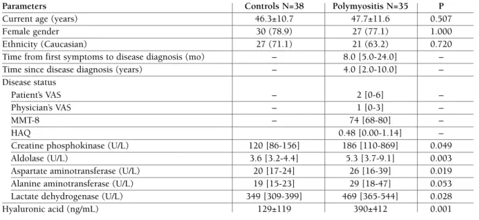
The demographic, clinical and laboratory characteris-tics of 35 patients with PM and 38 healthy individuals are shown in Table I. The average age, the percentage of females and ethnicity were comparable in both groups. The median time from first symptoms to di-sease diagnosis was 8 months [5.0-24.0] and the me-dian time since disease diagnosis was 4 years [2.0--10.0].
me-tAble I. deMogrAphIc, dIseAse stAtus pArAMeters And seruM level of the hyAluronIc AcId of pAtIents wIth polyMyosItIs And heAlthy control IndIvIduAls
Parameters Controls N=38 Polymyositis N=35 P
Current age (years) 46.3±10.7 47.7±11.6 0.507
Female gender 30 (78.9) 27 (77.1) 1.000
Ethnicity (Caucasian) 27 (71.1) 21 (63.2) 0.720
Time from first symptoms to disease diagnosis (mo) – 8.0 [5.0-24.0] –
Time since disease diagnosis (years) – 4.0 [2.0-10.0] –
Disease status
Patient’s VAS – 2 [0-6] –
Physician’s VAS – 1 [0-3] –
MMT-8 – 74 [68-80] –
HAQ 0.48 [0.00-1.14] –
Creatine phosphokinase (U/L) 120 [86-156] 186 [110-869] 0.049
Aldolase (U/L) 3.6 [3.2-4.4] 5.3 [3.7-9.1] 0.003
Aspartate aminotransferase (U/L) 20 [17-24] 26 [16-39] 0.019
Alanine aminotransferase (U/L) 19 [15-23] 29 [18-47] 0.053
Lactate dehydrogenase (U/L) 349 [309-399] 469 [365-544] 0.028
Hyaluronic acid (ng/mL) 129±119 390±412 0.001
VAS: Visual Analogic Scale; MMT: Manual Muscle Testing; HAQ: Health Assessment Questionnaire. Results are expressed as mean ± standard deviation (SD), median [interquartile 25th - 75th] or percentage (%).
dian patient VAS of 2 [0-6], physician VAS of 1 [0-3], MMT-8 of 74 [68-80] and HAQ of 0.48 [0.00-1.14]. Moreover, the serum level of muscle enzymes was slightly increased in patients with PM as shown in the Table I, but this increase was statistically significant in relation to the control group (p < 0.050).
The serum level of HA was also statistically increa-sed in patients with PM (390 ± 412 ng/mL) compared to healthy subjects (129 ± 119 ng/mL), p = 0,001.
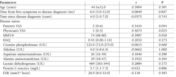
In addition, there was no correlation between se-rum levels of HA and the following continuous para-meters: age, the time from first symptoms to disease diagnosis, the time since disease diagnosis, HAQ, se-rum levels of muscle enzymes, C-reactive protein and erythrocyte sedimentation rate. However, there was a positive correlation between serum levels of HA and VAS (patient and physician), and a negative correla-tion between serum levels of HA and MMT-8.
The correlation between serum levels of HA ex-pressed in interquartile and categorical parameters analysed in the present study are shown in Table III. The data show that serum levels of HA did not corre-late with demographic data (gender and ethnicity) or autoantibodies. Moreover, the use of prednisolone and/or immunosuppressives (azathioprine 2-3
mg/kg/day, methotrexate 20-25 mg/week, cyclospori-ne 2-3 mg/kg/day and/or mycophenolate mofetil 2-3 g/day) did not affect the serum levels of HA.
The median disease duration until medication was started was 8 months [5.0-24.0]. The median dura-tion of using medicadura-tion at the time of HA analysis was 2 years [0.8-8.3].
dIscussIon
The present study showed, for the first time, the cor-relation between serum HA levels and patients with PM. The results showed a high level of serum HA in PM, with a tendency to correlate with the activity of PM disease.
HA plays an important regulatory role in the im-mune response by stimulating the expression of in-flammatory genes in several immunological cells pre-sent in lesion areas, promoting the recruitment of cells, cytokine release and cell migration5. Furthermore, HA
stimulates the release of factors that assist the action of fibroblasts in the inflammatory response, such as TNF-a and IL-1b6.
descri-tAble II. correlAtIon between deMogrAphIc, dIseAse stAtus pArAMeters of pAtIents wIth polyMyosItIs And seruM levels of the hyAluronIc AcId
Parameters r P
Age (years) 44.5±12.0 0.3904 0.391
Time from first symptoms to disease diagnosis (mo) 6.0 [3.0-12.0] 0.0849 0.847
Time since disease diagnosis (years) 4.0 [1.0-7.0] -0.0575 0.743
Disease status
Patient’s VAS 2 [0-6] 0.3424 0.044
Physician’s VAS 1 [0-3] 0.4073 0.015
MMT-8 74 [68-80] -0.3987 0.018
HAQ 0.43 [0.00-1.14] 0.2032 0.273
Creatine phosphokinase (U/L) 125.0 [72.0-275.0] -0.0615 0.600
Aldolase (U/L) 4.0 [4.8-6.5] -0.0662 1.000
Aspartate aminotransferase (U/L) 26 [16-39] 0.1840 0.298
Alanine aminotransferase (U/L) 29 [18-47] 0.1922 0.294
Lactate dehydrogenase (U/L) 469 [365-544] 0.2884 0.175
Protein C-reactive (mg/L) 3.7 [1.1-7.5] -0.023 0.896
ESR (mm/1sthour) 20.0 [8.0-33.0] -0.118 0.593
ESR: erythrocyte sedimentation rate; VAS: Visual Analogic Scale; MMT: Manual Muscle Testing; HAQ: Health Assessment Questionnaire. Results are expressed as mean ± standard deviation (SD), median [interquartile 25th- 75th].
tAble III. correlAtIon between seruM levels of the hyAluronIc AcId expressed In InterquArtIle And deMogrAphIc And treAtMent of the pAtIents wIth polyMyosItIs
AH1 AH2 AH3 AH4
N=9 N=9 N=9 N=8 P Female Gender 6 (66.7) 7 (77.8) 7 (77.8) 7 (87.5) 0.944 Caucasian 5 (55.6) 7 (77.8) 6 (66.7) 3 (37.5) 0.420 Disphagia 0 0 0 2 (22.3) Articular involvement 0 0 1 (11.1) 1 (11.1) Pulmonary involvement 2 (22.3) 1 (11.1) 1 (11.1) 0 Cardiac involvement 0 0 0 0 1.000 Antinuclear antibody 5 (55.6) 5 (55.6) 3 (37.5) 4 (44.5) Anti-Jo-1 antibody 2 (22.3) 0 1 (11.1) 0 Prednisolone Current using 4 (44.5) 6 (66.7) 6 (66.7) 3 (37.5) 0.593
Current doses > 20 mg/day 9 (100.0) 8 (88.9) 8 (88.9) 7 (87.5) 0.889 Immunosuppressives
One 2 (22.2) 6 (66.7) 5 (55.6) 3 (37.5) 0.253
Two 8 (88.9) 5 (55.6) 7 (77.8) 6 (75.0) 0.487
Three 8 (88.9) 9 (100.0) 8 (88.9) 8 (100.0) 1.000
Results are expressed as percentage (%). AH: hyaluronic acid expressed in interquartile (AH1: 0-72 ng/mL; AH2: 73-284 ng/mL; AH3: 285-731 ng/mL; AH4: 732-1651 ng/mL); Immunosuppressives: azathioprine 2-3 mg/kg/day, methotrexate 20-25 mg/week, cyclosporine 2-3 mg/kg/day, mycophenolate mofetil 2-3 g/day
bed in other autoimmune diseases, such as rheumatoid arthritis, systemic sclerosis, systemic lupus erythema-tosus, dermatomyositis and psoriatic arthritis7-11,19,20.
Regarding systemic lupus erythematosus, increased HA relates to disease activity as an immunomodula-tor, with specific molecular recruitment of immune cells to sites of inflammation7.
Serum HA levels were also significantly increased in patients with psoriatic arthritis11. However, the
cor-relation was significant only between HA levels and the degree of skin involvement. No correlation was de-monstrated with articular involvement11.
In dermatomyositis, few studies have been con-ducted, but the published ones correlated clinical ma-nifestations of this disease with mildly elevated values of HA10. Kubo et al4reported two patients who
pre-sented a positive correlation between disease activity and high levels of HA. The same authors observed, in 40 patients with dermatomyositis, that serum HA was elevated when compared to patients with systemic lu-pus erythematosus, rheumatoid arthritis and systemic sclerosis4. In DM, there is a correlation between
in-creased HA levels and cutaneous manifestations, such as photosensitivity. Therefore, it was concluded that the longitudinal measurement of the concentration of serum hyaluronate can be useful for estimating the ac-tivity of DM in patients whose initial serum hyaluro-nate is high10.
These studies show the strong correlation between HA and disease activity7-11,19,20. However, they did not
specifically analyse the role of immunosuppressives and/or glucocorticosteroid on the expression of HA.
Previous studies showed that cyclosporine21and
glu-cocorticosteroid22, for instance, could decrease the
le-vel of HA expression. Our study showed that the se-rum level of HA was independent of drug therapy.
In the present study, an increase in the serum level of HA in patients with PM was also noted and had a tendency to correlate with disease activity. It most li-kely was not possible to find a strong positive correla-tion between the serum level of HA and PM disease activity because the majority of our patients were re-latively stable in terms of clinical and laboratory data. Similarly to other systemic autoimmune diseases, HA could play a role in inflammatory regulation. Mo-reover, HA could stimulate the expression of inflam-matory genes in several immunological cells present in areas of muscle lesions, promoting the recruitment of cells, cytokine release and cell migration. Further-more, HA could be related to clinical manifestations of
autoimmune diseases that can cause impairment of proximal limb movement and cutaneous manifesta-tions, such as discoid lesions and shawl sign, in addi-tion to increased inflammatory response. However, more studies are needed to assess the mechanism of HA involved in the pathogenesis of PM, particularly its correlation with the clinical manifestations of the disease and their degree of severity because the de-mographic and laboratory parameters did not show a strong association with an increase in HA.
The small sample size is the major limitation of this study and it is due to our rigorous selection criteria for a relatively rare disease. Additionally, the inclusion of patients solely from a tertiary care centre may not re-present the full PM spectrum and could result in an overestimation of disease or drug complications of a more severe disease (bias). Therefore, the external va-lidity of our results needs to be confirmed.
conclusIons
It was observed that serum HA was elevated in patients with PM and showed a tendency to correlate with the activity of this disease. Additional studies are needed to assess this correlation, as well as to understand the mechanism involved in the pathogenesis of PM by HA and the possible prognostic role of HA in these pa-tients.
correspondence to
Shinjo SK
Av. Dr. Arnaldo, 455, 3 andar, sala 3150 São Paulo, Brasil
E-mail: samuel.shinjo@gmail.com
references
1. Dimachkie MM, Barohn RJ. Idiopathic inflammatory myopat-hies. Front Neurol Neurosci 2009;26:126-146.
2. Prieto S, Grau JM. The geoepidemiology of autoimmune mus-cle disease. Autoimmun Rev 2010;9:A330-4.
3. Gaubitz M. Epidemiology of connective tissue disorders. Rheu-matology (Oxford) 2006;45 Suppl 3:iii3-4.
4. Kubo M, Kikuchi K, Yazawa N, Fujimoto M, Tamaki T, Tama-ki K. Increased serum concentration of hyaluronate in derma-tomyositis patients. Arch Dermatol Res 1998; 290:579-581. 5. Dentener MA, Vernooy JH, Hendriks S, Wouters EF. Enhanced
levels of hyaluronan in lungs of patients with COPD: rela-tionship with lung function and local inflammation. Thorax 2005;60:114-119.
6. Jiang D, Liang J, Noble PW. Hyaluronan as an Immune Regu-lator in Human Diseases. Physiol Rev 2011;91:221-264. 7. Chang LM, Maheshwari P, Werth S, Schaffer L, Head SR,
gly-cosaminoglycans in cutaneous lupus erythematosus and der-matomyositis. J Histochem Cytochem 2011;59:336-345. 8. Yoshioka Y, Kozawa E, Urakawa H, Arai E, Futamura N, Zhuo
L, Kimata K, Ishiguro N, Nishida Y. Suppression of hyaluronan synthesis alleviates inflammatory responses in murine arthritis and in human rheumatoid synovial fibroblasts. Arthritis Rheum 2013;65:1160-1170.
9. Abignano G, Cuomo G, Buch MH, Rosenberg WM, Valentini G, Emery P, Del Galdo F. The enhanced liver fibrosis test: a cli-nical grade, validated serum test, biomarker of overall fibrosis in systemic sclerosis. Ann Rheum Dis 2014;73:420-427. 10. Kubo M, Ihn H, Matsukawa A, Kikuchi K, Tamaki K.
Derma-tomyositis with elevated serum hyaluronate. Clin Dermatol 1999;24:275-278.
11. Elkayam O, Yaron I, Shirazi I, Yaron M, Caspi D. Serum levels of hyaluronic acid in patients with psoriatic arthritis. Clin Rheu-matol 2000;19:455-457.
12. Bohan A, Peter JB. Polymyositis and dermatomyositis. N Engl J Med 1975;13:344-347.
13. Rider LG, Giannini EH, Harris-Love M, Joe G, Isenberg D, Pil-kington C, Lachenbruch PA, Miller FW; International Myosi-tis Assessment and Clinical Studies Group. Defining Clinical Improvement in Adult and Juvenile Myositis. J Rheumatol 2003;30:603-617.
14. Harris-Love MO, Shrader JA, et al.. Distribution and severity of weakness among patients with polymyositis, dermatomyosi-tis, and juvenile dermatomyositis. Rheumatology (Oxford) 2009;48:134-139.
15. Miller FW, Rider GL, Chung YL, et al. Proposed preliminary core set measures for disease outcome assessment in adult and juvenile idiopathic inflammatory myopathies. Rheumatology (Oxford) 2001;40:1262-1273.
16. Rider LG, Feldman BM, Perez MD, et al. Development of vali-dated disease activity and damage indices for the juvenile idio-pathic inflammatory myopathies: I. Physician, parent, and pa-tient global assessments. Juvenile Dermatomyositis Disease Ac-tivity Collaborative Study Group. Arthritis Rheum 1997;40:1976-1983.
17. Ekdahl C, Eberhardt K, Andersson SI, Svensson B. Assessing di-sability in patients with rheumatoid arthritis: use of a Swedish version of the Stanford Health Assessment Questionnaire. Scand J Rheumatol 1988;17:263-271.
18. Alexanderson H, Lundberg IE, Stenstrom CH. Development of the myositis activities profile -validity and reliability of a self-administered questionnaire to assess activity limitations in pa-tients with polymyositis/dermatomyositis. J Rheumatol 2002;29:2386-2392.
19. Volkova MV, Kunder EV. The criteria of differentiated diagnos-tics of early arthritis on the basis of analysis of serum hyaluro-nidase and deoxyribonuclease activity. Klin Lab Diagn 2012;10:22-26.
20. Levesque H, Baudot N, Delpech B, et al. Clinical correlations and prognosis based on hyaluronic acid serum levels in pa-tients with progressive systemic sclerosis. Br J Dermatol 1991;124:423-428.
21. Takagishi K, Itoman M, Goso Y, Kuwao S, Miyahara H, Kaiba-ra N. Effects of cyclosporin on serum hyaluronic levels in col-lagen arthritis. Inflamm Res 1995; 44: 79-82.
22. Smith TJ. Glucocorticoid regulation of glycosaminoglycan synt-hesis in cultured human skin fibroblasts: evidence for a recep-tor-mediated mechanism involving effects on specific de novo protein synthesis. Metabolism 1988; 37: 179-184.