Arlete Maria Lima Marques
Antimicrobial assessment of phages
entrapped in bio-based structures
Universidade do Minho
Escola de Engenharia
Outubro de 2016
Arle
te Maria Lima Mar
q
ues
Antimicrobial assessment of phages entrapped in bio-based s
tructures
Minho | 20
1
6
Universidade do Minho
Escola de Engenharia
Arlete Maria Lima Marques
Antimicrobial assessment of phages
entrapped in bio-based structures
Dissertação de Mestrado
Mestrado Integrado em Engenharia Biológica
Ramo Tecnologia Química e Alimentar
Trabalho efetuado sob a orientação de:
Doutora Sanna Maria Sillankorva
Doutor Miguel Ângelo Cerqueira
DECLARAÇÃO Nome: Arlete Maria Lima Marques
Título da dissertação:
Antimicrobial assessment of phages entrapped in bio-based structures
Análise antimicrobiana de bacteriófagos incorporados em estruturas de base biológica
Orientadores:
Doutora Sanna Maria Sillankorva
Doutor Miguel Ângelo Parente Ribeiro Cerqueira
Ano de conclusão: 2016
Designação do Mestrado: Mestrado Integrado em Engenharia Biológica
É AUTORIZADA A REPRODUÇÃO PARCIAL DESTA DISSERTAÇÃO, APENAS PARA EFEITOS DE INVESTIGAÇÃO, MEDIANTE DECLARAÇÃO ESCRITA DO INTERESSADO, QUE A TAL SE COMPROMETE.
Universidade do Minho, ____/_____/_________
v
A
GRADECIMENTOS
Após a finalização desta etapa, não poderia deixar de agradecer a todos os que durante estes anos me acompanharam, ajudaram e incentivaram.
Estou especialmente agradecida aos meus orientadores, Doutora Sanna Sillankorva e Doutor Miguel Cerqueira, pela oportunidade que me deram. Por todo o apoio, ajuda e também disponibilidade que demonstraram ao longo do desenvolvimento do trabalho.
Sou muito grata à Maria Costa pela amizade, por toda ajuda que me deu, pelo apoio incondicional, pela disponibilidade e dedicação que sempre demonstrou, pela paciência e também por todos os incentivos e descontração nos momentos mais complicados.
Também estou muito grata à Catarina Milho, que sempre se mostrou disponível para ajudar e colaborar durante o desenvolvimento do trabalho. Obrigada pelo apoio e dedicação.
Estou também muito agradecia ao Doutor Pablo Fuciños pela ajuda e contribuição com o seu conhecimento para o desenvolvimento do trabalho.
À professora Ana Paula Cunha, que se prontificou a ajudar na parte escrita do trabalho. À empresa Improveat por me ter dado a oportunidade de participar neste projecto Ao Iberian Nanotechnology Laboratory por me ter disponibilizado as instalações e os equipamentos necessários à realização de parte do trabalho.
Também quero agradecer ao Laboratório de Indústria e Processo e ao Laboratório de Microbiologia Aplicada, pelo apoio, pela amizade, pelos ensinamentos e pela ajuda que me deram durante estes meses.
Um especial agradecimento à minha família, ao meu pai Justino, às minhas irmãs Bibiana e Anita, e aos meus tios Elisa e Francisco por todo o amor, incentivo e por sempre me apoiarem. Aos meus primos pelo apoio e carinho.
Ao Bruno, pelo amor e carinho, pela paciência e por ser sempre um apoio incondicional. Às minhas amigas, pela amizade e pelo incentivo constantes durante estes anos. A todos aqueles que me ajudaram a concluir esta etapa, os meus sinceros agradecimentos.
vii
A
BSTRACT
Recently, edible packaging materials experienced a notable growth, being expected an important impact in the food industry in the next years. Also, bacteriophages have been recognized for their potential as biotherapeutic agents to control bacterial growth, and their use in food industry to control microbiogical growth has been presented as one of the solutions to maintain food quality. The main aim of this work was the functionalization of edible packaging materials with the use of phages. For the development of edible packaging, different types (Protanal SF 120RB; Protanal CR 8223; Protanal LFR 5/60; Protanal GP 1320 and Protanal Manugel) and concentrations (2.5 g/L, 5 g/L, 7.5 g/L, 10 g/L and 15 g/L) of sodium alginate were evaluated, being a concentration of 10 g/L of alginate CR 8223 and Manugel sellected. The produced films were crosslinked with different concentrations of CaCl2 (0 g/L, 10 g/L, 12.5 g/L and 15 g/L). The films were characterized in terms of moisture content, solubility, contact angle, isothermic adsorption, swelling behaviour, colour and opacity, and mechanical properties (TS and EB), being their chemical structure studied by Fourier Transform Infrared (FTIR) spectroscopy.
During characterization of CR 8223 and Manugel alginate films, it was possible to conclude that crosslinking has a significant effect on alginate structure and properties. Characterization of films allows to conclude that crosslinking decreases film thickness and the moisture content of films. It was also possible to notice that crosslinking does not change the films colour, but make them stronger and insoluble with a high swelling index. After the characterization, a concentration of 10 g/L of CR 8223 alginate was selected for phages incorporation, being crosslinked with 10 g/L of CaCl2. The choice was based on swelling and solubility films properties.
Antimicrobial activity of alginate films with entrapped phages showed that phage ΦIBB-PF7A can be incorporated in alginate-based films and maintain its activity, leading to a bacterial decrease of 4 log for the multiplicity of infection (MOI) of 10 in the first 24 h. It was also possible to confirm that phages are released onto chicken meat, leading to a decrease of Pseudomonas fluorescens contamination. Results show that alginate-based films with phages can be used to control bacterial growth in meat products, presenting a potential interest for the food industry.
Keywords: bacteriophage ΦIBB-PF7A, active packaging, antibacterial activity, Pseudomonas fluorescens, polysaccharide.
ix
R
ESUMO
As embalagens edíveis têm crescido notavelmente nos últimos anos, esperando-se nos próximos anos um grande impacto na indústria alimentar. Para além disso os bacteriófagos têm sido reconhecidos pelo seu potencial como agentes bio-terapêuticos no controlo do crescimento de bactérias e por isso têm vindo a ser apresentados como uma das soluções para o controlo do crescimento bacteriano na indústria alimentar.
O principal objetivo deste trabalho foi a funcionalização de embalagens com a incorporação de fagos em bio-estruturas. Para desenvolver as embalagens edíveis, diferentes tipos (Protanal SF 120RB; Protanal CR 8223; Protanal LFR 5/60; Protanal GP 1320 and Protanal Manugel) e concentrações (2,5 g/L, 5 g/L, 7,5 g/L, 10 g/L and 15 g/L) de alginato de sódio foram avaliados, tendo sido selecionados os alginatos CR 8223 e o Manugel com uma concentração de 10 g/L. Os filmes produzidos foram reticulados com diferentes concentrações de solução CaCl2 (0 g/L, 10 g/L, 12,5 g/L e 15 g/L). Os filmes foram caraterizados em termos de humidade, solubilidade, grau de inchamento, ângulos de contato, isotérmicas de adsorção, cor e opacidade e propriedades mecânicas (resistência à tração e elongação à rutura), as estruturas quimícas foram estudadas através da espectroscopia por infravermelho com transformada de Fourier (FTIR). Durante a caracterização dos filmes de alginato CR 8223 e Manugel, foi possível concluir que a reticulação tem um efeito significativo na estrutura e propriedades destes. A caracterização dos filmes permitiu concluir que a reticulação diminui a espessura dos filmes, bem como a humidade. Também foi possível concluir que a reticulação não altera a cor dos filmes, mas torna-os mais fortes, insolúveis e com uma grande capacidade de absorção. Depois de caracterizados, o alginato CR 8223 com concentração de 10 g/L foi selecionado para incorporação de fagos, e foi reticulado com CaCl2 com concentração de 10 g/L. A escolha deste foi baseada nas propriedades do filme como o grau de inchamento e a solubilidade.
A atividade antimicrobiana dos filmes com fagos incorporados mostrou que o fago ΦIBB-PF7A pode ser incorporado em filmes de alginato, mantendo a sua actividade após libertado levando a uma redução de 4 log para uma multiplicidade de infeção (MOI) de 10 em 24 h. Também foi possível confirmar que os fagos libertam-se para a carne levando a uma diminuição da contaminação pela bactéria Pseudomonas fluorescens. Os resultados mostram que os filmes de alginato com fagos incorporados pode ser usados no controlo do crescimento bacteriano em produtos cárneos, mostrando assim um potencial interesse para a indústria alimentar.
x
Palavras-chave: bacteriófago ΦIBB-PF7A, embalagens funcionais, Pseudomonas fluorescens, atividade antimicrobiana, polissacarídeos.
xi
T
ABLE OF
C
ONTENTS
Agradecimentos ... v
Abstract ...vii
Resumo ... ix
General nomenclature ... xiii
List of Figures ... xv
List of Tables ... xix
1. Introduction ... 3 1.1 Motivation ... 3 1.2 Aims ... 3 1.3 Thesis Overview ... 4 2. State-of-the-art ... 7 2.1 Edible Packaging ... 7
2.1.1 Materials used in edible films and coatings production ... 9
2.1.2 Plasticizers ... 14
2.2 Bacteriophages ... 15
2.2.1 Bacteriophages discovery ... 15
2.2.2 Classification of bacteriophages ... 16
2.2.3 Life cycle of bacteriophages ... 18
2.3 Pseudomonas fluorescens ... 20
3. Materials And Methods ... 25
3.1 Sodium alginate films ... 25
3.1.1 Films crosslinking ... 25
3.2 Characterization of sodium alginate films... 26
3.2.1 Moisture content ... 26
3.2.2 Water solubility ... 26
3.2.3 Sorption isotherms ... 27
3.2.4 Colour and opacity ... 27
3.2.5 Fourier transform infrared (FTIR) ... 28
xii 3.2.7 Mechanical properties ... 29 3.2.8 Swelling ... 29 3.2.9 Contact angles ... 29 3.3 Bacteriophage production ... 30 3.3.1 Bacteriophage titration ... 30 3.4 Pseudomonas fluorescens ... 31 3.4.1 Bacteria titration ... 31
3.5 Alginate films with entrapped phages ... 32
3.5.1 Films with entrapped phages titration ... 32
3.6 Antimicrobial activity of alginate films with entrapped phage ... 33
3.7 Antimicrobial activity of alginate films with entrapped phage in chicken meat 33 3.8 qPCR to quantify phages, as an alternative to the PFUs quantification ... 34
4. Results And Discussion ... 37
4.1 Moisture content ... 37
4.2 Water solubility ... 38
4.3 Sorption isotherms ... 40
4.4 Colour and opacity ... 43
4.5 Fourier transform infrared (FTIR) spectroscopy ... 44
4.6 Film thickness ... 46
4.7 Mechanical properties ... 48
4.8 Swelling ... 51
4.9 Contact angles ... 53
4.10 Antimicrobial activity of alginate films with entrapped phages ... 54
4.11 Antimicrobial activity of alginate films with entrapped phages in chicken meat 58 4.12 qPCR to quantify phages, as an alternative to the PFUs quantification ... 59
5. Conclusions ... 65
5.1 Conclusion and Future work ... 65
xiii
G
ENERAL NOMENCLATURE
EB – elongation-at-break
M/G – manuronic to guluronic ratio M – initial mass of the film
M0 – mass of the film after drying
MC – moisture content SW – swelling index S1 – weight of wet film
S0 – initial weight of the film
RH – relative humidity
R 2 – coefficient determination
TS – tensile strength t – time
Yb– black standard plate
Yw – white standard plate
Greek symbol θ – contact angle Abbreviations
FTIR – fourier transform infrared O2 – oxygen
CO2 – carbon dioxide CaCl2– calcium chloride CFU – colony forming unit PFU – plaque forming unit DNA – deoxyribonucleic acid RNA – ribonucleic acid mRNA – messenger RNA ss – single strand ds – double strand
xiv
ATR – attenuated total reflectance Rpm – rotation per minute
qPCR – quantitative polymerase chain reaction Ct – threshold cycle
ΔRn – magnitude of the generated signal in qPCR ICTV – International Committee on Taxonomy of Viruses MW – molecular weight
v - volume
LB – lysogeny broth medium
LBA – lysogeny broth medium with agar OD – optical density
xv
L
IST OF
F
IGURES
Figure 2-1 - Functional properties of edible films and coatings on food product (Adapted from (Lin
& Zhao, 2007)). ... 8
Figure 2-2 Alginate structure. A) mannuronic and guluronic acid structure; B) M and G blocks (adapted from (Andersen, Auk-Emblem, & Dornish, 2015)). ... 10
Figure 2-3 Alginate production methods. A) Calcium alginate process; B) Alginic acid process (adapted from (McHugh, 2003)). ... 11
Figure 2-4 Ca2+ crosslinking with alginate (adapted from (Kühbeck et al., 2015)) ... 12
Figure 2-5 Glycerol structure (adapted from (Sigma, 2016)). ... 15
Figure 2-6 Representation/morphology of a phage (adapted from (Hanlon, 2007)). ... 16
Figure 2-7 Schematization of lytic life cycle (adapted from (Melo et al., 2012)) ... 18
Figure 2-8 Schematization of lysogenic life cycle (adapted from (Melo et al., 2012)). ... 19
Figure 2-9 Pseudomonas fluorescens (adapted from (Boresi, 2009)). ... 20
Figure 3-1 CIELab colour space (adapted from (CIELab, 1976))... 27
Figure 3-2 Phage enumeration using the small drop plaque assay (adapter from (Oliveira et al., 2013)). ... 31
Figure 4-1 Moisture content of 10 g/L of CR 8223 alginate-based films for increasing concentrations of CaCl2 (Different letters in same column indicate a statistically significant difference, p<0.05). ... 37
Figure 4-2 Moisture content of 10 g/L of Manugel alginate-based films for increasing concentrations of CaCl2 (Different letters in same column indicate a statistically significant difference, p<0.05). ... 38
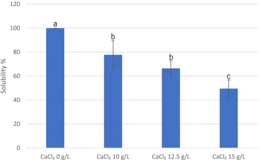
Figure 4-3 Solubility of 10 g/L of CR 8223 alginate-based films for increasing concentrations of CaCl2 (Different letters in same column indicate a statistically significant difference, p<0.05). .. 39
Figure 4-4 Solubility of 10 g/L of Manugel alginate-based films for increasing concentrations of CaCl2 (Different letters in same column indicate a statistically significant difference, p<0.05). .. 40
Figure 4-5 Experimental data and Flory-Huggins model for the adsorption isotherms for 10 g/L alginate Manugel films. (line – experimental data, dots – adjusted model values). ... 42
Figure 4-6 Experimental data and Flory-Huggins model for the adsorption isotherms for 10 g/L alginate CR 8223 films. (line – experimental data, dots – adjusted model values). ... 43
xvi
Figure 4-7 FTIR spectra of 10 g/L of CR 8223 alginate-based films with increasing concentrations of CaCl2. ... 44 Figure 4-8 FTIR spectra of 10 g/L of Manugel alginate-based films with increasing concentrations of CaCl2. ... 45 Figure 4-9 Thickness of 10 g/L of CR 8223 alginate-based films for increasing concentrations of CaCl2 (Different letters in same column indicate a statistically significant difference, p<0.05). .. 46 Figure 4-10 Thickness of 10 g/L of Manugel alginate-based films for increasing concentrations of CaCl2 (Different letters in same column indicate a statistically significant difference, p<0.05). .. 47 Figure 4-11 Elongation-at-break of 10 g/L of CR 8223 alginate-based films for increasing concentrations of CaCl2 (Different letters in same column indicate a statistically significant difference, p<0.05). ... 49 Figure 4-12 Tensile strength of 10 g/L of CR 8223 alginate-based films for increasing concentrations of CaCl2 (Different letters in same column indicate a statistically significant difference, p<0.05). ... 49 Figure 4-13 Elongation-at-break of 10 g/L of Manugel alginate-based films for increasing concentrations of CaCl2 (Different letters in same column indicate a statistically significant difference, p<0.05) ... 49 Figure 4-14 Tensile strength of 10 g/L of Manugel alginate-based films for increasing concentrations of CaCl2 (Different letters in same column indicate a statistically significant difference, p<0.05). ... 51 Figure 4-15 Swelling index of 10 g/L of CR 8223 alginate-based films for increasing concentrations of CaCl2 (Different letters in same column indicate a statistically significant difference, p<0.05). * - films that dissolved in water, it was not possible to determine swelling index. ... 52 Figure 4-16 Swelling index 10 g/L of Manugel alginate-based films for increasing concentrations of CaCl2 (Different letters in same column indicate a statistically significant difference, p<0.05). * - films that dissolved in water, it was not possible to determine swelling index. ... 52 Figure 4-17 Water contact angle for 10 g/L of CR 8223 alginate-based films for increasing concentrations of CaCl2 (Different letters in same column indicate a statistically significant difference, p<0.05). ... 53 Figure 4-18 Water contact angle for 10 g/L of Manugel alginate-based films for increasing concentrations of CaCl2 (Different letters in same column indicate a statistically significant difference, p<0.05). ... 53
xvii
Figure 4-19 Log reduction of bacteria during time, MOI of 10, at room temperature (22 °C). ... 55
Figure 4-20 Log reduction of bacteria during time, MOI of 100, at room temperature (22 °C). . 55
Figure 4-21 Variations of PFUs during experiment (24h) for MOI of 10 and MOI of 100 at room temperature (22 °C). ... 56
Figure 4-22 Log reduction of bacteria during time, MOI of 10, at refrigerated temperature (4 °C). ... 56
Figure 4-23 Log reduction of bacteria during time, MOI of 100, at refrigerated temperature (4 °C). ... 56
Figure 4-24 Variations of PFUs during experiment (24h) for MOI of 10 and MOI of 100 at refrigerated temperature (4 °C). ... 57
Figure 4-25 CFUs/mL per meat gram in films with and without phage. ... 59
Figure 4-26 PFUs/mL in alginate films with phage. ... 59
Figure 4-27 qPCR amplification plot. ... 61
xix
L
IST OF
T
ABLES
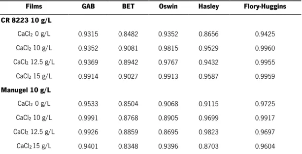
Table 2-1 Phages families (adapted from (Hanlon, 2007; Melo et al., 2012)). ... 17 Table 3-1 Primers for ΦIBB-PF7A phage and their characteristics... 34 Table 4-1 R 2 values for each isotherm model for CR 8223 10 g/L and Manugel 10 g/L
alginate-based films. ... 41 Table 4-2 – A and B constants for Flory-Huggins model ... 42 Table 4-3– L, a and b values, opacity of alginate films. ... 44
1
Chapter 1
1.INTRODUCTION
3
1.
I
NTRODUCTION
1.1
Motivation
Food industry is one of the most important industries in the world, with great relevance for human health. To ensure and improve food safety and quality, new technologies arise every day. New approaches appear to reduce consumer’s health risks and to comply sanitary legislation, for national and international markets. Health safety is one of the major concerns of the present and future. Currently, one in ten people become sick due to food contaminations with pathogens, specialy pathogenic bacteria such as: Salmonella, Listeria, Escherichia coli and Campylobacter. Deaths resulting from food poisoning are alarming, reaching values of 420 000 deaths per year . Smart packaging trough the development of new methodologies and with the combination of different systems (e.g modified and controled atmosphere, incorporation of functional compounds) is a promising solution for food related health problems, since they are able to ensure the quality, traceability (allow pathogen identification in the moment of consumption) and safety (avoiding microbial growth), allowing their transportation all around the world any time of year. Recently, consumers changed their demands, not only about safe products, but also with their sources and their functional properties. Today, the regular consumer desires natural food products, with no chemical additives and slightly processed, and at the same time that are ready for consumption and easily prepared (Gouvêa, Mendonça, Soto, & Cruz, 2015). Smart and active packaging market is growing each year, and due to the major advantages related to food quality and safety, it is expected to become a field of major impact in food industry and consequently more explored.
1.2
Aims
The main objective of the dissertation was the functionalization of packaging materials with entrapped phages in bio-based structures. Other objectives were:
- Physicochemical characterization of alginate-based films;
- Study of the influence of crosslinking on alginate-based films properties; - Evaluation and optimization of phages’ incorporation on alginate-based films; - Evaluation of the phage release behaviour from alginate-based films;
ANTIMICROBIAL ASSESSMENT OF PHAGES ENTRAPPED IN BIO-BASED STRUCTURES
4
- Evaluation of the antimicrobial effect of phage-loaded structures against contaminated meat;
- Study of an alternative method to quantify phages.
1.3
Thesis Overview
The dissertation structure is divided in five chapters. In this chapter are described the motivation of the work, research main objectives and the outline of the dissertation. From chapter 2 to 5 they are distributed as follows:
Chapter 2 presents the stat-of-the-art, where is present an overview on the edible packaging, focusing on the sodium alginate, that is the material used for the development of the bio-structure that was used to entrappe phages. Also, in this chapter it is presented an overview on phages, especially the phage ΦIBB-PF7A, that is specific to Pseudomonas fluorescens.
Chapter 3 presents the material and methods used in the production and characterization of the alginate films; it is also described the phage production method and the methodology used for their entrapment in alginate-based films. Also, in this chapter, are presented the approaches used to evaluate the antimicrobial activity of the active alginate-based films against bacteria P. fluorescens, both in vitro and in chicken meat.
Chapter 4 presents the results and discussion of the characterization of alginate-based films without phages and their antimicrobial activity with entrapped phages.
Finally, the last chapter, shows the overall conclusions, recommendations and suggestions for future work.
The results from this project already resulted in a poster exhibition in IUFOST 2016 congress and two papers, that are being written for publication.
5
Chapter 2
2.STATE-OF-THE-ART
7
2.
S
TATE
-
OF
-
THE
-
ART
2.1
Edible Packaging
Edible packaging experienced a notable growth in recent years and an important impact in the food industry. This growth is due to an increase in the interest, research and development of new packaging materials using natural polymers to replace the non-biodegradable, synthetic materials used in synthetic packaging. Factors like the impact on sustainability, the increased interest in renewable resources and biodegradable materials with no ecological impact on the environment are influencing the growth perspectives on the use of these materials (Cerqueira, Teixeira, & Vicente, 2016; Cerqueira, 2010).
The main objective of the food packaging is to protect and transport food products in a cost-effective way that satisfies the industry, consumer’s desires and, at the same, maintain quality and safety of the food products. Furthermore, packaging provides food protection from chemical, biological and physical external effects (Marsh & Bugusu, 2007). Like synthetic materials, edible materials can guarantee some of these packaging functionalities such as: protection of food from external influences (relative humidity, oxygen and carbon dioxide), separation and protection of food from exposure to the environment, protection from microbial contaminations and consequently delaying food deterioration, increasing shelf-life, which also allows food long distance transport, maintaining the quality and safety parameters for human consumption (Cerqueira, Costa, Rivera, & Ramos, 2014; Pascall & Lin, 2013).
The main difference between synthetic and edible packaging is the type of materials used in the packaging composition of each product. Synthetic packaging uses non-biodegradable materials like petroleum-based plastic, glass and metal, while edible packaging only uses biopolymers like proteins, lipids, polysaccharides and waxes, and other materials approved to be eaten by the consumers (Cerqueira et al., 2016).
It is possible to distinguish two types of edible packaging, coatings and films. Coating is when the film solution is applied by several methods such as dipping, spraying, brushing and panning directly on the surface of the food product, and after dried it forms a thin film that involves and protects the food. A film is when the biopolymer solution is prepared and dried separately from the food product, that is later applied on it by wrapping. In general, its thickness is less than 0.3 mm (Bourtoom, 2008; Cerqueira et al., 2016; Lacroix, 2009; Pavlath & Orts, 2009).
ANTIMICROBIAL ASSESSMENT OF PHAGES ENTRAPPED IN BIO-BASED STRUCTURES
8
The application of edible coatings and films to extend shelf life of fresh and minimally processed food products has been highlighted due to the demand for minimal food processing and storage technologies to protect foods from environmental effects. One important property is their barrier capacity against moisture, oxygen, carbon dioxide and other volatile compounds transfer in a food system (Figure 2-1), that turns possible the improvement of food quality and extension of its shelf life. Furthermore, it is possible to add extra functionalities through the incorporation of functional ingredients in their composition such as antioxidants, antimicrobials, nutrients, and flavour to improve food stability, quality and safety (Lin & Zhao, 2007).
Overall, the main advantages and benefits of the use of edible films and coatings are their capacity to act as a:
barrier to moisture: decreasing the moisture loss of food products, which can cause changes in texture, flavour and appearance;
barrier to oxygen: control oxygen gas exchange between food and the surrounding atmosphere, delaying deterioration and enzymatic oxidation;
barrier to volatile compounds: restrict the exchange of volatile compounds, preventing loss of natural volatile flavours of food;
barrier to mechanical stress: protect from physical damages caused by mechanical impact, pressure, vibration, and other mechanical factors;
Figure 2-1 - Functional properties of edible films and coatings on food product (adapted from (Lin & Zhao, 2007)).
2.STATE-OF-THE-ART
9 carrier: it can act as a carrier of functional ingredients, such as: antimicrobial and antioxidant agents, nutraceuticals, colour and flavour ingredients for reducing microbial loads, delaying oxidation and discoloration and also to improve food quality (Lin & Zhao, 2007; Tharanathan, 2003).
2.1.1 Materials used in edible films and coatings production
Edible films and coatings can be produced by different edible materials such as: polysaccharides, protein, and lipids, with the possible addition of plasticizers and/or surfactants (Dhanapal et al., 2012). Their performance is directly related with the material composition and the environmental conditions. For example, the relative humidity (RH) will affect films performance, since the capacity of the film to act as a barrier to moisture and other gases decreases with the increase of RH (G. I. Olivas & Barbosa-Cánovas, 2008). Also food water activity is an important factor when selecting the edible films materials, since that different materials can exhibit different functional properties depending on relative humidity conditions (G. Olivas & Barbosa-Cánovas, 2009; G. I. Olivas & Barbosa-Cánovas, 2008).
Polysaccharides
Polysaccharides are natural polymers composed by monosaccharide residues that are binded by O-glycosidic linkages and they can be negatively, positively or neutrally charged, depending on their source. They can act as an energy reserve in plants and animals, having also structural functions in plant cell walls and in the insect’s exoskeleton (Cerqueira, Souza, Teixeira, & Vicente, 2012; Nelson & Cox, 2000). Polysaccharides are innumerous due to different monosaccharides composition, linkage types and patterns, chain shapes, and degree of polymerization, influencing their physicochemical properties (Cerqueira et al., 2012). Although polysaccharides have hydrophilic properties that provide strong barriers to CO2 and O2 under certain conditions, on the other hand, they have a poor moisture barrier and mechanical properties (Miguel A. Cerqueira et al., 2011; Pascall & Lin, 2013). In the case of poor water barrier properties, the addition of fat, to get emulsified films, is assumed to significantly reduce water transfer through the film (Pastrana et al., 2016).
ANTIMICROBIAL ASSESSMENT OF PHAGES ENTRAPPED IN BIO-BASED STRUCTURES
10
The most common polysaccharides used as edible films and coatings are starch and its derivatives, carrageenan, alginate, pullulan, gellan gum, xanthan gum, cellulose and derivatives, chitosan and pectin (Cerqueira et al., 2011; Dhanapal et al., 2012).
Alginate
Alginates are hydrophilic colloidal carbohydrates extracted from various species of brown seaweeds, which belongs to the Phaeophyceae. Alginates are linear water-soluble biopolymers formed by sequences of -(1-4)-linked units of -ᴅ-mannuronic acid (M blocks) and -L-guluronic (G blocks) residues in different proportions and different distribution in the chain (Figure 2-2), being the carboxylic groups from uronic acids responsible for their negative charge. The chemical composition (e.g. molecular weight) and sequence of the M and G residues affects the physicochemical properties of alginates, being the ratio of mannuronic and guluronic acid residues dependent on the biological source and the plant maturation state (Azevedo, Bourbon, Vicente, & Cerqueira, 2014; Lee & Mooney, 2013; Milani & Maleki, 2012; Nieto, 2016; Pastrana et al., 2016). Alginate is presented in the cell walls of brown algae as calcium, magnesium and sodium salts of alginic acid.
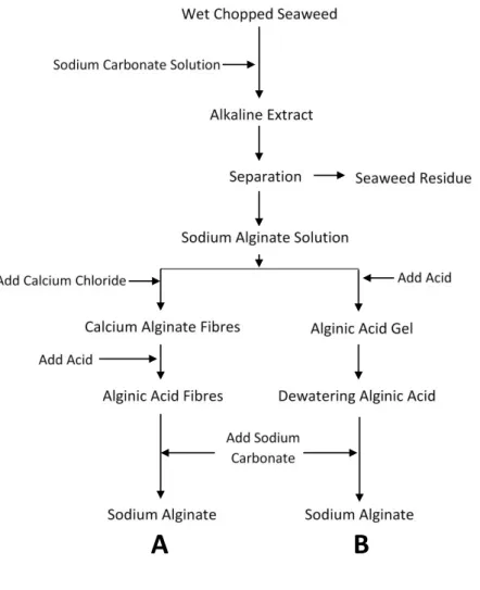
Alginate production process starts with wet chopped seaweeds that are subjected to an alkaline extraction, where the residues are removed by filtration and the alginate in the aqueous solution is further processed to produce the final sodium alginate in two ways as presented in
Figure 2-2 Alginate structure. A) mannuronic and guluronic acid structure; B) M and G blocks (adapted from (Andersen, Auk-Emblem, & Dornish, 2015)).
2.STATE-OF-THE-ART
11 Figure 2-3. In the calcium alginate process (Figure 2-3 A), the calcium chloride is added to the initial alginate extract to form fibers of calcium alginate that are insoluble in water. Then, acid is added to the fibers of calcium alginate to convert it into alginic acid. After that, the alginic acid fibers slowly react with sodium carbonate, being converted into sodium alginate. Sodium alginate is sometimes extruded into pellets, which are then dried and milled (McHugh, 2003; Nieto, 2016).
In the alginic acid method (Figure 2-3 B), acid is added to the alginate solution to form alginic acid, which is separated from the water once it forms an insoluble gel. To remove some water from this gel, alcohol is added to the alginic acid, followed by sodium carbonate that converts the alginic acid into sodium alginate. Sodium alginate does not dissolve in the mixture of alcohol and water, and is therefore, precipitated out and afterwards dried and milled to an appropriate particle size (McHugh, 2003; Nieto, 2016).
Alginate is non-toxic and has unique colloidal properties, such as thickening, stabilizing, suspending, film forming, gel producing and emulsion stabilizing (Dhanapal et al., 2012). Films
Figure 2-3 Alginate production methods. A) Calcium alginate process; B) Alginic acid process (adapted from (McHugh, 2003)).
ANTIMICROBIAL ASSESSMENT OF PHAGES ENTRAPPED IN BIO-BASED STRUCTURES
12
formed by alginate are uniform, transparent, and exhibit poor water resistance because of their hydrophilic nature. They are good oxygen barriers that can delay lipid oxidation in food products (Azevedo et al., 2014; Dhanapal et al., 2012; Lin & Zhao, 2007).
Alginates have the ability to react with di- and trivalent cations, specifically calcium ions. In the presence of Ca2+, alginate acquires extreme toughness and resilience. Calcium-induced gelation has been demonstrated to result from specific and strong interactions between Ca2+ with G blocks of alginate, where a specific link forms a row of reversible and strong electrostatic crosslinks called the “egg-box” structure (Figure 2-4). (Nieto, 2016; J.-W. Rhim, 2004; Yoon, Oh, Jo, Lee, & Hwang, 2014). G blocks and M blocks respond differently to calcium ions, and the G blocks react to calcium crosslinking faster than the M blocks. This happens because of G block three-dimensional “egg-box” molecular conformation, which binds with Ca2+ ions to form salt bridges. Furthermore, M blocks associate through Ca2+ salt bridges, forming more elastic gels with good heat stability (Nieto, 2016).
The crosslinking of alginate films with calcium increases their tensile strength, showing that film structure forms better without the charge repulsion. Also, crosslinked calcium-alginate always yields a strong, clear film that is insoluble in water, compared to sodium alginate film (Cathell & Schauer, 2007; Galus, Uchański, & Lenart, 2013; Liling et al., 2016; Nieto, 2016; Remuñán-López & Bodmeier, 1997; Russo, Malinconico, & Santagata, 2007; Ying, 2006).
2.STATE-OF-THE-ART
13 Proteins
Proteins are essentially high-molecular-weight linear polymers composed of a chain of amino acids. The composition and the sequence of amino acids determine the secondary, tertiary and quaternary structure of these macromolecules (Han & Gennadios, 2005; Souza & Carneiro-da-Cunha, 2016). Proteins can exist as fibrous and globular structures. Fibrous proteins are water insoluble and work as the main structural material of animal tissues. They are fully extended and associated closely with each other in parallel structures, generally through hydrogen bonding, to form fibers. On the other hand, globular proteins, such as wheat gluten, corn zein, soy protein and whey protein are soluble in water and acids, bases or salt aqueous solutions. These proteins bend into complex spherical structures held together by a combination of hydrogens, ionic, hydrophobic and covalent bonds. Their chemical and physical properties are dependent on the amount of amino acid residues and their placement along the protein polymer chain (Bourtoom, 2008; Cuq, Gontard, Cuq, & Guilbert, 1996; Pastrana et al., 2016; Wittaya, 2012).
Both types of proteins, globular and fibrous, can form films. Generally, in film solutions, proteins need denaturation by heat, acid, base and/or solvent, to form more extended structures, which is required to form films. Protein films are mostly formed from solutions or dispersions of the protein as the solvent/carrier evaporates, which is generally limited to water, ethanol or ethanol-water mixture (Bourtoom, 2008; Kester & Fennema, 1986). Once extended, protein chains can unite through hydrogen, ionic, hydrophobic and covalent binding. The chain-to-chain interaction that produces cohesive films is influenced by the degree of chain extension and the nature and sequence of amino acid residues. Uniform distribution of polar, hydrophobic, and/or thiol groups along the polymer chain increases the likelihood of the respective interactions. Higher polymer chain-to-chain interactions results in films that are stronger but less flexible and less permeable to gases, vapours and liquids (Kester & Fennema, 1986). Polymers with groups that can associate through hydrogen or ionic bonding form films that are excellent oxygen barriers but are affected by moisture (Salame, 1986). Contrarily, those polymers where the hydrophobic groups are dominant are poor oxygen barrier but excellent moisture barriers (Pastrana et al., 2016). Protein films at low RH are expected to be good oxygen barriers. Often, low molecular weight plasticizers are added to protein films to increase flexibility by reducing chain-to-chain interactions, although their incorporation also increases film permeability (Pastrana et al., 2016).
ANTIMICROBIAL ASSESSMENT OF PHAGES ENTRAPPED IN BIO-BASED STRUCTURES
14
Lipids and waxes
Lipid compounds include neutral lipids of glycerides, esters of glycerol and fatty acids, like sunflower oil, coconut oil, palm oil and cocoa butter. Waxes are composed by esters of long-chain monohydric alcohols and fatty acids, and some of the examples are beeswax, candelilla wax, carnauba wax and jojoba wax (Aguirre-Joya et al., 2016; Pastrana et al., 2016).
Lipids are generally used in edible films to reduce water vapour permeability, which is possible due to their relative low polarity. However, waxes, due to their high content in esters of long-chain alcohols and fatty acids, as well as long-chain alkanes, are more efficient to reduce the permeability to water vapour. Lipid-based films are thicker and more brittle due the hydrophobicity of lipids. Consequently, they must be associated with film forming agents such as polysaccharides or proteins, to provide mechanical strength. Generally, in this films, water vapour permeability decreases with the increase of the hydrophobicity phase concentration (Aguirre-Joya et al., 2016; Bourtoom, 2008; Pastrana et al., 2016).
2.1.2 Plasticizers
Plasticizers are additives that can be added to the film solution to increase the plasticity or fluidity of the material (Krull, Patel, Li, Bilgili, & Davé, 2016). The incorporation of a plasticizer that is miscible with the film polymer will partially eliminate the interactions between the polymer chains that are responsible for mechanical cohesion. The interaction between the polymer and the plasticizer will increase the free space and the mobility of the polymer chains. As a consequence, rigidity and brittleness of structures are reduced and material flexibility increased. The plasticizer used in films will affect their mechanical properties, and normally leads to a reduction of the glass transition temperature, a decrease of tensile strength and increase of elongation-at-break of the films (Guillard, Guillaume, Kurek, & Gontard, 2016; Jarray, Gerbaud, & Hemati, 2016; Krull et al., 2016; G. I. Olivas & Barbosa-Cánovas, 2008).
The addition of a plasticizer modifies the local segmental motions, causing an indirect effect on the transport parameters leading to an increase of gas permeability that increases with the plasticizer amount. This incorporation also affects the sensitivity of biopolymers to moisture that is not just due to the increase of the free volume and chain mobility, that usually favors water diffusion in the polymer network, but also due to the affinity of the plasticizer to the water leading to an increase of the amount of absorbed water (Guillard et al., 2016).
2.STATE-OF-THE-ART
15 To choose a good plasticizer is important to consider the compatibility of the plasticizer with the polymer, to produce stable and homogeneous coatings and films. This is possible with a high miscibility between the plasticizer and the polymer. If polymer – plasticizer incompatibility occurs, mechanical properties and the release of incorporated materials will be affected (Jarray et al., 2016). One of the most known and used plasticizer is glycerol.
Glycerol (C3H8O3, 1,2,3 – propanetriol)(Figure 2-5), generally known as glycerine, is the simplest triol. It is a colourless, odourless, viscous and hygroscopic liquid substance with a slightly sweet taste. Glycerol can be found in all natural fats and oils, such as fatty esters, and is an important intermediate in the metabolism of living organisms (Kong, Aroua, & Daud, 2016; Ralf, Bernd, Udo, Wolfgang, & Reetta, 2012).
2.2
Bacteriophages
2.2.1 Bacteriophages discovery
Bacteriophages (phages) are viruses that only infect bacteria and multiply inside of their specific hosts, causing bacterial lyses (Brovko, Anany, & Griffiths, 2012; Parreira, 2002). They were discovered in 1915 by F. W. Twort, who had an important role in the concept of the nature of the bacteriophages. However, he did not continue this research (Adams, 1959). In 1917, Felix d’Herelle, who was working at the Pasteur Institute, in Paris, detected that emulsions of human feces in solid media resulted in bacterial growth in all the plate, but with some small circular spots showing no bacterial growth, which he named taches vierges (plates). Then, he isolated those plates and gave them the name “bacteriophages” which means “eater of bacteria”, because of their ability to lyse bacteria (Adams, 1959; Brovko et al., 2012; Melo, Sillankorva, & Azeredo, 2012). D’ Herelle’s isolated phages were produced in large scale in his laboratory in Paris, now
ANTIMICROBIAL ASSESSMENT OF PHAGES ENTRAPPED IN BIO-BASED STRUCTURES
16
known as L’Oréal. They were produced at least five types of phages for commercial proposes, and these phages were named as Bacté-coli-phage, Bacté-rhinophage, Bacté-intesti-phage, Bacté-pyo-phage and Bacté-staphy-Bacté-pyo-phage (Melo et al., 2012). However, twenty years later, it was discovered the first antibiotic, the penicillin. This reason, allied with some early clinical failures and theoretical concerns, conducted to the abandonment of the phage therapy in USA and in Western Europe. Nonetheless, in Eastern Europe, the research and application of phage therapy continued, and it remains to be regarded as a good method to treat bacterial infectious diseases (Choinska-Pulit, Mitula, Sliwka, Laba, & Skaradzinska, 2015; Melo et al., 2012).
2.2.2 Classification of bacteriophages
Phages are the largest group of viruses, being also the most abundant life forms on Earth. They are ten times more numerous in the environment than bacteria, and it is estimated that there are 1032
phages in our planet, including in water, soil and air (Brovko et al., 2012; Hanlon, 2007; Kutateladze & Adamia, 2010). Phages have a wide morphology variety and their genomes can be either double stranded (ds) or single stranded (ss) DNA or RNA. The International Committee on Taxonomy of Viruses (ICTV) has classified phages into 13 families (Table 2-1) (Choinska-Pulit et al., 2015; Hanlon, 2007; Melo et al., 2012; Parreira, 2002).
2.STATE-OF-THE-ART
17
Table 2-1 Phage families (adapted from (Hanlon, 2007; Melo et al., 2012)).
Family name Morphology Genome
Corticoviridae Icosahedral capsid
with lipid layer dsDNA
Cystoviridae Enveloped, icosahedral capsid, lipids dsRNA Fuselloviridae Pleomorphic, envelope, lipids, no capsid dsDNA
Inoviridae Rod-shaped with
helical symmetry ssDNA
Leviviridae Quasi-icosahedral
capsid ssRNA
Lipothrixviridae Enveloped filaments,
lipids dsDNA
Microviridae Icosahedral capsid ssDNA
Myoviridae Contractile tail dsDNA
Plasmaviridae
Pleomorphic, envelope, lipids, no
capsid
dsDNA
Podoviridae Short, non-contractile
tail dsDNA
Rudiviridae Helical rods dsDNA
Siphoviridae Long, non-contractile
tail dsDNA
Tectiviridae
Icosahedral capsid with inner lipoprotein
vesicle
ANTIMICROBIAL ASSESSMENT OF PHAGES ENTRAPPED IN BIO-BASED STRUCTURES
18
In the Caudovirales order, the most abundant order of phages, these are composed of a capsid, a protein shell that has frequently the shape of icosahedron that protects the genetic information (Figure 2-6), a tail, which length can be different between phage families and can have either contractile or non-contractile structure, and tail fibers, which are connected to the tail and contain the phage receptors, which are responsible for recognizing specific attachment sites on the bacterial host surface (Azeredo, 2013; Hanlon, 2007).
2.2.3 Life cycle of bacteriophages
The interaction between the phage and the bacterial host is highly specific, and the recognition is done by proteins that are at the end of the tail or tail fibers. After recognition, first there is a reversible connection to the receptors, and once the base of the tail is positioned correctly, an irreversible connection between the phage and the receptor in the bacteria occurs. Usually, the receptors are lipopolysaccharides, pilli or outer membrane proteins in Gram-negative bacteria, and in Gram-positive bacteria the receptors are commonly teichoic and lipoteichoic acids and associated proteins (J. Azeredo, 2013). After the irreversible binding between the phage and the host, the phage transfers its genetic material into the bacteria. The injection of the genome into the host cell depends on a variety of mechanisms based on the morphology of the virus, but, in the most cases, it involves contraction of the tail and formation of a hole in the bacterial wall. From here, and depending of its biology, phages take one of two types of life cycle – the lytic cycle, where virulent phages are responsible for a fast lyses and death of the host, and the lysogenic cycle, of temperate phages, where phages stay in a quiescent state in the host genome interior (Hanlon, 2007; Melo et al., 2012).
Lytic life cycle
In the lytic life cycle (Figure 2-7), the viral genome is transcribed by the bacterial cell RNA polymerase, producing mRNA, and taking over of the host’s metabolic machinery, and this processes ends up producing new phages that will lyse the bacterial cell from the inside and restart a new cycle infecting new cells (J. Azeredo, 2013; Hanlon, 2007; Melo et al., 2012; Parreira, 2002).
2.STATE-OF-THE-ART
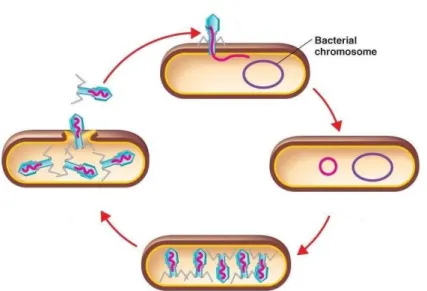
19 Lysogenic life cycle
In the lysogenic cycle (Figure 2-8), the phage starts also by injecting its genome into the bacterial cell, but, unlike the lytic cycle, at the end of the cycle the bacterial host is not lysed. The phage genome is integrated into the bacterial DNA and when the cell replicates, the genome of the phage is passed to each daughter cell genome (prophages). Cells can do some rounds of division, but eventually one of the cells will suddenly be lysed and will release phage progeny.
Figure 2-8 Schematization of lysogenic life cycle (adapted from (Melo et al., 2012)). Figure 2-7 Schematization of lyic life cycle (adapted from (Melo et al., 2012)).
ANTIMICROBIAL ASSESSMENT OF PHAGES ENTRAPPED IN BIO-BASED STRUCTURES
20
Phage ΦIBB-PF7A
Phage ΦIBB-PF7A, a specific phage for Pseudomonas fluorescens, was isolated from raw sewage and characterized previously (S. Sillankorva, Neubauer, & Azeredo, 2008). This phage belongs to the Podoviridae family, which is characterized by phages with a short non-contractile tail. It has also an icosahedral capsid with 63 nm of diameter and a tail size of13 nm x 8 nm, tapering, and genomically belongs to the T7likeviruses. This phage has a latent period of 10 minutes and a burst size of 153 plaque forming units per infected cell (S. Sillankorva et al., 2011, 2008).
2.3
Pseudomonas fluorescens
P. fluorescens belongs to the Pseudomonadaceae family and to the Pseudomonas genus. This bacterium has the form of a straight or curved bacillus, with a size between 0.5 μm to 1.0 μm by 1.5 μm to 4.0 μm (Figure 2-9). Pseudomonas are Gram-negative, motile due to polar flagella. They do not form spores and have a respiratory metabolism (never fermentative). They are aerobic, however, they can grow in an anaerobic environment, using in this case nitrate instead of oxygen as an electron acceptor in the respiratory chain (Sá-Correia, 2000; Silva, 2014). This bacterium belongs to the group of fluorescent bacteria, producing a water soluble pigment, called pyoverdin, which is yellowish and fluorescent when radiated with ultraviolet light. This pigment is a siderophore that is located in the cytoplasmic membrane and operates in the seizure of iron (Silva, 2014).
Pseudomonas has the particularity of having very simple nutritional requirements because it uses various organic compounds as carbon and energy sources, it can grow chemotrophically at
2.STATE-OF-THE-ART
21 neutral pH and has an optimum growth temperature between 25 °C and 30 °C (Sá-Correia, 2000; Silva, 2014).
P. fluorescens is very commonly present in water and soil, and can grow at temperatures as low as 4 °C. They also have the ability to hydrolyse lipids and proteins, and consequently, are responsible for spoilage of refrigerated foods such as meat, milk, eggs and seafood. Due to their ability to use a variety of carbon sources, including simple nitrogen compounds that are present in food products, P. fluorescens is able to synthesize growth factors and produce oxidized compounds that negatively affect the smell and taste of food. Furthermore, this bacterium is also very resistant to many disinfectants that are used in the food industry (Sá-Correia, 2000; Silva, 2014). This microorganism is rarely pathogenic, because of its difficulty of growing at 37 °C, however, it can be a source of blood and blood products contamination when they are refrigerated (Sá-Correia, 2000; Silva, 2014).
23
Chapter
3
M
ATERIALS
A
ND
3. MATERIALS AND METHODS
25
3.
M
ATERIALS
A
ND
M
ETHODS
3.1
Sodium alginate films
Film forming solutions were prepared with different types of sodium alginate (Protanal SF 120RB; Protanal CR 8223; Protanal LFR 5/60; Protanal GP 1320 and Protanal Manugel) using different concentrations (2.5 g/L, 5 g/L, 7.5 g/L, 10 g/L and 15 g/L). The films were prepared in distilled water by adding the sodium alginate and under stirring until its complete solubilization, which took approximately 18 h. Afterwards, 5 g/L of glycerol was added, and was left stirring overnight at room temperature (approximately 22 °C). The film-forming solutions were cast in polystyrene petri dishes with the respective volumes: 13 mL to 45 mm petri plates, 28 mL to 96 mm petri plates and 71 mL to 140 mm petri plates. Then, plates were stored in an oven at 30 °C during 48 h to dry (J.-W. Rhim, 2004). Based on the processability of the films and their capability to form stand-alone and resistant films, two types of sodium alginate were chosen.
Alginate CR 8223 (FMC BioPolymer) with M/G ratio of 65/35 and a molecular weight (MW) of 300 kDa, and Manugel (FMC BioPolymer) with M/G ratio 30/70 and a MW < 200 kDa, were selected and produced for subsequent crosslinking and characterization.
3.1.1 Films crosslinking
Solutions of calcium chloride (CaCl2) with different concentrations 2.5 g/L, 5 g/L, 7.5 g/L, 10 g/L, 12.5 g/L and 15 g/L, were used to crosslink alginate-based films, and each solution was prepared by slowly adding CaCl2 powder in a constantly stirred distilled water solution. SF120RB, CR8223, LFR5/60, GP1320 and Manugel films were prepared in order to analyze their behaviour when crosslinked with different concentrations of CaCl2, and to select the most suitable to form the pretended films. After several tests, the selected films were alginate CR 8223 and Manugel with a concentration of 10 g/L, crosslinked with CaCl2 with concentrations of 10 g/L, 12.5 g/L and 15 g/L.
The film’s crosslinking process was adapted from the method described by (J.-W. Rhim, 2004). Briefly, the solution of CaCl2 was added to the films in the petri plates using a pipette, being used 1.900, 4.092 and 10.400 mL of calcium chloride in the petri plates of 45, 96 and 140 mm of diameter, respectively. The volume of CaCl2 solution used to crosslink films was determined based on preliminary studies, aiming a total volume that covered the film. Alginate films were left
ANTIMICROBIAL ASSESSMENT OF PHAGES ENTRAPPED IN BIO-BASED STRUCTURES
26
with thecrosslinkersolution for 5 min, and after that the CaCl2 was removed and films were left to dry at room temperature overnight. The dried films were then preconditioned in a desiccator containing a satured solution of Mg(NO3)2.6H2O at 53 % of RH and room temperature (≈ 22 °C) before being analyzed.
3.2
Characterization of sodium alginate films
3.2.1 Moisture content
Moisture content was determined according to the method described by (J. W. Rhim, Gennadios, Weller, Carole Cezeirat, & Hanna, 1998). Each treated film was cut in a circle with 2 cm of diameter and conditioned again in the desiccator at 53 % of RH for more 24 h. Three samples of each film were weighed and dried at 105 °C for 24 h, and after that they were weighed again. Moisture content (MC) was determined in percentage using the following equation 3.1:
= − × ( 3.1 )
where M0 is the initial mass of the film and M is the mass of the film after drying. Moisture content
was measured at least in triplicate for each type of film, and an average was taken as a result.
3.2.2 Water solubility
For water solubility, film samples were cut in circles with 2.6 cm of diameter, dried at 105 °C during 24 h and placed in beakers containing 50 mL of distilled water. After sealing beakers with parafilm, these were placed in an orbital agitator at 150 rpm at room temperature for 24 h. Insolubilized dry pieces collected from the beakers were dried at 105 °C for 24 h and weighed (J. W. Rhim et al., 1998).
The solubility of films was determined based on the equation 3.2, where M0 is the initial
mass of the film and M is the mass of the insoluble pieces of the film after drying. Water solubility was calculated in triplicate (at least) for each type of film, and an average was taken as a result.
3. MATERIALS AND METHODS
27 3.2.3 Sorption isotherms
Sorption isotherms of alginate films were determined based on protocols (Ortman, n.d.) and (Kou, 1999). Initially, films were cut in a shaped circle and were conditioned in a desiccator containing silica with a RH of 0 g/L, at room temperature, for 24 h. Afterwards, sample cups were prepared with saturated salt solutions of LiCl, MgCl2, Mg(NO3)2, NaCl and K2SO4 for 0.11, 0.23, 0.53, 0.76 and 0.97 of RH, respectively.
Sorption isotherms were determinate using the equipment Aqualab (Aqualab 4TE, Pullman, Washington) at a 25 °C, where, on top of the receptacle containing 5 mL of salt solutions were placed a circle sample of the alginate film. After the stabilization of the film to each salt solution, samples were weighed and the value of the aw was observed. Adsorption test was done
for each type of film starting with the salt solution with a lower RH (LiCl – 0.11 RH) to the salt with a highest RH (K2SO4 – 0.97 RH).
3.2.4 Colour and opacity
Alginate films colour and opacity were measured using a colourimeter (CHROMA METER CR-400/410, AQUATEKNICA, SA, Konica Minolta, Japan). Calibration of the equipment was performed on a white standard plate (Y=93.9; x=0.3158; y=0.3321) that was also used as a background to determine the colour of each alginate films. L*, a* and b* parameters were evaluated by reflectance measurements (Cerqueira, 2010). The model of CIELab is demonstrated in Figure 3-1, where the a axis extends from green (-a) to red (+a) and the b axis from blue (-b) to yellow (+b); the brightness (L) increases from the bottom (L=0, black) to the top (L=100, white) of the three-dimensional model (CIELab, 1976). The values of a and b approaching zero means neutral colours, and as the colour becomes more chromatic and saturated values increase.
The RGB colour of each film was obtained based on L*, a* and b* values and was calculated using MATLAB, where the function lab2rgb([L a b],‘OutputType’,’unit8’) was introduced, and the RGB values were obtained. With the RGB values it was possible to determine the colour code on the site: http://www.rapidtables.com/web/color/RGB_Color.htm, and to know the exact colour of the film the code was inserted in the website http://www.color-hex.com/color/f7f3ec.
ANTIMICROBIAL ASSESSMENT OF PHAGES ENTRAPPED IN BIO-BASED STRUCTURES
28
Also, the opacity of each film was determined. The opacity of a material indicates how much light passes through it, so the more opaque the film is lower is the quantity of light that can pass through it. The opacity was determined according to the Hunter Lab method, that relates the opacity of each sample on a black standard plate (Yb) and the opacity of the same sample on a
white standard plate (Yw) (equation 3.3). Ten measurements were performed for each film and the
mean values were used to determinate each parameter.
� � % = � � ( 3.3 )
3.2.5 Fourier transform infrared (FTIR)
The films were characterized by Fourier Transform Infrared (FTIR) Spectroscopy on a Bruker FT-IR VERTEX 80/80v (Boston, USA) in Attenuated Total Reflectance mode (ATR) with a platinum crystal accessory between 400 and 4000 cm-1, using 16 scans at a resolution of 4 cm-1. Before analysis, an open bean background spectrum was recorded as a blank.
3.2.6 Film thickness
The thickness of each film was measured with a micrometer (No. 293-5, Mitutoyo, Japan). Measurements were taken on ten different places on rectangular strips of 10×2 cm, and the average thickness value was calculated. At least six measures were made for each film.
3. MATERIALS AND METHODS
29 3.2.7 Mechanical properties
The mechanical properties of the films were measured following the methodology described by ASTM D882-10 and using the texturometer (TA.HD plus, Stable Micro Systems, United Kingdom) with the software Exponent. The samples were conditioned at room temperature and with a relative humidity of 53 %. Each film strip was clamped between grips with an initial distance of 100 mm. The force and deformation were analysed during extension at 50 mm min-1. Results of tensile strength (TS) and elongation-at-break (EB) were obtained in MPa and percentage, respectively. At least six strips of each film were measured.
3.2.8 Swelling
The swelling index (SW) of films was determined as described by (Cao, Fu, & He, 2007) with some modifications. The films were cut in squares with 4 cm2 of size, and after conditioned in a desiccator containing a saturated solution of Mg(NO3)2·6H2O at 53 % RH at room temperature (≈ 22 °C) during 24 h, the films were weighed. Afterwards they were immersed in glass beakers with 100 mL of distilled water for 24 h at room temperature. Then, samples were wiped with paper filter to remove liquid excess and were weighed again. The quantity of absorbed water in percentage was calculated using equation 3.4, where S1 is the weight of the wet film and S0 is the initial weight
of the film:
�� = � − �� × ( 3.4 )
Swelling index was calculated in triplicate for each type of film, and an average was taken as a result.
3.2.9 Contact angles
The contact angle of films was measured by the drop method based on protocol of (Galus & Kadzińska, 2016) in the equipment (System OCA20 DATAPhysics, Germany). Briefly, a droplet of ultrapure water was placed on the film surface using a syringe Hamilton with a volume of 2 µL with a drip rate of 1 µL/s. Then, the angle formed between the film and the water drop was calculated with the image/video analysis software of the equipment. For each film ten measurements were performed.
ANTIMICROBIAL ASSESSMENT OF PHAGES ENTRAPPED IN BIO-BASED STRUCTURES
30
3.3
Bacteriophage production
Phage production was performed according to well established protocols (Sambrook & Russell, 2001). Briefly, 100 µL of the overnight grown bacteria and 3 mL of top agar (LB with 6 g/L of agar) were added to petri dishes (96 mm) containing LB medium with 15 g/L of agar (LBA). After the top agar layer was dry, 10 µL of phage ΦIBB-PF7A was added and spread with the help of a sterile paper strip. After overnight incubation at room temperature, 3–5 mL of SM buffer (5.8 g L-1 NaCl, 2 g L-1MgSO
4.7H2O, 50 mL L-1 1M TRIS ((HOCH2)3CNH2, pH 7.5) were added to each plate and incubated under agitation (90 rpm) at 4 °C during 18 h–24 h. The SM buffer with the eluted phages was collected to Erlenmeyers and 5.84 g NaCl were added per each 100 mL of mixture. After the NaCl was completely dissolved by stirring gently, the mixtures were incubated during 1h, at 4°C and 90 rpm. The obtained mixture was centrifuged at 9000 × g, per 10 min, at 4 °C, to precipitate non-lysed bacteria, and the supernatant was collected. Ten grams of polyethylene glycol 8000 were added per each 100 mL of solution, and this was incubated with agitation at 90 rpm, during approximately 24 h at 4 °C. After this, phage particles were recovered in the pellet after centrifugation (10 min, 9000 × g at 4°C) and ressuspended in approximately 7 mL of SM buffer. The phage solution was purified from bacterial debris by adding chloroform in a proportion of 1:4. After adding the chloroform, the mixture was homogenized during 30 seconds by vortexing, and then centrifuged for 5 min, at 4 °C, 3500 × g. The aqueous phase containing pure phage particles was transferred to a sterile Falcon.
3.3.1 Bacteriophage titration
To determine the phage titre in a sample, the tear drop plaque assay was used (Oliveira, Milho, & Vilas boas, 2013). Briefly, serial dilutions (1:10) of the sample were prepared in SM buffer. An overnight bacterial culture, grown at 30 C (100 µL), and 3–5 mL of top agar were poured onto LBA, and the petri dishes were swirled to evenlyspread the bacteria. The petri dishes were left to dry, and then 10 µL of each dilution was dropped on the agar and the dishes were tilted at 45 until the tear drop reached close to the end of the dish. The petri dishes were allowed to dry and were incubated overnight at room temperature. In the next day, the plaques forming units (PFUs) in the drop (Figure 3-2) containing 3–30 plaques were counted. The phage titre was calculated using Equation 3.5.
3. MATERIALS AND METHODS
31
Number of phages PFUs/mL = �� × � � ( 3.5 )
3.4
Pseudomonas fluorescens
To relate the bacterial growth of P. fluorescens with the optical density during time, first a fresh inoculum was prepared and it was left stirring overnight at 120 rpm and 30 °C. Optical density was measured at 620 nm at different time points and the number of colony forming units (CFUs/mL) was also measured. With the results from optical density and bacterial concentrations over time it was possible to draw the calibration curve of P. fluorescens growth, being equation 3.6 determined:
��
= × × − 5 × ( 3.6 )
3.4.1 Bacteria titration
To determine the number of colony forming units (CFUs) of P. fluorescens in a sample, the tear drop technique assay was used. First, serial dilutions of 1:10 of the sample were prepared (900 µL of NaCl 9 g/L +100 µL of sample). Then, in plates with solid LBA, 10 µL of each dilution was dropped on the agar and the petri dishes were tilted at 45 until the tear drops reached close to the end of the dish. After drying, the plates were incubated overnight at room temperature, or at
ANTIMICROBIAL ASSESSMENT OF PHAGES ENTRAPPED IN BIO-BASED STRUCTURES
32
30 °C, and in the next day the colony forming units (CFUs) in the drop containing 3-30 colonies were counted. The bacteria concentration was calculated using equation 3.7.
Number of viable bacteria CFUs/mL = �� × � � ( 3.7 )
3.5
Alginate films with entrapped phages
A solution of 10.8 g/L concentration of sodium alginate was prepared with distilled water, to obtain a final concentration (with phage) of 10 g/L. This solution was stirred at room temperature until it was completely dissolved. After this, it was added glycerol in a concentration of 5 g/L, and it was left stirring overnight. Then, the phage sample was added to this solution (1 mL per each 12 mL of sodium alginate solution) and stirred for 30 min before it was poured into petri dishes. To dry, the plates were incubated at 30 °C for 48 h.
For the crosslinking of alginate films with incorporated phages, a CaCl2 solution of 10.8 g/L was prepared by adding slowly the CaCl2 powder to a stirred distilled water solution at room temperature. After dissolving the calcium chloride, the solution of phages (1 mL of phage sample per each 12 mL of calcium chloride solution) was added, and after stirring during approximately 15 minutes, it was added to the films on the petri plates using a pipette. It was used 1.900 mL and 4.092 mL of CaCl2 in the plates with films of 45 and 96 mm, respectively. Alginate films were left with thecross linkersolution for 5 minutes, and after that, the CaCl2 was removed and films were left to dry at room temperature, overnight.
3.5.1 Films with entrapped phages titration
To determine the number of phages in alginate films crosslinked with CaCl2 the films were placed in a Falcon with 20 mL of SM buffer and stirred for 45 min at 250 rpm, in order to release all the phages into the buffer. Then, the procedure described in section 3.3.1. was followed.
3. MATERIALS AND METHODS
33
3.6
Antimicrobial activity of alginate films with entrapped phage
The evaluation of the antimicrobial performance of the alginate films with entrapped phages was performed using a modification of the standard 206 JIS Z 2801 (JIS Z 2801, 2010). The test was performed at different temperatures (room temperature, 22 °C, and refrigerated temperature, 4 °C) and at different periods of time (0 h, 4 h, 8 h, 12 h, 24 h). The films were cut in 2×2 cm squares and on top of them it was added 50 µL of P. fluorescens with different concentrations. Alginate films without phages but with bacteria were used as control for each time and temperature. Concentration of bacteria was calculated based on equation 3.6 that relate OD with concentration of bacteria.
After each period of time the different films with bacteria were placed in Falcons with 2 mL of SM buffer and stirred at 250 rpm for 15 min, and vortexed during 30 s. To relate the decrease of the bacteria concentration with the release and action of phages, the concentration of P. fluorescens and phages was determined as described in section (3.3.1) and (3.4.1).
3.7
Antimicrobial activity of alginate films with entrapped phage in
chicken meat
To evaluate the capacity of ΦIBB-PF7A phage to control P. fluorescens in chicken meat, CR 8223 10 g/L alginate films with incorporated phages were prepared as described in 3.5. After this, the concentration of phage in the prepared films was determined as described in section 3.3.1. As a control, calcium-alginate films without phages were prepared as described in section 3.1. Then, it was prepared a fresh inoculum of P. fluorescens with LB medium, that grew overnight at 30 °C, with an agitation of 120 rpm. Later, chicken meat was cut in little cubes, and these were immersed in ethanol 70 % (v/v) for 30 s and let to dry. In a petri dish it was poured 100 µL of bacteria inoculum and a cube of meat was steeped in it, until all the liquid was absorbed by the meat sample. At the bottom of sample cups, it was placed a film, with or without phages (control), and on top of each film it was placed a piece of chicken meat. All samples were kept at 4 °C.
To analyse the effect of phages present in the films on P. fluorescens growth during time, at 0 h, 0.5 h, 1 h, 2 h, 3 h, 4 h, 6 h, 8 h, 12 h, 22 h and 24 h timepoints it was determined the CFUs/mL in the samples of chicken meat, as described in 3.4.1, and the PFUs/mL of each film (with phage) as described in 3.3.1.