Original article doi:10.12980/JCLM.3.2015J5-11 ©2015 by the Journal of Coastal Life Medicine. All rights reserved.
Effect of ions on the activity of brain acetylcholinesterase from tropical ish
Caio Rodrigo Dias Assis1*
, Amanda Guedes Linhares1
, Vagne Melo Oliveira1
, Renata Cristina Penha França1
, Juliana Ferreira Santos2 , Elba Verônica Matoso Maciel Carvalho3, Ranilson Souza Bezerra1, Luiz Bezerra Carvalho Jr1
1
Laboratory of Enzymology-LABENZ, Biochemistry Department and Laboratory of Immunopathology Keizo Asami, Federal University of Pernambuco, Recife-PE, Brazil
2
Academic Unit of Serra Talhada, Federal Rural University of Pernambuco, Serra Talhada-PE, Brazil
3
Laboratory of Glycoproteins, Biochemistry Department, Federal University of Pernambuco, Recife-PE, Brazil
Journal of Coastal Life Medicine
*Corresponding author: Caio Rodrigo Dias de Assis, Laboratory of Enzymology-LABENZ, Biochemistry Department and Laboratory of Immunopathology Keizo Asami, Federal University of Pernambuco, Recife-PE, Brazil.
Tel: + 55 81 21268540 Fax: + 55 81 21268576 E-mail: [email protected]
Foundaton Project: Supported by Fundação de Apoio à Ciência e Tecnologia do Estado de Pernambuco (FACEPE) for financial support (Grant numbers: IBPG-1301-2.08/08 FACEPE, IBPG-0523-2.08/11 FACEPE, BFP-0036-2.08/13 FACEPE and BFP-0111-2.08/13).
1. Introduction
Acetylcholinesterase (AChE, EC 3.1.1.7) is a crucial enzyme for the development and functioning of the nervous system and play an important role in hematopoietic differentiation and neural development[1]. Its classical function is to modulate the nerve impulse through the hydrolysis of the neurotransmitter acetylcholine in the synaptic cleft[2]. AChE inhibition is the mechanism of action of organophosphorus and carbamate pesticides, as well as the mode
of action of the drugs used in treatment of Alzheimer’s disease[2]. Therefore, AChE has been also used for monitoring these pesticide exposures in vivo[3] and in vitro[4] and even as a biocomponent of biosensors[5].
The investigation of AChE inhibitors and interfering substances is relevant to identify the usefulness of this enzyme as a tool in environmental and food monitoring[5-7]. Monitoring at biochemical level can specifically detect the presence of contaminants in the environment before they reach higher organizational levels[8].
Several studies reported inhibition of AChE activity by ions[9-11]. AChE activation by Ca2+, Mg2+, Al3+ has also been reported[12,13]. Therefore, high content of these ions in water samples from rivers, lakes and other environments can influence the detection of anticholinesterasic pesticides. These findings must be taken into account when biosensors based on AChE activity are proposed to analyze pesticide presence under some environment conditions. This fact can lead to false positives or negatives and misinterpretations in the analysis of results. Cholinesterase inhibition has been assayed in several species, including aquatic organisms, since the event effectively A RT I C L E I N F O A B S T R AC T
Objective:To investigate the effect of ions on brain acetylcholinesterase (AChE; EC 3.1.1.7)
activities from economic important fish [pirarucu, Arapaima gigas; tambaqui, Colossoma
macropomum; cobia, Rachycentron canadum (R. canadum) and Nile tilapia, Oreochromis
niloticus (O. niloticus)] comparing with a commercial enzyme from electric eel [Electrophorus
electricus (E. electricus)].
Methods: The in vitro exposure was performed at concentrations ranging from 0.001 to 10
mmol/L (except for ethylene diamine tetraacetic acid; up to 150 mmol/L). Inhibition kinetics
on R. canadum and O. niloticus were also observed through four methods (Michaelis-Menten,
Lineweaver-Burk, Dixon and Cornish-Bowden plots) in order to investigate the type of inhibition produced by some ions.
Results: Hg2+ , As3+
, Cu2+ , Zn2+
, Cd2+
caused inhibition in all the species under study. Ca2+ , Mg2+ and Mn2+
induced slight activation in R. canadum enzyme while Pb2+ , Ba2+
, Fe2+ , Li+
inhibited the AChE from some of the analyzed species. The lowest IC50 and Ki values were estimated
for E. electricus AChE in presence of Hg2+
, Pb2+ , Zn2+
. Under our experimental conditions, the results for R. canadum and O. niloticus, As3+
, Cu2+ , Cd2+
, Pb2+
and Zn2+
showed a non-competitive/mixed-type inhibition, while Hg2+
inhibited the enzyme in a mixed/competitive-like manner.
Conclusions: E. electricus AChE activity was affected by ten of fifteen ions under study showing that this enzyme could undergo interference by these ions when used as pesticide biosensor in environmental analysis. This hindrance would be less relevant for the crude extracts.
Article history:
Received 9 Feb 2015
Received in revised form 28 Feb 2015
Accepted 22 Apr 2015 Available online 9 Jun 2015
Keywords:
Ions
Acetylcholinesterase Biomarkers Fish
mirrors environmental impact even when these compounds are not present in the water due to the fact that they frequently remain attached to the enzyme.
This study investigated the effect of different ions (Al3+ , As3+
, Ba2+, Ca2+, Cd2+, Cu2+, EDTA2-, Hg2+, K+, Li+, Fe2+, Mg2+, Mn2+, Pb2+ and Zn2+) that could influence/interfere on the activity of brain AChE from three freshwater species of economic importance in aquaculture: Nile tilapia [Oreochromis niloticus (O. niloticus)], tambaqui [Colossoma macropomum (C. macropomum)], pirarucu
[(Arapaima gigas (A. gigas)]; one saltwater farmed species: cobia
[Rachycentron canadum (R. canadum)] and a commercial enzyme
from electric eel [Electrophorus electricus (E. electricus)], providing information about their inhibitory behaviour and their potential interference in the use of AChE from these species as a biomarker for the presence of anticholinesterase compounds. In our previous studies, AChE from the same species was physicochemical and kinetically characterized and used to investigate the effect of organophosphorus and carbamate pesticides showing sensitivity comparable to a commercial and purified enzyme[14].
2. Materials and methods
2.1. Materials
AChE from electric eel E. electricus type VI-S, Acetylthiocholine iodide, bovine serum albumin, 5,5’-dithiobis(2-nitrobenzoic) acid (DTNB), tris (hydroxymethyl) aminomethane e magnesium sulphate were purchased from Sigma-Aldrich (St. Louis, MO, USA). Hydrogen chloride, aluminium chloride, barium chloride, calcium chloride, lithium chloride and sodium arsenite were obtained from Merck (Darmstadt, Germany). Cadmium chloride, copper chloride, ferrous chloride, manganese chloride, lead chloride and zinc chloride were acquired from Vetec (Rio de Janeiro, Brazil). Disodium EDTA, mercuric chloride and potassium chloride were from Reagen (Rio de Janeiro, Brazil). The microplate spectrophotometer used was Bio-Rad xMark™ (Hercules, CA, USA) whereas the tissue disrupter was IKA RW-20 digital (Staufen, Germany). The juvenile specimens of C. macropomum [(30.0 ± 4.2) cm; (512.5 ± 123.7) g], A. gigas [(76.8 ± 8.7) cm; (4 118.0 ±
207.9) g] and O. niloticus [(12.0 ± 3.0) cm; (7.9 ± 1.2) g] were supplied
by the Department of Fisheries and Aquaculture of the Universidade Federal Rural de Pernambuco (Recife, PE, Brazil). R. canadum [(51.67 ± 1.50) cm; (1 575.0 ± 329.6) g] was supplied by Aqualider Ltda. (Recife, PE, Brazil).
2.2. Enzyme extraction
The juvenile fishes were cultured under appropriate conditions and were sacrificed in ice bath (0 °C). The whole brains were immediately removed, pooled (from 5 per pool for R. canadum to 30 per pool
for O. niloticus) and homogenized in 0.5 mol/L Tris-HCl buffer,
pH 8.0, maintaining a ratio of 20 mg of tissue per mL of buffer. The homogenates were centrifuged for 10 min at 3 320 r/min (4 °C) and the supernatants (crude extracts) were frozen at -20 °C for further assays.
2.3. Enzyme activity and protein determination
Enzyme activity was evaluated using an adaptation of Ellman’s method according to Assis et al.[14]. Briefly, 0.25 mmol/L DTNB
(200 μL) prepared in 0.5 mol/L Tris-HCl buffer pH 7.4 was added to the crude extract (20 μL), and the reaction started by the addition of 62 mmol/L acetylthiocholine iodide (20 μL) except for the C.
macropomum assays (125 mmol/L). Enzyme activity was determined
by reading the absorbance increase at 405 nm for 180 seconds. A unit of activity (IU) was defined as the amount of enzyme capable of converting 1 μmol/L of substrate per min. A blank was prepared with the buffer instead crude extract sample. Protein content was estimated according to Sedmak and Grossberg[15], using bovine serum albumin as the standard.
2.4. Activity in presence of ions
AChE activity was assayed at 25 °C in presence of fifteen ions: Al3+
(AlCl3), Ba 2+
(BaCl2), Ca 2+
(CaCl2), Cd 2+
(CdCl2), Cu 2+
(CuCl2 and CuSO4), Fe
3+
(FeCl3), Hg 2+
(HgCl2), K +
(KCl), Li+ (LiCl), Mg2+ (MgSO4), Mn2+ (MnCl2), As3+ (NaAsO2), Pb2+ (PbCl2 and Pb(C2H3O2)2), Zn2+ (ZnCl2) and the complex chelating ion EDTA 2-as C10H14N2Na2O8. The ions were diluted to five concentrations ranging from 0.001 to 10 mmol/L (each concentration 10-fold higher than the previous one) excepting EDTA2- which was assayed in concentrations up to 150 mmol/L. The ions solutions (10 μL) were incubated with crude extract (10 μL) for 40 min[6]. In order to minimize false negatives through thiobis-nitrobenzoate (TNB) and thiocholine reactions with some inhibitory ions, the incubation were performed only with the ions and the enzymatic extract and the blanks were performed with buffer instead of enzymatic preparation subtracting these interferences and spontaneous substrate hydrolysis. After the incubation, DTNB (200 μL) was added right before the
substrate acetylthiocholine (20 μL) and the mixture was read at 405 nm for 180 second. The controls were performed with distilled water in the incubation instead of the ions solutions. The activity in the absence of the ions was considered as 100%.
Some assays were also carried out with activator ions in order to verify false positive occurrence by an eventual binding to DTNB: before DTNB and substrate addition, 10 μL of the samples were incubated
for 40 min with 10 μL of 10 mmol/L neostigmine bromide (a total cholinesterase inhibitor) and with 10 μL of each of these ions (10 mmol/L). Blanks were performed replacing the samples by buffer and following the same procedure.
2.5. Inhibition kinetics
Samples of O. niloticus and R. canadum preparations were incubated with the most inhibitory ions (As3+, Cu2+, Cd2+, Hg2+, Pb2+ and Zn2+) at six concentrations (0 to 10 mmol/L) and hyperbola model curves were produced with fourteen substrate concentrations ranging from 0 to 20.83 mmol/L to obtain the kinetic parameters in presence or absence of ions (kmapp, Vmapp and km, Vmax, respectively). Then, data were transformed to double reciprocal (1/vvs 1/s), Dixon (1/v vs i) and Cornish-Bowden (s/v vs i) plots in order to investigate the kinetic behaviour of the ions towards AChE and to distinguish unambiguously the types of inhibition[16,17].
The dissociation constant of the enzyme-inhibitor complex (ki) was estimated for competitive, mixed and non-competitive inhibitor ions using the intersection of linear regression curves from different concentrations of substrates in the Dixon plots[17] and also using Cheng and Prusoff equation[18]:
Ki= IC50
1+
km [S]
Where IC50 is the concentration capable of inhibiting 50% of enzyme activity. [S] represents substrate concentration and km is the Michaelis-Menten constant.
from the intersection of linear regression curves generated from different concentrations of substrates in the Cornish-Bowden plots[16].
2.6. Statistical analysis
In the previous sections, means of treatments and means of kinetic parameters in presence or absence of inhibitory ions were statistically analyzed using One-way ANOVA followed by Tukey’s test. In section 2.4., data were fitted to linear and non-linear regression through sigmoidal (Boltzmann) or exponential decay (P < 0.05) modelling using MicroCal襆 Origin襆 version 8.0 in order to estimate the concentration capable to inhibit enzyme activity in 50% (IC50).
3. Results
3.1. Activity in presence of ions
Table 1 reports the results referring to 1 mmol/L concentration of ions. Activation effect was only observed for the ions Mg2+
(13%) and Mn2+
(38%) on the R. canadum AChE. Ca2+
induced an increase of approximately 30% in R. canadum AChE activity at 10 mmol/L (data not shown). The inhibitions found here for Cu2+ and Zn2+ at 1 mmol/ L were, respectively, 75% and 23% (R. canadum), 75% and 78%
(E. electricus). Cu2+
inhibited (A. gigas) 23% of enzymatic activity, behaviour not induced by Zn2+ in this species. Zn2+ induced 35 (C.
macropomum) and 29% (O. niloticus) inhibition. At 1 mmol/L, Pb2+
was able to inhibit the enzyme from A. gigas (32%), R. canadum
(15%), E. electricus (71%). Cadmium induced inhibitions of 33% (R.
canadum), 49% (E. electricus) and 35% (O. niloticus). As3+ inhibited
C. macropomum (57%), R. canadum (63%), E. electricus (57%) and
O. niloticus (61%) enzyme activities at 1 mmol/L. Ba2+
, Fe2+ and Li+ induced, under our experimental conditions, similar pattern (Table 1) and only E. electricus was significantly sensitive to these ions at 1 mmol/L. The chelating ion EDTA2- only inhibited R. canadum (6%) and
E. electricus (28%) at 1 mmol/L. The enzymes from the other species
under study were significantly inhibited only on the range of 50-100 mmol/L by this ion.
Among the fifteen ions analyzed, the most inhibitory ion was Hg2+, which completely inactivated AChE from all the species under study when they were exposed to 1 mmol/L. However, the enzyme activity from A. gigas was less inhibited (71%) than the others.
No statistical difference was observed between activator ions (Al3+, Ca2+
, Mg2+ , Mn2+
and K+
) action on brain AChE from O. niloticus, C.
macropomum, A. gigas and R. canadum incubated with neostigmine
bromide and their respective blanks in order to investigate interferences in the colorimetric readings by them.
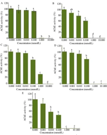
Figure 1 displays an example of typical inhibitions plots showing the effect of Hg2+ ion on AChE of the species under study and from which were estimated the IC50 values for this ion as well as all other ions.
120
100
80
60
40
20
0
120
100
80
60
40
20
0
120
100
80
60
40
20
0
120
100
80
60
40
20
0 120
100
80
60
40
20
0
A
ChE acti
vity (%)
A
ChE acti
vity (%)
A
ChE acti
vity (%)
A
ChE acti
vity (%)
A
ChE acti
vity (%)
A
C D
E a
a a
a
a
a
b
b a
a a
b a
a a a ab
b
b
b
b
c
c
c c
c c
d
d
d 0.000 0.001 0.010 0.100 1.000 10.000
Concentration (mmol/L) B
Figure 1. AChE activity from five species exposed to Hg2+ .
A: O. niloticus; B: C. macropomum; C: A. gigas; D: R. canadum; E: E. electricus; Data were compared using ANOVA and Tukey’s test (P < 0.05).
0.000 0.001 0.010 0.100 1.000 10 .000 Concentration (mmol/L)
0.000 0.001 0.010 0.100 1.000 10 .000 Concentration (mmol/L) 0.000 0.001 0.010 0.100 1.000 10.000
Concentration (mmol/L)
0.000 0.001 0.010 0.100 1.000 10.000 Concentration (mmol/L)
3.2. Inhibition kinetics
Table 1 also shows the IC50 related to the ions towards the species under study. E. electricus was the most sensitive species presenting the lowest values for Cu2+, Hg2+, Pb2+, Zn2+ and was the only species here to present this parameter for Ba2+, EDTA2-, Fe2+ and Li+. C.
macropomum and R. canadum presented low IC50 values for As
3+ and
Table 1
Inhibition and IC50 of AChE activity by ions and heavy metals from several freshwater and marine species at 1 mmol/L.
Species O. niloticus C. macropomum A. gigas E. electricus R. canadum
Inhibition (%)
IC50 (mmol/L)
Inhibition (%)
IC50 (mmol/L)
Inhibition (%)
IC50 (mmol/L)
Inhibition (%)
IC50 (mmol/L)
Inhibition (%)
IC50 (mmol/L) Al3+
ne - ne - ne - ne - 13
-As3+ 61 0.58 57 0.32 ne - 57 0.98 63 0.21
Ba2+
ne - 25 - ne - 60 0.05 ne
-Cd2+
35 - ne 6.30 ne - 50 1.00 33 1.10
Cu2+
ne - ne 4.13 23 5.77 75 0.05 75 0.37
EDTA2- ne - ne - ne - 28 21.25 6
-Fe2+
ne - 20 - 15 - 43 1.16 ne
-Hg2+
100 0.24 100 0.13 71 0.38 100 0.01 100 0.12
K+
ne - ne - ne - 26 - ne
-Li+ ne - ne - ne - 57 0.38 ne
-Mn2+
ne - ne - ne - 24 - 38*
-Pb2+
ne - ne - 32 - 71 0.01 15
-Zn2+
23 35 ne 78 23
ne: No effect at 1 mmol/L (P< 0.05); –: No IC50 estimated at 1 mmol/L; *
Hg2+ whereas brain AChE activity from A. gigas was less affected by the ions. The ki values using Cheng and Prusoff equation[18]
followed the same trend on Table 2 where the lowest values occurred with E. electricus exposed to Hg2+
, Pb2+ and Zn2+
. From now on the results are related to only two species: R. canadum and O. niloticus. The other species behaved similarly.
Tables 3 and 4 show kinetic parameters km, Vmax and their inhibited analogues kmapp and Vmapp based on hyperbola model for brain AChE activity from R. canadum and O. niloticus. Using this model, Hg2+ showed a competitive-like trend of behaviour for both species. Cu2+ and Cd2+
presented non-competitive inhibition whereas As3+ , Pb2+
and Zn2+
showed mixed-type inhibition.
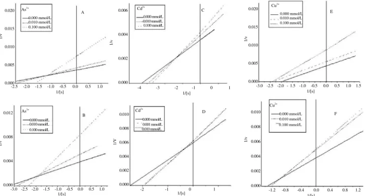
Figures 2 and 3 allow comparison between inhibitory effect of ions
on R. canadum and O. niloticus brain AChE activity using
Lineweaver-Burk regression plots. These results corroborate Tables 3 and 4 in relation to As3+, Pb2+ and Zn2+. However, Cd2+, Cu2+ and Hg2+ ions showed mixed-type inhibition according to Lineweaver-Burk plots. The behaviour was similar between both species, excepting Cu2+
.
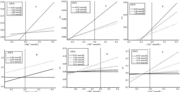
Figures 4-7 present a comparison between Dixon and Cornish-Bowden regression plots in which the types of inhibitory effects were confirmed by both graphical methods. In these figures, Hg2+ presents a competitive-like inhibitory effect. Table 5 provides another estimate of
Table 2
ki (μmol/L)* of fifteen ions and heavy metals from several freshwater and marine species.
Species Al3+
As3+ Ba2+
Ca2+ Cd2+
Cu2+
EDTA 2-Fe2+
Hg2+ K+
Li+ Mg2+
Mn2+ Pb2+
Zn2+
O. niloticus - 38.76 - - - 16.00 - - - 408.32
C. macropomum - 24.59 - - 484.00 317.30 - - 10.00 - - - 301.18
A. gigas - - - 433.80 - - 28.57 - - -
-E. electricus - 78.50 4.01 - 100.90 4.01 1 702.50 92.90 0.80 - 30.40 - - 0.80 0.80
R. canadum - 25.37 - - 132.90 44.70 - - 14.50 - - - 759.90
ki: The dissociation constant of the enzyme-inhibitor complex; *: Estimated by Cheng and Prusoff equation (1973); –: No k
i estimated in the range of concentration under study.
Table 3
Kinetic parameters of AChE from R. canadum concerning several concentrations of six inhibitory ions using hyperbola model.
Concentration (mmol/L)
Hg2+
Cu2+
Zn2+
Pb2+
Cd2+
As3+
Km (mmol/L) Vmax
(mIU/mg protein)
Km (mmol/L) Vmax
(mIU/mg protein)
Km (mmol/L) Vmax
(mIU/mg protein)
Km (mmol/L) Vmax
(mIU/mg protein)
Km (mmol/L) Vmax
(mIU/mg protein)
Km (mmol/L) Vmax
(mIU/mg protein)
0.000 0.731 ± 0.104a 230.039 ± 5.082ab 0.373 ± 0.081a 251.481 ± 6.286a 0.509 ± 0.031a 204.087 ± 1.748a 0.309 ± 0.052a 226.95 ± 4.055a 0.371 ± 0.091a259.181 ± 7.589a 0.307 ± 0.073a 258.556 ± 6.891a
Concentration (mmol/L)
Kmapp (mmol/L) Vmapp
(mIU/mg protein)
Kmapp (mmol/L) Vmapp
(mIU/mg protein)
Kmapp (mmol/L) Vmapp
(mIU/mg protein)
Kmapp (mmol/L) Vmapp
(mIU/mg protein)
Kmapp (mmol/L) Vmapp
(mIU/mg protein)
Kmapp (mmol/L) Vmapp
(mIU/mg protein) 0.001 1.023 ± 0.174b 242.374 ± 8.012b 0.440 ± 0.072a 198.893 ± 4.613b 0.642 ± 0.130b 219.66 ± 7.179b 0.497 ± 0.104ab205.584 ± 5.586b0.475 ± 0.052a225.974 ± 6.207b0.564 ± 0.129ab223.114 ± 7.252b
0.010 0.964 ± 0.106b 254.282 ± 5.284b 0.494 ± 0.090a 208.317 ± 5.147b
- - 0.429 ± 0.092ab199.394 ± 4.588b0.221 ± 0.161a210.687 ± 10.142b
-
-0.100 0.714 ± 0.121a 224.005 ± 6.275a 0.226 ± 0.107a 198.949 ± 7.282b - - 0.474 ± 0.069ab199.131 ± 3.829b0.605 ± 0.117a212.462 ± 7.265b0.706 ± 0.148ab131.415 ± 4.477c
1.000 - - 0.187 ± 0.121a 106.905 ± 4.656c0.506 ± 0.123ab144.686 ± 4.631d0.497 ± 0.093ab178.530 ± 4.558c0.410 ± 0.081a182.720 ± 4.449c1.640 ± 0.577b 109.380 ± 8.790d
10.000 - - 0.197 ± 0.324a 32.749 ± 3.769d 1.126 ± 0.105d 75.425 ± 1.620e 0.579 ± 0.052b 134.952 ± 7.979d
- - 2.601 ± 0.890b 71.761 ± 6.767e
Possible classification
Competitive-like Non-competitive Mixed Mixed Non-competitive Mixed
Kmapp: Michaelis-Menten constant in presence of inhibitors; Vmapp: Maximum rate of substrate hydrolysis in presence of inhibitors; Lowercase letters in column indicate significant
differences (P< 0.05) using ANOVA and Tukey’s test.
0.020
0.015
0.010
0.005
0.000
0.012
0.008
0.004
0.000
0.006
0.004
0.002
0.000
0.020
0.015
0.010
0.005
0.000
0.010
0.008
0.006
0.004
0.002
0.000 0.010
0.008
0.006
0.004
0.002
0.000
1/v
1/v
1/v 1/v
1/v
1/V
A C E
F
B D
As3+ Cd2+ Cu
2+
Cu2+
Cd2+
As3+
-2.5 -2.0 -1.5 -1.0 -0.5 0.0 0.5 1.0 -4 -3 -2 -1 0 1 -3.0 -2.5 -2.0 - 1.5 -1.0 -0.5 0.0 0.5 1.0 1.5
-2 -1 0 1 -1.2 -0.8 -0.4 0.0 0.4 0.8 1.2 -3.0 -2.5 -2.0 -1.5 -1.0 -0.5 0.0 0.5 1.0
1/[s] 1/[s] 1/[s]
1/[s]
1/[s] 1/[s]
Figure 2. Double reciprocal regression plots of brain AChE activity from R. canadum (A, C and E plots) and O. niloticus (B, D and F plots) exposed to several concentrations of inhibitory ions (As3+, Cd2+ and Cu2+).
0.000 mmol/L 0.010 mmol/L 0.100 mmol/L
0.000 mmol/L 0.010 mmol/L 0.100 mmol/L
0.000 mmol/L 0.010 mmol/L 0.100 mmol/L
0.000 mmol/L 0.010 mmol/L 0.100 mmol/L 0.000 mmol/L
0.001 mmol/L 0.010 mmol/L 0.000 mmol/L
0.05
0.04
0.03
0.02
0.01
0.0
0.10
0.08
0.06
0.04
0.00
0.10
0.05
0.00
-0.05
-0.10
-0.15
-0.20 0.06
0.04
0.02
0.00
0.4
0.2
0.0
-0.2 0.3
0.2
0.1
0.0
-0.1
1/v
1/v
s/v
1/v
s/v
s/v
0.83 mmol/L 0.83 mmol/L
0.83 mmol/L
0.83 mmol/L 0.83 mmol/L
0.83 mmol/L ASCh
ASCh
ASCh
ASCh ASCh ASCh
A C E
F
B D
-0.1 0.0 0.1 0.2 0.3 0.4 0.5 -0.1 0.0 0.1 0.2 0.3 0.4 -0.2 -0.1 0.0 0.1 0.2 0.3 0.4
-2.0 -1.6 -1.2 -0.8 -0.4 0.0 0.4 -1.0 -0.8 -0.6 -0.4 -0.2 0.0 0.2 0.4 -0.6 -0.4 -0.2 0.0 0.2 0.4
i (As3+-mmol/L) i (Cd2+
-mmol/L) i (Cu2+
-mmol/L)
i (Cu2+
-mmol/L) i (As3+
-mmol/L) i (Cd2+
-mmol/L)
1.66 mmol/L 1.66 mmol/L
1.66 mmol/L
1.66 mmol/L 1.66 mmol/L
1.66 mmol/L
2.08 mmol/L 2.08 mmol/L
2.08 mmol/L
2.08 mmol/L 4.16 mmol/L
2.08 mmol/L
16.6 mmol/L 4.16 mmol/L
16.6 mmol/L
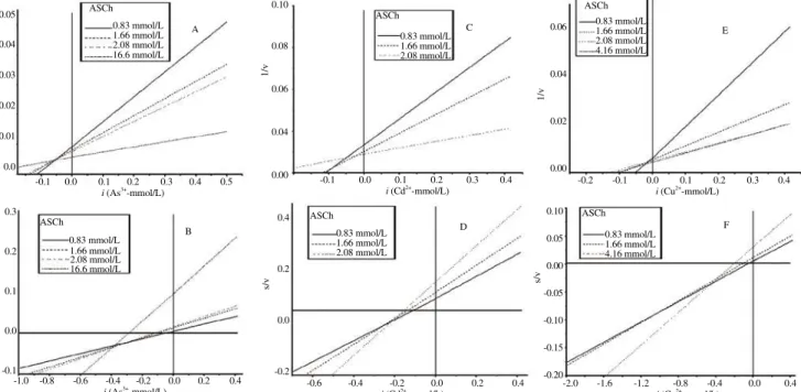
Figure 4. Dixon (A, C and E) and Cornish-Bowden (B, D and F) regression plots of brain AChE activity from R. canadum exposed to several concentrations of inhibitory ions (As3+, Cd2+ and Cu2+).
ASCh: Acetylthiocholine iodide. Table 4
Kinetic parameters of AChE from O. niloticus concerning several concentrations of five inhibitory ions using hyperbola model.
Concentration (mmol/L)
Hg2+
Cu2+
Zn2+
Cd2+
As3+
Km (mmol/L) Vmax
(mIU/mg protein)
Km (mmol/L) Vmax
(mIU/mg protein)
Km (mmol/L) Vmax
(mIU/mg protein)
Km (mmol/L) Vmax
(mIU/mg protein)
Km (mmol/L) Vmax
(mIU/mg protein) 0.000 0.856 ± 0.093a 194.497 ± 3.784a 0.477 ± 0.153a224.490 ± 9.559a 0.617 ± 0.086a196.170 ± 4.170a 0.786 ± 0.115a 210.869 ± 7.447a 0.092 ± 0.077a 220.897 ± 7.179a
Concentration (mmol/L)
Kmapp(mmol/L) Vmapp
(mIU/mg protein)
Kmapp(mmol/L) Vmapp
(mIU/mg protein)
Kmapp(mmol/L) Vmapp
(mIU/mg protein)
Kmapp(mmol/L) Vmapp
(mIU/mg protein)
Kmapp(mmol/L) Vmapp
(mIU/mg protein)
0.001 0.781 ± 0.090a 188.803 ± 3.906a 0.736 ± 0.128a192.806 ± 5.575b 0.649 ± 0.097a199.061 ± 4.853a 0.952 ± 0.202a 196.964 ± 10.168ab 0.481 ± 0.076ab 196.529 ± 3.968b
0.010 0.812 ± 0.163a 190.900 ± 6.977a - - 0.683 ± 0.113a195.810 ± 5.427a 0.798 ± 0.159a 186.694 ± 6.725b 0.482 ± 0.107ab 191.680 ± 5.455b
0.100 3.905 ± 0.692b 192.573 ± 10.353a 0.679 ± 0.101a161.086 ± 4.037c 0.952 ± 0.248a191.231 ± 9.397a 0.686 ± 0.161a 178.677 ± 6.725b 0.591 ± 0.142b 127.205 ± 4.090c
1.000 - - 0.424 ± 0.072a109.233 ± 3.011d 0.978 ± 0.286a164.343 ± 9.492b - - 1.443 ± 0.268c 97.529 ± 4.145d
10.000 - - 0.411 ± 0.216a 22.615 ± 1.385e 1.541 ± 0.442b 91.084 ± 5.831c 0.918 ± 0.153a 153.044 ± 5.003c
-
-Possible classification Competitive-like Non-competitive Mixed Non-competitive Mixed
Kmapp: Michaelis-Menten constant in presence of inhibitors; Vmapp: Maximum rate of substrate hydrolysis in presence of inhibitors; Lowercase letters in column indicate significant
differences (P< 0.05) using ANOVA and Tukey’s test.
0.010
0.008
0.006
0.004
0.002
0.000
0.10
0.08
0.06
0.04
0.02
0.00
0.03
0.02
0.01
0.00
0.020
0.015
0.010
0.005
0.000 0.008
0.006
0.004
0.002
0.000 0.03
0.02
0.01
0.00
1/v 1/v 1/v
1/v
1/v
1/v
0.000 mmol/L
0.000 mmol/L 0.000 mmol/L
0.00 mmol/L
0.00 mmol/L 1.00 mmol/L
1.00 mmol/L 10.0 mmol/L
10.0 mmol/L 0.000 mmol/L
A
C
E
F
B D
Hg2+
Pb2+ Zn
2+
Pb2+
Hg2+
-1.5 -1.0 -0.5 0.0 0.5 1.0 -2.5 -2.0 -1.5 -1.0 -0.5 0.0 0.5 1.0 1.5 -2.0 -1.5 -1.0 -0.5 0.0 0.5 1.0 1.5
-1.5 -1.0 -0.5 0.0 0.5 1.0 -3 -2 -1 0 1 -1.5 -1.0 0.5 0.0 0.5 1.0
1/[s] 1/[s] 1/[s]
1/[S] 1/[s] 1/[s]
0.010 mmol/L
0.010 mmol/L 0.100 mmol/L
0.010 mmol/L 0.001 mmol/L 0.100 mmol/L
0.100 mmol/L 1.000 mmol/L
0.100 mmol/L
Figure 3. Double reciprocal regression plots of brain AChE activity from R. canadum (A, C and E plots) and O. niloticus (B, D and F plots) exposed to several concentrations of inhibitory ions (Hg2+, Pb2+ and Zn2+).
0.10
0.08
0.06
0.04
0.02
0.00
0.012
0.008
0.004
0.00
0.08
0.06
0.04
0.02
0.00
0.2
0.0
-0.2 0.4
0.2
0.0
-0.2
0.2
0.0
-0.2
1/v
1/v
1/v
s/v
s/v
s/v
0.83 mmol/L 0.83 mmol/L 0.83 mmol/L
0.83 mmol/L 0.83 mmol/L 0.83 mmol/L
1.66 mmol/L 2.08 mmol/L 10.41 mmol/L 1.66 mmol/L
2.08 mmol/L
2.08 mmol/L
4.16 mmol/L
4.16 mmol/L
16.6 mmol/L
-0.1 0.0 0.1 0.2 0.3 0.4 -2.0 -1.5 -1.0 -0.5 0.0 -0.1 0.0 0.1 0.2 0.3 0.4
-0.2 0.0 0.2 0.4 -1.2 -0.8 -0.4 0.0 0.4 -0.2 0.0 0.2 0.4 i (As3+
-mmol/L) i (Cd2+
-mmol/L) i (Cu2+
-mmol/L)
i (As3+
-mmol/L) i (Cd2+
-mmol/L) i (Cu2+
-mmol/L)
ASCh ASCh ASCh
ASCh ASCh ASCh
1.66 mmol/L 1.66 mmol/L 1.66 mmol/L
2.08 mmol/L 2.08 mmol/L 2.08 mmol/L
4.16 mmol/L 10.41 mmol/L
A C E
F
B D
Figure 6. Dixon (A, C and E) and Cornish-Bowden (B, D and F) regression plots of brain AChE activity from O. niloticus exposed to several concentrations of inhibitory ions (As3+
, Cd2+ and Cu2+
). ASCh: Acetylthiocholine iodide.
Table 5 ki* and k’
i
** (μmol/L) of ions on brain AChE activity from
O. niloticus and R. canadum.
Species As3+ Cd2+ Cu2+ Hg2+ Pb2+ Zn3+
ki k’i ki k’i ki k’i ki k’i ki k’i ki k’i
O. niloticus 32.25 116.0b
482.4 920.5 16.5 58.5c
4.14 - 105.3 554.7 167.42 124.5c
77.1 96.5 187.9
R. canadum 50.00 276.3b 37.8a 210.4 63.1 133.5c 3.29 - 426.1 612.0 120.20 117.5c
379.0 93.6 211.0 148.1
*
ki: the dissociation constant of the enzyme-inhibitor complex estimated by Dixon plots (1953); a: Substrate concentration from 2.08 mmol/L; k’i: The dissociation constant of the enzyme-inhibitor-substre complex estimated by Cornish-Bowden plots (1974); b: Substrate concentration from 16.60 mmol/L; c: Substrate concentration from 4.16 mmol/L.
0.25
0.20
0.15
0.10
0.05
0.00
0.012
0.010
0.008
0.006
0.004
0.03
0.02
0.01
0.00
0.3
0.2
0.1
0.0
-0.1
-0.2 0.15
0.10
0.05
0.00
0.05
-0.10 0.2
0.1
0.0
-0.1
1/v 1/v
s/v
s/v
0.83 mmol/L 0.83 mmol/L
0.83 mmol/L
0.83 mmol/L 0.83 mmol/L
1.66 mmol/L
2.08 mmol/L 2.08 mmol/L
4.16 mmol/L 4.16 mmol/L
10.4 mmol/L 10.4 mmol/L
ASCh ASCh ASCh
ASCh ASCh ASCh
A C E
B D
F -0.2 0.0 0.2 0.4
-0.2 0.0 0.2 0.4
-0.4 -0.2 0.0 0.2 0.4 -0.4 -0.2 0.0 0.2 0.4
-0.6 -0.4 -0.2 0.0 0.2 0.4 -0.2 0.0 0.2 0.4 i (Hg2+
-mmol/L)
i (Hg2+
-mmol/L)
i (Pb2+
-mmol/L) i (Zn2+
-mmol/L)
i (Pb2+
-mmol/L) i (Zn2+
-mmol/L)
1.66 mmol/L 1.66 mmol/L
1.66 mmol/L
1.66 mmol/L
2.08 mmol/L 2.08 mmol/L
2.08 mmol/L
2.08 mmol/L 10.4 mmol/L 4.16 mmol/L
Figure 5. Dixon (A, C and E) and Cornish-Bowden (B, D and F) regression plots of brain AChE activity from R. canadum exposed to several concentrations of inhibitory ions (Hg2+
, Pb2+ and Zn2+
). ASCh: Acetylthiocholine iodide.
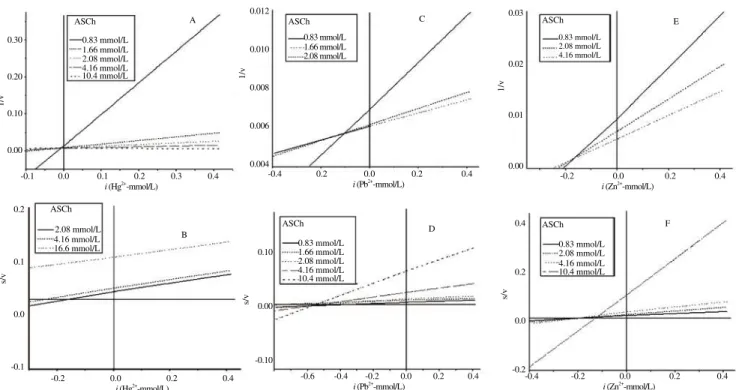
s/v
the ki values from Dixon plots and the enzyme-substrate-inhibitor complex (k’i) from Cornish-Bowden plots for the inhibitory ions. The values for k’i were higher than ki in all situations (excepting competitive inhibition in which k’i does not exist) in both species.
4. Discussion
Some studies pointed to the influence of ions on the AChE activity by binding to peripheral sites promoting conformational modifications or changing the hydration state of the active center which alters the rate of substrate hydrolysis by the enzyme[12,19,20]. Hughes and Bennett[12] working with E. electricus AChE reported three classes of metal ion effects on AChE activity; activation by Ca2+, Mg2+ and Al3+; inactivation by Cu2+, Na+, Pb2+ and Zn2+ and a non-specific effect of Li+
whereas Tomlinson et al.[13] using the same species divided the effect of ions into two groups: the first performs an activating action comprising Ca2+, Mg2+, Mn2+ and Na+; the second one exerts inhibitory effects and is formed by Cd2+
, Cu2+ , Hg2+
, Ni2+ , Pb2+
and Zn2+
. Our results differ from these groups only in relation to Al3+ and Li+.
The main peripheral anionic site in AChE is described as a region near the rim of the gorge where the active center is located[21]. Nevertheless, there are binding sites for positively charged activators and inactivators far from the active site of the enzyme which are different for organic and inorganic molecules[13]. Roufogalis and Quist[22] called α-site the anionic sub-site of the active center (choline binding site). According to them, the β-site is located bordering the gorge while the γ-site is the one far from the active site. Rosenberry[23] named the same sites, respectively as C, P1 and P2. In addition, this author reported the sites P3 and P4 which are binding sites for inorganic cations and hydrophobic organic cations as well as P2 and, according to Roufogalis and Wickson[24], these sites can cause allosteric disturbances in enzymatic activity. The β-site (or P1) is known to bind metal ion of the Tomlinson’s
first group such as Ca2+, Mg2+, Mn2+, polar cations and is considered an accelerator site whose occupancy can even stabilize the activated conformation of the enzyme[13]. Furthermore, Mg2+
binds to α and β-sites and can act as competitive inhibitor at low substrate concentrations or low affinity substrates (“poor” substrates). At high substrate concentrations, it probably is displaced from α-site to only occupy the β-site, therefore causing activation[22]. In the present work, only R. canadum AChE was positively affected by the activator ions Mn2+
, Mg2+ and Ca2+
(this last in concentrations above 1 mmol/L) whereas E. electricus AChE was inhibited by Mn2+ at 0.01 mmol/L. This occurred possibly by the fact that, according to Tomlinson et al.[13] working with E. electricus, activation is not well demonstrable in high ionic strength conditions as in the present work and R. canadum is a sea water species which, in other words, means that their enzymes may have evolved under conditions of higher salt fluctuations compared to freshwater species and could mirror activation effects even under high ionic strength conditions. These findings may seem contrasting with the influence of Ca2+ and Mg2+
on E. electricus AChE activity in other studies. Tomlinson
et al.[20] and Hughes and Bennet[12] found activations of 60% and
40%, respectively by Ca2+, while Tomlinson et al.[20] reported an increase of about 60% in their work with Mg2+
. However, the activation reported for Ca2+
and Mg2+
by these authors occurred at high substrate concentrations and low ionic strength buffers. In relation to Mn2+
, the results here corroborate other works[25,26]. Al3+ didn’t appear among the groups of ions reported by Tomlinson et al.[13] and the results in literature are contradictory. In some works, Al3+ inhibited AChE from bovine brain and from electric organ of E. electricus and this inhibition occurred in an ionic strength-dependent manner[27,28]. These studies advocated the existence of an interaction between this ion and the residue Glu in the active site of the enzyme. However, such residue can only provide a weak interaction, confirmed by the findings for the active site of butyrylcholinesterase (BChE) (which also present this
0.30
0.20
0.10
0.00
0.012
0.010
0.008
0.006
0.004
0.03
0.02
0.01
0.00
0.4
0.2
0.0
-0.2 0.10
0.00
-0.10 0.2
0.1
0.0
-0.1
1/v
1/v
1/v
s/v
s/v
s/v
0.83 mmol/L 0.83 mmol/L 0.83 mmol/L
2.08 mmol/L 4.16 mmol/L
1.66 mmol/L 1.66 mmol/L
2.08 mmol/L 2.08 mmol/L
2.08 mmol/L
0.83 mmol/L 0.83 mmol/L
1.66 mmol/L
2.08 mmol/L 2.08 mmol/L
4.16 mmol/L 4.16 mmol/L
10.4 mmol/L 10.4 mmol/L
10.4 mmol/L 4.16 mmol/L
4.16 mmol/L 16.6 mmol/L
i (Hg2+-mmol/L) i (Pb2+
-mmol/L) i (Zn2+
-mmol/L)
i (Hg2+-mmol/L) i (Pb2+-mmol/L) i (Zn2+-mmol/L)
ASCh ASCh ASCh
ASCh
ASCh ASCh
A C E
F
B D
-0.1 0.0 0.1 0.2 0.3 0.4 -0.4 0.2 0.0 0.2 0.4 -0.2 0.0 0.2 0.4
-0.2 0.0 0.2 0.4 -0.6 -0.4 -0.2 0.0 0.2 0.4 -0.4 -0.2 0.0 0.2 0.4
Figure 7. Dixon (A, C and E) and Cornish-Bowden (B, D and F) regression plots of brain AChE activity from O. niloticus exposed to several concentrations of inhibitory ions (Hg2+
, Pb2+ and Zn2+
residue in its catalytic triad) by Sarkarati et al.[29]. Here, the ionic strength conditions are different and Al3+
did not affect AChE from any species under study.
The activator ions (Ca2+, Mg2+ and Mn2+) plus Al3+ and K+ presented no complexation or interaction with protein or colorimetric reagents capable of increase absorbance in the assay causing false positives when using the enzyme as biomarker. Besides binding to the anionic sites of AChE, these activator ions can interact with “hard bases” side chains such as carboxylate groups in the sample preparation[13]. However, this effect did not appear to be important in our experimental conditions.
Cu2+ , Zn2+
and Cd2+
are part of the second group defined by Tomlinson and co-workers[13] and are known as strong inhibitors of AChE. The action of ligands on P2, P3 and P4 sites allows allosteric disturbances at the catalytic site inhibiting enzymatic activity[23]. The inhibitions exerted by Cu2+
and Zn2+
found in literature are varied: Tomlinson et al.[20] which observed inhibition of 100% by Cu2+ and Zn2+ in the activity of E. electricus AChE at 1 mmol/L and the value reported by Hughes and Bennett[12]
who found 20% inhibition promoted by Cu2+
for the same species and ion concentration. Nemcsók et al.[30] observed an inhibition of 69% at 0.36 mmol/L of Cu2+
and no effect for Zn2+
whereas Bocquené et al.[31] reported an inhibition of 100% in two marine species (Scomber scomber and Pleuronectes platessa) under Cu2+ exposure at 1 mmol/L. These last authors found for the same species, respectively, 57.4% and 70% at 1 mmol/L for Zn2+
. In the present study, E. electricus AChE was the most sensitive enzyme for Cu2+, Zn2+ and Cd2+ presenting IC50 of 0.05, 0.01 and 1.0 mmol/ L, respectively. Silva et al.[6] reported IC50 values for Cu
2+ , Zn2+ and Cd2+
of 2.10, 2.57 and 6.14 mmol/L, respectively, using Cichla
ocellaris AChE. Olson and Christensen[19] observed IC50 values by
Cu2+ , Zn2+
and Cd2+
of 0.16, 10.0 and 0.57 mmol/L, respectively, on the activity of AChE from Pimephales promelas (P. promelas). Ba2+ and Fe2+ induced, under our experimental conditions, a similar pattern of inhibition. Only E. electricus AChE presented IC50 for these ions. Unlike the other species under study, Fe
2+
inhibited significantly E. electricus enzyme at concentrations such as 0.1 mmol/L and Ba2+ did it at 0.01 mmol/L. Ba2+ and Fe2+ only exerted inhibitory effects on C. macropomum, A. gigas and R.
canadum at 1 mmol/L or higher concentrations. Similarly, Li+
only inhibited significantly E. electricus enzyme (IC50 of 0.38 mmol/ L) and seems to compete for the same site with Pb2+
although they present different valences, as evidenced by the study of Hughes and Bennett[12] which reported a relief of Pb2+ inhibition effects by the presence of Li+. These authors described a non-defined effect of Li+
alone and in fact for A. gigas, C. macropomum, O. niloticus
and R. canadum no positive or negative effect was observed.
The chelating ion EDTA2- only inhibited R. canadum and E.
electricus AChE presenting for the latter an IC50 value of 21.25
mmol/L. The enzymes from the other species in study were significantly inhibited only above 50 mmol/L by this ion. Such results (except for E. electricus) are in accordance with Tomlinson
et al.[13], and enable this chelating agent to protect against divalent
metallic interferences when using AChE from these species as biomarker for anticholinesterase agents analyzes.
According to Olson and Christensen[19], the ion As3+
(from AsO 2-) exhibits much more significant inhibitory activity, as compared to As5+. These authors reported a 50% inhibition at 0.03 mmol/ L using P. promelas. Here, we used As3+
, which induced values of IC50 in concentrations lower than 1 mmol/L for C. macropomum,
R. canadum, E. electricus and O. niloticus. Another report about
exposure to arsenic in S. scomber and P. platessa describes 33 and 31% of inhibition, respectively at 1 mmol/L[31]. Recently, Silva
et al.[6] reported an IC50 of 0.1 mmol/L by this ion on C. ocellaris
AChE. Hg2+
ion completely inactivated AChE from C. macropomum, R.
canadum, E. electricus and O. Niloticus, when these enzymes were
exposed to 1 mmol/L or lower concentrations. The AChE activity from A. gigas was the most resistant . These values are not too discrepant from those reported by Olson and Christensen[19], who found 50% inhibition at 1.6 mmol/L for P. promelas. Gill et al.[32], using AChE from Puntius conchonius, observed 67% of inhibition at 0.001 mmol/L. Here, Pb2+
was able to inhibit the enzymes from
A. gigas, R. canadum and induced an IC50 of 0.01 mmol/L on the
activity of E. electricus AChE. Hughes and Bennett[12] observed an inhibition of about 100% with E. electricus at 1mmol/L, while Olson and Christensen[19] reported 50% inhibition at 7.1 mmol/L
for P. promelas AChE.
Hg2+
and Pb2+ belongs to the Tomlinson’s second group of ions and according to Valle and Ulmer[33], inhibit a large number of enzymes by strongly interacting with their functional sulfhydryl groups. AChE was in the past included among such enzymes although no free sensitive sulfhydryl group are present in its structure except the one described in the Torpedo californica
electric organ. It was noted that most of these enzymes present such groups in form of disulfide bonds (e.g.: E. electricus AChE) or only one in a position (not conserved) buried or accessible through the solution but not always capable to react with thiol agents[10,34,35]. Investigations in the binding sites of Hg2+
to human BChE, observed no mercury bound to sulfhydryl groups in crystal structure and the only free accessible cysteine was persulfured (Cys-S-SH) and not easily susceptible to reduction[10]. Moreover, arsenic was not listed in the second group of Tomlinson but was also regarded as a free -SH ligand. According to Mounter and Whittaker[36], As-S link is readily hydrolyzed in alkaline solutions. However, in the present work the enzymes remained inhibited by As3+
in basic conditions evidencing the binding of this metal with sites other than free sulfhydryl groups. In other words, the classical explanation about the action of inhibitory ions on free sulfhydryl groups of enzymes is not sufficient for the inhibition of cholinesterases activity[36,37]. Tomlinson et al.[13], working with AChE from E. electricus
reported that Hg2+
and Pb2+
complex with the product of Ellman method, thiocholine (TCh), interfering in the assay. Nevertheless, in the same work it was found that Hg2+ strongly inhibited the enzyme when using p-nitrophenyl acetate as substrate. Additionally, they demonstrated that this ion decreased the rate of carbamoylation of the enzyme active site by M7C (7-(dimethylcarbamoyloxy)-N-methylquinoline iodide), which proves the tight binding of Hg2+ to the peripheral sites of AChE and their interference on the active site. The same was observed in the work of Frasco et al.[9] in which was reported the binding of TCh and thiobis-nitrobenzoate ion (TNB) with not only Hg2+
, but also Cd2+ , Cu2+
and Zn2+
Hg2+ and Pb2+ precipitates were not observed during the assays. Here, according to inhibition kinetic analyzes using hyperbola model with R. canadum and O. niloticus, Cu2+
behaved as non-competitive inhibitor and Zn2+ as mixed-type. Double reciprocal plot analyzes also showed Zn2+ as mixed-type inhibitor for both species whereas showed Cu2+
as non-competitive (with R. canadum) or mixed (with O. niloticus) inhibitor. Dixon and Cornish-Bowden plots compared analyzes can provide disambiguation on kinetic behaviour[16] and, in the present work, suggest that these two ions act as mixed-type inhibitors. In addition, Cd2+
behaved as non-competitive inhibitor towards AChE from R. canadum and O.
niloticus when analyzing with hyperbola model. Nevertheless, all
the other three graphic approaches such as double reciprocal, Dixon and Cornish-Bowden pointed this ion as a mixed-type inhibitor. These results for Cu2+ and Cd2+ are corroborated by Sarkarati et al.[29] using human serum BChE, while Hughes and Bennett[12]
regarded Cu2+ and Zn2+ as non-competitive inhibitor exposing AChE from E. electricus to these metals.
Kinetic analysis of the inhibitory behaviour of As3+ and Pb2+
on
R. canadum and O. niloticus brain AChE pointed to mixed-type
inhibitors in all models used (hyperbola, double reciprocal, Dixon and Cornish-Bowden). For the ions classified as mixed or non-competitive inhibitors in this study (As3+
, Cd2+ , Cu2+
, Pb2+ and Zn2+
), the values of k’i were higher than that of ki which means that the enzyme-substrate-inhibitor complex is the limiting step in the rate of substrate hydrolysis confirming the type of their inhibitory behavior. In the present work, Hg2+ ion seems to present a competitive-like inhibitory action using all the models excepting double reciprocal plots which showed a mixed-type behavior. However, Frasco et
al.[10] reported that no Hg2+
ion was attached to the anionic sub-site (choline binding sub-site or ammonium binding sub-site) of the AChE active center in the three-dimensional structure of the enzyme and therefore could not be a competitive inhibitor. Nevertheless, the effects of Hg2+ binding could present competitive-like consequences. The same authors state that the first and main binding site of Hg2+ to AChE is located at the omega loop (cysteine loop or W-loop) behind the choline binding site of the active center. These two regions are mutually responsive to ligand-dependent conformational changes and it was proposed by other authors that occupancy of peripheral site induces movements in the loop which in turn modify the orientation of the key tryptophan residue present in the choline binding site of the active center[23,38-40]. It suggested that conformational alterations from the binding of Hg2+
to the loop and the other three peripheral mercury-binding sites reported by Frasco
et al.[10] could allosterically be transmitted to the choline binding
locus of the active center interfering in substrate binding and, inversely, the substrate binding to peripheral (substrate inhibition site) and active sites, in increasing concentrations, could hinder Hg2+ binding to AChE loop since the position of residues in this region undergo a modification becoming more exposed to the solution after peripheral site occupancy[39,40]. The ion would be displaced from the loop and not from the active center therefore mimicking the behavior of a competitive inhibitor. This is corroborated by our results of Cornish-Bowden analysis of Hg2+ action that presented no k’i value. It means that no enzyme-substrate-inhibitor complex was perceptible as in the case of competitive inhibition. Further studies are necessary to directly confirm such behavior.
These results suggest that AChE from the species under study could be useful as biomarker of Hg2+
ion, according to the type of effluent discharged in a given area. For the other ions, in most of
cases they have little potential to interfere on the enzyme activity in samples not associated with mine and industrial effluents.
R. canadum AChE was responsive to activator ions (Ca2+
, Mg2+ and Mn2+) in the high ionic strength conditions of the present work. In addition, the commercial enzyme from E. electricus was strongly influenced by the majority of the ions analyzed (unlike the other enzymes proposed in this paper) which is an undesirable feature for a biomarker of anticholinesterasic agents. In contrast with E.
electricus enzyme, AChE from A. gigas was the most resistant to the
ions. Nevertheless, EDTA can be used to protect the enzyme activity against divalent metallic cations since only exerted interfering effects on R. canadum and E. electricus enzyme at 1 mmol/L. Inhibition kinetic analyzes in the present experimental conditions classified As3+, Cd2+, Cu2+, Pb2+ and Zn2+ as mixed or non-competitive inhibitors of R. canadum and O. niloticus brain AChE. The most reactive ion was Hg2+
, which strongly inhibited the AChE from the five species. This ion presented competitive-like features of inhibitory behavior, even without binding to the active center of the enzyme (as demonstrated in other studies) probably due to its interaction with regions responsive to peripheral site occupancy. Further studies are required to elucidate such competitive mimicking effect.
Ions and heavy metals may arise as probable contaminants in samples from different sources, and can cause false positives or negatives in the analyses of pesticides or other anticholinesterasic agents. On the other hand, analyzing the inhibition produced by these substances along with other methods, it is possible to use the enzyme also as a biomarker for the presence of some heavy metals, according to the waste composition from a given area.
Conflict of interest statement
We declare that we have no conflict of interest.
Acknowledgment
The authors would like to dedicate this study to Dr. Patrícia Fernandes de Castro (in memoriam) for her invaluable help and to thank Financiadora de Estudos e Projetos (FINEP/RECARCINE), Petróleo do Brasil S/A (PETROBRAS), Secretaria Especial de Aqüicultura e Pesca (SEAP/PR), Conselho Nacional de Pesquisa e Desenvolvimento Científico (CNPq) and Fundação de Apoio à Ciência e Tecnologia do Estado de Pernambuco (FACEPE) for financial support (Grant numbers: IBPG-1301-2.08/08 FACEPE, IBPG-0523-2.08/11 FACEPE, BFP-0036-2.08/13 FACEPE and BFP-0111-2.08/13). Our gratitude also goes to Universidade Federal Rural de Pernambuco and Aqualider, for providing fish juvenile specimens.
References
[1] Campanha HM, Carvalho F, Schlosser PM. Active and peripheral anionic sites of acetylcholinesterase have differential modulation effects on cell proliferation, adhesion and neuritogenesis in the NG108-15 cell line. Toxicol Lett 2014; 230: 122-31.
[2] Karczmar AG. Cholinesterases (ChEs) and the cholinergic system in ontogenesis and phylogenesis, and non-classical roles of cholinesterases - a review. Chem Biol Interact 2010; 187: 34-43. [3] Y l d i r i m N C , Y l d i r i m N , D a n a b a s D , D a n a b a s S . U s e o f
Uzuncayir Dam Lake (Tunceli, Turkey). Environ Toxicol Pharmacol
2014; 37: 1169-76.
[4] Sogorb MA, González-González I, Pamies D, Vilanova E. An alternative in vitro method for detecting neuropathic compounds based on acetylcholinesterase inhibition and on inhibition and aging of neuropathy target esterase (NTE). Toxicol In Vitro 2010; 24: 942-52. [5] Valdés-Ramírez G, Fournier D, Ramírez-Silva MT, Marty JL. Sensitive
amperometric biosensor for dichlorovos quantification: application to detection of residues on apple skin. Talanta 2008; 74: 741-6.
[6] Silva KC, Assis CR, Oliveira VM, Carvalho LB Jr, Bezerra RS. Kinetic and physicochemical properties of brain acetylcholinesterase from the peacock bass (Cichla ocellaris) and in vitro effect of pesticides and metal ions. Aquat Toxicol 2013; 126: 191-7.
[7] Rodríguez-Fuentes G, Armstrong J, Schlenk D. Characterization of muscle cholinesterases from two demersal flatfish collected near a municipal wastewater outfall in Southern California. Ecotoxicol Environ Saf 2008; 69: 466-71.
[8] Monserrat JM, Geracitano LAG, Bianchini A. Current and future perspectives using biomarkers to assess pollution in aquatic ecosystems. Comments Toxicol 2003; 9: 255-69.
[9] Frasco MF, Fournier D, Carvalho F, Guilhermino L. Do metals inhibit acetylcholinesterase (AChE)? Implementation of assay conditions for the use of AChE activity as a biomarker of metal toxicity. Biomarkers
2005; 10: 360-75.
[10] Frasco MF, Colletier JP, Weik M, Carvalho F, Guilhermino L, Stojan J, et al. Mechanisms of cholinesterase inhibition by inorganic mercury.
FEBS J 2007; 274: 1849-61.
[11] Reddy GR, Basha MR, Devi CB, Suresh A, Baker JL, Shafeek A, et al. Lead induced effects on acetylcholinesterase activity in cerebellum and hippocampus of developing rat. Int J Dev Neurosci 2003; 21: 347-52.
[12] Hughes RJ, Bennett J. Effect of metal ions on the activity of acetylcholinesterase. Biochem Soc Trans 1985; 13: 219-20.
[13] Tomlinson G, Mutus B, McLennan I. Activation and inactivation of acetylcholinesterase by metal ions. Can J Biochem 1981; 59: 728-35. [14] Assis CR, Linhares AG, Oliveira VM, França RC, Carvalho
EV, Bezerra RS, et al. Comparative effect of pesticides on brain acetylcholinesterase in tropical fish. Sci Total Environ 2012; 441: 141-50.
[15] Sedmak JJ, Grossberg SE. A rapid, sensitive and versatile assay for protein using Coomassie brilliant blue G250. Anal Biochem 1977; 79: 544-52.
[16] Cornish-Bowden A. A simple graphical method for determining the inhibition constants of mixed, uncompetitive and non-competitive inhibitors. Biochem J 1974; 137: 143-4.
[17] Dixon M. The determination of enzyme inhibitor constants. Biochem J
1953; 55: 170-1.
[18] Cheng Y, Prusoff WH. Relationship between the inhibition constant (Ki) and the concentration of inhibitor which causes 50 per cent inhibition (I50) of an enzymatic reaction. Biochem Pharmacol 1973; 22(23): 3099-108.
[19] Olson DL, Christensen GM. Effects of water pollutants and other chemicals on fish acetylcholinesterase (in vitro). Environ Res 1980; 21: 327-35.
[20] To m l i n s o n G , M u t u s B , M c L e n n a n I . M o d u l a t i o n o f acetylcholinesterase activity by peripheral site ligands. Mol Pharmacol
1980; 18: 33-9.
[21] Eastman J, Wilson EJ, Cerveñansky C, Rosenberry TL. Fasciculin 2 binds to the peripheral site on acetylcholinesterase and inhibits substrate hydrolysis by slowing a step involving proton transfer during
enzyme acylation. J Biol Chem 1995; 270: 19694-701.
[22] Roufogalis BD, Quist EE. Relative binding sites of pharmacologically active ligands on bovine erythrocyte acetylcholinesterase. Mol Pharmacol 1972; 8: 41-9.
[23] Rosenberry TL. Acetylcholinesterase. Adv Enzymol Relat Areas Mol Biol 1975; 43: 103-218.
[24] Roufogalis BD, Wickson VM. Acetylcholinesterase - specific inactivation of allosteric effects by a water soluble carbodiimide. J Biol Chem 1973; 248: 2254-6.
[25] M a r t i n e z H , B o n i l l a E . Wa t e r i n t a k e a n d b r a i n c h o l i n e -acetyltransferase and acetylcholinesterase activities in manganese treated rats. Neurobehav Toxicol Teratol 1981; 3: 277-80.
[26] Santos D, Milatovic D, Andrade V, Batoreu MC, Aschner M, Marreilha dos Santos AP. The inhibitory effect of manganese on acetylcholinesterase activity enhances oxidative stress and neuroinflammation in the rat brain. Toxicology 2012; 292: 90-8. [27] Moraes MS, Leite SR. Inhibition of bovine brain acetylcholinesterase
by aluminum. Braz J Med Biol Res 1994; 27: 2635-8.
[28] Sharp TR, Rosenberry TL. Ionic strength dependence of the inhibition of acetylcholinesterase activity by Al3+
. Biophys Chem 1985; 21: 261-4.
[29] Sarkarati B, Çokuğraş AN, Tezcan EF. Inhibition kinetics of human
serum butyrylcholinesterase by Cd2+ , Zn2+
and Al3+
: comparison of the effects of metal ions on cholinesterases. Comp Biochem Physiol C Pharmacol Toxicol Endocrinol 1999; 122: 181-90.
[30] Nemcsók J, Németh Á, Buzás ZS, Boross L. Effects of copper, zinc
and paraquat on acetylcholinesterase activity in carp (Cyprinus carpio
L.). Aquat Toxicol 1984; 5: 23-31.
[31] Bocquené G, Galgani F, Truquet P. Characterization and assay conditions for use of AChE activity from several marine species in pollution monitoring. Mar Environ Res 1990; 30: 75-89.
[32] Gill TS, Tewari H, Pande J. Use of the fish enzyme system in monitoring water quality: effects of mercury on tissue enzymes. Comp Biochem Physiol C 1990; 97: 287-92.
[33] Valle BL, Ulmer DD. Biochemical effects of mercury, cadmium and lead. Ann Rev Biochem 1972; 41: 91-128.
[34] MacPhee-Quigley K, Vedvick TS, Taylor P, Taylor SS. Profile of the disulfide bonds in acetylcholinesterase. J Biol Chem 1986; 261: 13565-70.
[35] Steinberg N, Roth E, Silman I. Torpedo acetylcholinesterase is inactivated by thiol reagents. Biochem Int 1990; 21: 1043-50. [36] Mounter LA, Whittaker VP. The effect of thiol and other
group-specific reagents on erythrocyte and plasma cholinesterases. Biochem J 1953; 53: 167-73.
[37] Ali N, Hoque MA, Haque A, Salam KA, Karim MR, Rahman A, et al. Association between arsenic exposure and plasma cholinesterase activity: a population based study in Bangladesh. Environ Health 2010; 9: 36-44.
[38] Barak D, Ordentlich A, Bromberg A, Kronman C, Marcus D, Lazar A, et al. Allosteric modulation of acetylcholinesterase activity by peripheral ligands involves a conformational transition of the anionic subsite. Biochemistry 1995; 34: 15444-52.
[39] Simon S, Le Goff A, Frobert Y, Grassi J, Massoulie J. The binding sites of inhibitory monoclonal antibodies on acetylcholinesterase.
Identification of a novel regulatory site at the putative “back door”. J Biol Chem 1999; 274: 27740-6.