Structural, functio nal and
im m uno lo gical studie s o n a
po lym e ric bacte rial pro te in
1Instituto de Estudios de la Inmunidad Humoral, Facultad de Farmacia y Bioquímica, Universidad de Buenos Aires, Buenos Aires, Argentina
2Department of Natural Sciences, Bowie State University, Bowie, MD, USA 3Instituto de Investigaciones Bioquímicas (Fundación Campomar, IIBBA-CO NICET, FCEN-UBA), Buenos Aires, Argentina
P.C. Baldi1, C.A. Velikovsky1, B.C. Braden2, G.H. Giambartolomei1, C.A. Fossati1 and F.A. Goldbaum3
Abstract
The characterization of proteins from Brucella spp, the causative agent of brucellosis, has been the subject of intensive research. We have described an 18-kDa cytoplasmic protein of Brucella abortus and shown the potential usefulness of this protein as an antigen for the serologic diagnosis of brucellosis. The amino acid sequence of the protein showed a low but significant homology with that of lumazine synthases. Lumazine is an intermediate product in bacterial riboflavin biosynthesis. The recombinant form of the 18-kDa protein (expressed in E. coli) folds like the native Brucella protein and has lumazine-synthase enzymatic activity. Three-dimensional analysis by X-ray crystallography of the homolog Bacillus subtilis lumazine synthase has revealed that the enzyme forms an icosahedral capsid. Recombi-nant lumazine synthase from B. abortus was crystallized, diffracted X rays to 2.7-Å resolution at room temperature, and the structure suc-cessfully solved by molecular replacement procedures. The macromo-lecular assembly of the enzyme differs from that of the enzyme from B. subtilis. The Brucella enzyme remains pentameric (90 kDa) in its crystallographic form. Nonetheless, the active sites of the two en-zymes are virtually identical at the structural level, indicating that inhibitors of these enzymes could be viable pharmaceuticals across a broad species range. We describe the structural reasons for the differ-ences in their quaternary arrangement and also discuss the potential use of this protein as a target for the development of acellular vaccines.
Co rre spo nde nce
F.A. Goldbaum Instituto de Investigaciones Bioquímicas, Fundación Campomar Av. Patricias Argentinas, 435 Buenos Aires 1405 Argentina
Fax: + 54-114-865-2246 E-mail: goldbaum@ iib.uba.ar
Presented at the XIV Annual Meeting of the Federação de Sociedades de Biologia Experimental, Caxambu, MG, Brasil, August 25-28, 1999.
Research supported by grant BID 802/O C-AR-PICT 00084 from Agencia Nacional de Promoción Científica y Tecnológica (ANPCyT) and by grant PIP 0764/98 from Consejo Nacional de Investigaciones Científicas y Técnicas (CO NICET). B.C. Braden was supported by NIH grant
1R15AI44790-01and National Aeronautics and Space Administration grant NCC5-232 (Model Institutes for Excellence).
Received January 7, 2000 Accepted March 10, 2000
Ke y wo rds
·Brucella lumazine synthase
·X-ray structure
·Immunogenicity
·Brucellosis detection
·Bacillus subtilis lumazine synthase
Intro ductio n
The sequence, structure and function of protein components of microorganisms rep-resent a field of increasing interest to both microbiologists and immunologists. The char-acterization of proteins from pathogenic bac-teria can help to understand the interaction
between the bacterium and the host and to study the humoral and cellular immune re-sponses elicited during the infection. More-over, proteins can be useful as specific anti-gens for the serologic diagnosis of bacterial infections and as specific targets for rational drug design of chemotherapeutic agents.
Brucella spp, the causative agent of brucel-losis, has been the subject of intensive re-search, but only a few proteins have been characterized at the structural and functional level. Human and animal brucellosis still constitutes an important health problem in many developing countries. The disease is caused by different species of Brucella which can be distinguished by their preferential host and also by the type of lipopolysaccha-ride (LPS) present on their surface. Species expressing smooth LPS include B. abortus, B. suis and B. melitensis, which infect mainly cows, pigs and goats and sheep, respectively. Species expressing rough LPS include B. canis and B. ovis, which infect mainly dogs and sheep, respectively. Serology has proven to be an important tool for the diagnosis, prognosis and management of this infection. However, the most widely used tests (mainly agglutination tests) rely on the detection of antibodies directed at Brucella LPS, in spite of several studies showing the diagnostic drawbacks associated with the measurement of this response (1,2). Because of the exist-ence of shared epitopes between the LPS from Brucella and that from another Gram-negative bacterium, false-positives hinder the serologic diagnosis of brucellosis. In addition, the development of high anti-LPS titers in vaccinated animals makes it difficult to differentiate vaccinated from infected cattle by means of agglutination assays (3).
D iagno stic use fulne ss o f Bruce lla cyto plasmic pro te ins
In view of the diagnostic problems men-tioned above, the main goal of our group has been to obtain Brucella cytoplasmic pro-teins free from LPS and to test their diagnos-tic usefulness by ELISA. Our first approach was to prepare an LPS monoclonal anti-body (termed BC68) which was coupled to a gel matrix to obtain an immunosorbent. The cytoplasmic fraction of B. abortus was passed through this immunosorbent so that the LPS
was retained by the attached antibody while the proteins eluted. This complex mixture of LPS-free cytosolic proteins (CP) was used as antigen in an indirect ELISA to test the reactivity of sera from different hosts. We have found serum reactivity to CP in brucel-losis patients (4) and also in cows (5), sheep, goats, pigs and dogs (6) infected with differ-ent Brucella species, which suggests that the internal antigens are common to all the Bru-cella species regardless of the type of LPS. Anti-CP antibodies proved to be useful for differentiating active from inactive human brucellosis (4). In addition, a serologic fol-low-up performed on patients with different outcomes of the disease showed that the kinetics of the antibody response to proteins is correlated with the clinical outcome of patients (4).
IgG antibodies against this antigen (Figure 1). In cattle, detection of antibodies to the 18-kDa protein proved useful to distinguish between animals vaccinated with strain 19 of B. abortus and pregnant heifers experi-mentally infected with a virulent strain of B. abortus (5). While the latter had high levels of antibodies against the 18-kDa protein, the vaccinated cattle developed only a low and transient antibody response that was no longer detected at 90 days post-vaccination (a time when anti-LPS antibodies were still detected at high levels). The 18-kDa protein and the CP antigen were also useful for the diagnosis of canine brucellosis (6,8). The antibody reactivity to cytoplasmic proteins of Bru-cella was investigated by ELISA in sera from 30 dogs having confirmed or suspected brucellosis. Antibodies to the 18-kDa pro-tein were found in 26 animals which were also positive for anti-CP antibodies and for anti-LPS antibodies (the latter detected by the slide agglutination test). The overall cor-relation between the two anti-protein ELISAs reached 93.3% (8).
Ide ntity o f the 18-kD a pro te in
The 18-kDa protein was purified by af-finity chromatography using the mAb BI24 and the sequence of three internal peptides was determined (7). Later, Hemmen et al. (9) cloned a gene encoding for a 17-kDa Bru-cella protein whose deduced amino acid se-quence showed homology with the internal peptides previously described by us, sug-gesting that the two proteins were identical. These investigators found this protein to be useful for diagnosing ovine and bovine bru-cellosis. Others identified a gene encoding a homologue of the 17-kDa Brucella protein in Rhodococcus spp. In turn, database search-ing revealed that the sequence of both the Brucella and the Rhodococcus proteins had a low but significant homology with that of lumazine synthases involved in bacterial ri-boflavin biosynthesis (10). To further
char-acterize the Brucella protein and to deter-mine whether it is a lumazine synthase we decided to produce it in recombinant form based on the published nucleotide sequence.
Re co mbinant e xpre ssio n o f the Bruce lla 18-kD a pro te in
The 18-kDa protein from Brucella abor-tus was successfully expressed in the pET vector as inclusion bodies in BL21(DE3) cells. Attempts to obtain soluble expression of the protein were unsuccessful. In the ab-sence of dithiothreitol (DTT) no refolded material was obtained. The refolded protein was purified by anion exchange chromatog-raphy under reducing conditions. The appro-priate folding of the recombinant 18-kDa protein was confirmed by testing the reactiv-ity of sera from human and animal brucello-sis against both the native and the recombi-nant protein. Although the purified 18-kDa recombinant protein was soluble in solu-tions containing 1 mM DTT, it aggregated in
A
nt
ib
od
y
tit
er
(1
/d
ilu
tio
n)
6400
3200
1600
800
400
200
100
0
0 5 10 15 20 25 30 35 40 45 50 55 60 Time after initial symptoms (w eeks)
Anti-CP IgM Anti-CP IgG
Anti-18 kDa IgM Anti-18 kDa IgG
the absence of a reducing agent. Since the protein contains a single cysteine, this be-havior was thought to reflect the formation of intermolecular associations by means of disulfide bridges. This hypothesis was con-firmed later when the aggregation in non-reducing media was prevented by treatment with iodoacetamide under reducing condi-tions. Gel-exclusion chromatography re-vealed that the iodoacetamide-treated pro-tein has a molecular weight of about 90 kDa, indicating the presence of a pentamer of the 18-kDa protein.
In immunoblotting assays, the recombi-nant protein was recognized by the mAb BI24 previously used by us to characterize the 18-kDa protein, confirming the identity between the protein described by us (7) and that cloned by Hemmen et al. (9).
Functio nal studie s o n the Bruce lla 18-kD a pro te in
E. coli cells containing the plasmid en-coding the 18-kDa protein were grown and induced to express recombinant protein. For the determination of 6,7-dimethyl-8-ribityl-lumazine synthase activity, the appropriate reaction mixture was prepared and the cell extract from recombinant bacteria was added. Aliquots were taken at regular intervals and the reaction was stopped by the addition of
trichloroacetic acid. The concentration of 6,7-dimethyl-8-ribityllumazine was deter-mined by high performance liquid chroma-tography coupled with fluorometric moni-toring. The 6,7-dimethyl-8-ribityllumazine synthase activity found in the cell extract was approximately 6-fold higher than the activity displayed by control bacteria, clearly indicating that the recombinant Brucella 18-kDa protein is an enzyme with lumazine synthase activity (11).
Crystallo graphic structure o f Bruce lla lumazine synthase
Crystallographic studies were undertaken to further characterize the 18-kDa protein. Recombinant lumazine synthase from Bru-cella abortus was crystallized at room tem-perature by the hanging-drop vapor-diffu-sion method, yielding crystals that diffracted to 2.7-Å resolution at 300o K. Data were collected at the LNLS protein facility in Campinas, SP, Brazil.
The structure of the Brucella protein was successfully solved by molecular replace-ment procedures using the lumazine syn-thase subunits from the Bacillus subtilis struc-ture modeled as poly-alanines. The tertiary structure of the monomeric protein and the pentameric assembly of the monomers closely resemble those found in the B. subtilis lumazine synthase (12). The most striking difference between the structures of lumazine synthase from Brucella and B. subtilis is the non-icosahedral nature of the Brucella en-zyme assembly (13).
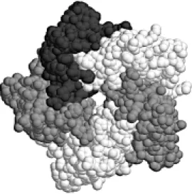
The pentameric assemblies of the en-zymes from B. subtilis and Brucella abortus indicate no large-scale difference in struc-ture (Figure 2).
As the active site of lumazine synthase is formed by the interface between two adja-cent monomers of the pentameric assembly, the Brucella enzyme maintains the active site of the B. subtilis enzyme. When the lumazine substrate analog (5-nitroso-6-Figure 2 - Space-fill model of the
ribitylamino-2,4(1H,3H) pyrimidindione) from the 2.4-Å capsid structure of B. subtilis (12) is modeled into the Brucella enzyme binding site, it is clear that the main-chain atoms responsible for recognition and bind-ing of the substrate are topologically well preserved. The only structural difference in the recognition site is the substitution of a tryptophan residue in the B. abortus protein for a phenylalanine in the B. subtilis protein. As the function of this side chain apparently consists of substrate binding to the substrate pyrimidine ring via stacking interactions, the Phe and Trp side chains appear to be func-tionally equivalent.
We further analyzed the structural rea-sons for divergence in macromolecular as-sembly between pentameric and icosahedral enzymes. The C-terminal a-helix of the B. subtilis protein begins at residue 121 for nearly two turns but deviates into a 5-residue loop before continuing to the C-terminus as an a-helix. This 5-residue kink (GTKAG) contributes a number of contacts to a neigh-boring pentamer. On the other hand, the C-terminal a-helix in the B. abortus structure begins at residue 122 and is a continuous helix, unable to form this potential capsid-stabilizing loop.
On the basis of sequence alignment, Mörtl et al. (14) suggested that a four-residue in-sertion in the yeast Saccharomyces cerevi-siae lumazine synthase compared to B. subtilis protein was the basis for the lack of capsid assembly for the yeast enzyme. These residues, however, are located just after the kink in the B. subtilis structure and would not necessarily disrupt the C-terminal a -helix. Glycine residues initiate and termi-nate the kink in the B. subtilis structure. Moreover, a consensus sequence of GT(or G)KAG occurs in all of the lumazine syn-thase molecules which form icosahedral 60-mer capsids (Figure 3), namely B. subtilis (12), E. coli (14) and spinach (15). Lumazine synthases which have been identified as pentameric, B. abortus (herein) and yeast
(14,16) do not contain the glycine residues that originate and terminate the kink. Gly-cine exhibits a broader range of conforma-tional stability than amino acids with ß-car-bons which in this case may destabilize the a-helical conformation, allowing the helix to bend. While the C-terminal a-helix of the B. subtilis enzyme is bent as a result of this kink, the helix in the B. abortus enzyme is relatively straight and represents yet another reason for the lack of capsid formation as a result of steric contacts with a potential pentamer neighbor.
Co ncluding re marks
Previous reports have shown the poten-tial usefulness of Brucella lumazine syn-thase in the serologic diagnosis of human and animal brucellosis (5,7,9,17). Interest-ingly, the determination of the antibody re-sponse to this protein yields results equiva-lent to those obtained with a complex mix-ture of cytoplasmic proteins of Brucella (5,7,8).
The expression system and refolding pro-cedures allowed us to obtain the protein as a soluble pentamer, with antigenic properties similar to those of the native Brucella pro-tein. The enzymatic activity displayed by the purified recombinant Brucella abortus lumazine synthase protein confirms that the biological activity of this protein is that of lumazine synthase.
As stated by Mörtl et al. (14), bacteria are devoid of an uptake system for riboflavin. They are therefore dependent on internal riboflavin synthesis and should be vulner-able to inhibitors of riboflavin synthesis. Since this enzyme is not present in mam-mals, knowledge about its three-dimensional structure could serve as a basis for the ration-al design of enzyme inhibitors with thera-peutic activity.
analogue B. subtilis enzyme complexed with an analog substrate. The protein structure of B. subtilis revealed that the enzyme forms a particle with a molecular weight close to 1,000 kDa (12). This particle comprises 60 ß subunit monomers (lumazine synthase) ar-ranged in 12 pentamers, forming an icosahe-dral capsid. It contains also 3 a subunits (riboflavin synthase) enclosed inside the capsid. The entire structure is called lumazine synthase-riboflavin synthase complex. In contrast, the lumazine synthase of E. coli is not physically associated with another en-zyme of the riboflavin pathway (14).
Comparison with the structure of Bru-cella lumazine synthase has revealed the mechanism of divergent macromolecular assembly while maintaining a conserved binding site structure. It appears that the
formation of the substrate binding site and the assembly of the enzyme are related. While the Brucella and yeast enzyme share a pentameric assembly they also feature a Trp residue at the binding site (Figure 3), the bulk of the side chain compensating for a more open site than found in the Bacillus enzyme. The capsid-forming lumazine syn-thases (from E. coli, B. subtilis and spinach) are sequence homologous in the C-terminal a-helix and contain a phenylalanine inter-acting with the substrate. As a consequence, the lumazine synthase from Photobacterium leiognathi, Haemophilus influenzae, Acti-nobacillus pleuropneuminiae, Photobacte-rium phosphoreum, tobacco and Arabidop-sis, all sharing the GT (or G) KAG sequence within the C-terminal helix and a phenylala-nine at the substrate binding site, are there-fore potential capsid structures, whereas the enzyme from Rhodococcus (DYFST in the C-terminal helix and Trp in the substrate binding site), like the Brucella and yeast enzymes, will be pentameric in its active form. Conservation of the main features of the active site among species would indicate that inhibitors of these enzymes could be used as viable pharmaceuticals across a broad species range.
Recently, Persson et al. (18) determined the three-dimensional structures of the capsid-forming lumazine synthase from spin-ach and of the pentameric enzyme from the fungus Magnaporte grisea. They found a different structural reason for macromolecu-lar divergence, indicating the heterogeneity of the phylogenetic evolution of this en-zyme.
Even though Brucella abortus lumazine synthase does not form the icosahedral par-ticle enzyme, its polymeric nature could ex-plain, at least in part, the immunogenic na-ture of this protein. Interestingly, as in the B. subtilis protein, the 8-10 amino-terminal resi-dues of the Brucella protein are not well defined in their crystallographic structure. The lack of defined electron density for these Figure 4 - Structural basis for the
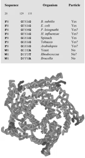
use of Brucella lumazine syn-thase as a carrier for displaying other immunogenic peptides from bacterial proteins. The a -carbon backbone of the penta-mer (light grey) and the structur-ally defined N-terminal residues of each monomer (dark grey) are show n.
Figure 3 - Conservation of critical residues for capside-forming and pentameric lumazine synthases from different species.
Sequence Organism Particle
20 129 133
FN GTKAG B. subtilis Yes
FN GTKAG E. coli Yes
FN GTKMG P. leiognathi Yes?
FN GTKAG H. influenzae Yes?
FN GGKAG Spinach Yes
FN GGKAG Tobacco Yes?
FN GGKAG Arabidopsis Yes?
WN GIDEA Yeast No
WH DYFST Rhodococcus No?
residues suggests that this portion of the protein is not essential for the overall folding of the pentamer. In principle, these amino-terminal residues could be changed by pep-tides pertaining to other immunogenic Bru-cella proteins (Figure 4). Construction and expression of chimeric proteins using the pentameric structure of this Brucella protein
as a carrier and presenting different peptides to the immune system in a pentavalent man-ner and within an immunogenic context could be the basis for the development of acellular vaccines. Experimental tests of this hypo-thesis are needed in order to ascertain the usefulness of Brucella lumazine synthase as an immunogenic carrier protein.
Re fe re nce s
1. Alton GG, Jones LM , Angus RD & Verger JM (1988). Techniques for the Brucellosis Laboratory. Institut National de la Recher-che Agronomique, Paris.
2. Diaz R & M oriyón I (1989). Laboratory techniques in the diagnosis of human bru-cellosis. In: Young EJ & Corbel M J (Edi-tors), Brucellosis: Clinical and Laboratory Aspects. CRC Press Inc., Boca Raton. 3. Nicoletti P, Jones LM & Berman DT
(1978). Comparison of the subcutaneous and conjunctival route of vaccination w ith Brucella abortus strain 19 vaccine in adult cattle. Journal of the American Veterinary M edical Association, 173: 1450-1456. 4. Goldbaum FA, Rubbi CP, Wallach JC,
M iguel SE, Baldi PC & Fossati CA (1992). Differentiation betw een active and inac-tive human brucellosis by measuring anti-protein humoral immune responses. Jour-nal of Clinical M icrobiology, 30: 604-607. 5. Baldi PC, Giambartolomei GH, Goldbaum
FA, Abdón LF, Velikovsky CA, Kittelberger R & Fossati CA (1996). Humoral immune response against LPS and cytoplasmic proteins of Brucella in cattle vaccinated w ith Brucella abortus S19 or experimen-tally infected w ith Yersinia enterocolitica 0:9. Clinical and Diagnostic Laboratory Immunology, 3: 472-476.
6. Baldi PC, Wanke M M , Loza M E & Fossati CA (1994). Brucella abortus cytoplasmic proteins used as antigens in an ELISA potentially useful for the diagnosis of ca-nine brucellosis. Veterinary M icrobiology, 41: 127-134.
7. Goldbaum FA, Leoni J, Wallach JC & Fossati CA (1993). Characterization of an 18-kilodalton Brucella cytoplasmic protein w hich appears to be a serological marker of active infection of both human and
bo-vine brucellosis. Journal of Clinical M icro-biology, 31: 2141-2145.
8. Baldi PC, W anke M M , Loza M E, M onachesi N & Fossati CA (1997). Diag-nosis of canine brucellosis by detection of IgG antibodies against an 18 kDa cyto-plasmic protein of Brucella spp. Veteri-nary M icrobiology, 57: 273-281. 9. Hemmen F, Weynants V, Scarcez T,
Letesson J-J & Saman E (1995). Cloning and sequence analysis of a new ly identi-fied Brucella abortus gene and serological evaluation of the 17-kilodalton antigen that it encodes. Clinical and Diagnostic Laboratory Immunology, 2: 263-267. 10. De M ot R, Nagy I, Schoof s G &
Vanderleyden J (1996). Identification of a Rhodococcus gene cluster encoding a ho-molog of the 17-kDa antigen of Brucella and a putative regulatory protein of the AsnC-Lrp family. Current M icrobiology, 33: 26-30.
11. Goldbaum FA, Velikovsky CA, Baldi PC, M örtl S, Bacher A & Fossati CA (1999). The 18 kDa cytoplasmic protein of Bru-cella species - an antigen useful for diag-nosis - is a lumazine synthase. Journal of M edical M icrobiology, 48: 833-839. 12. Ritsert K, Huber R, Turk D, Ladenstein R,
Schmidt-Base K & Bacher A (1995). Stud-ies on the lumazine synthase/riboflavin synthase complex of Bacillus subtilis: crystal structure analysis of reconstituted, icosahedral ß-subunit capsids w ith bound substrate analogue inhibitor at 2.4 Å reso-lution. Journal of M olecular Biology, 253: 151-167.
13. Braden BC, Velikovsky CA, Cauerhff AA, Polikarpov I & Goldbaum FA (2000). Diver-gence in macromolecular assembly: X-ray crystallographic structure analysis of
lumazine synthase from Brucella abortus. Journal of M olecular Biology, 297: 1031-1036.
14. M örtl S, Fischer M , Richter G, Tack J, Weinkauf S & Bacher A (1996). Biosyn-thesis of riboflavin: lumazine synthase of Escherichia coli. Journal of Biological Chemistry, 271: 33201-33207.
15. Jordan DB, Bacot KO, Carlson TJ, Kessel M & Viitanen PV (1999). Plant riboflavin biosynthesis. Cloning, chloroplast loca-tion, expression, purificaloca-tion, and partial characterization of spinach lumazine syn-thase. Journal of Biological Chemistry, 274: 22114-22121.
16. Garcia-Ramirez JJ, Santos M A & Revuelta JL (1995). The Saccharomyces cerevisiae RIB4 gene codes for 6,7-dimethyl-8-ribityllumazine synthase involved in ribo-flavin biosynthesis. M olecular character-ization of the gene and purification of the encoded protein. Journal of Biological Chemistry, 270: 23801-23807.
17. Letesson J-J, Tibor A, Van Eynde G, Wansard V, Weynants V, Denoel P & Saman E (1997). Humoral immune re-sponses of Brucella-infected cattle, sheep and goats to eight purified recombinant Brucella proteins in an indirect enzyme-linked immunosorbent assay. Clinical and Diagnostic Laboratory Immunology, 4: 556-564.