*e-mail: [email protected]
HPMA and HEMA Copolymer Bead Interactions with Eukaryotic Cells
Cristina D.Vianna-Soaresa*, Cherng-Ju Kimb, Kadriye Ciftcic, Michael R. Borensteinc
aUniversidade Federal de Minas Gerais,Faculdade de Farmácia,
31270-010 Belo Horizonte - MG, Brazil
bLoma Linda University, 92354-2741 Loma Linda - CA
cTemple University School of Pharmacy, Department of Pharmaceutical Sciences,
19140 Philadelphia - PA, United States
Received: December 27, 2003; Revised: May 24, 2004
Two different hydrophilic acrylate beads were prepared via aqueous suspension polymerization. Beads produced of a hydroxypropyl methacrylate (HPMA) and ethyleneglycol methacrylate (EDMA) copolymer were obtained using a polyvinyl alcohol suspending medium. Copolymers of 2-hydroxyethyl methacrylate (HEMA), methyl methacrylate (MMA) and ethyleneglycol meth-acrylate (EDMA) beads were obtained using magnesium hydroxide as the suspending agent. Fol-lowing characterization by scanning electron microscopy (SEM), nitrogen sorption analysis (NSA) and mercury intrusion porosimetry (MIP), the beads were cultured with monkey fibroblasts (COS-7) to evaluate their ability to support cell growth, attachment and adhesion. Cell growth behavior onto small HPMA/EDMA copolymer beads and large HEMA/MMA/EDMA copolymer beads is evaluated regarding their hidrophilicity/hidrophobicity and surface roughness.
Keywords:cell attachment, adhesion, hydrophilic/hydrophobic beads, acrylate polymer
1. Introduction
Extensive research has been carried out to attain biocompatibility and/or to modify the surface interactions between cells and artificial polymeric materials. Cellular behavior such as adhesion, morphological change and pro-liferation on various polymeric materials has been investi-gated by several groups1-4. It has been observed that the sur-face conditions for cell attachment and proliferation change with the cell type and the material composition. The role of scaffolding has received increased attention recently due to the issues involved with cell attachment and compatibility when using biocompatible materials for organ replace-ments2.
Interactions between polymeric materials and eukaryotic cells have been demonstrated in previous studies, in which the effect on the cell growth, function and metabolism were assessed2-5. These observations laid the framework to study and design better materials for contact, implant, scaffold media, membrane or tissue engineering. For instance, Koyano, Minoura, Nagura and Kobayashi4 demonstrated that the attachment and growth of cultured mouse fibroblasts was dependent on the chitosan polymer concentration of
hydrogels. In a similar study, the effect of surface rough-ness, the chemical structure and the contact angle of poly-meric films on cell growth was evaluated in mouse fibroblasts5. The authors observed a greater influence of the film texture on the cell growth compared to contact angle of the film. The surface texture of the biomaterial control-led cell adhesion, shape, proliferation and function.
Ma et al.3 discussed the effect of porosity, apparent pore
volume and distance between adjacent fibers on the cell pro-liferation of skin fibroblasts of a polymeric fibrous matrix. They reported that the porosity of the matrix was an impor-tant structural parameter for regulation, distribution and growth of cells forming the tissue scaffolding architecture. Kaufmann et al.2 evaluated the interaction of
biodegrad-able highly porous polymer sponges with hepatocytes. They showed that both cell-cell and cell-matrix interactions may be potentially regulated to optimize the phenotype of cul-tured cells. In addition, they investigated the role of these variables in hepatocyte gene expression.
co-prepared by using a free radical suspension polymerization in an aqueous medium with the use of an organic suspend-ing agent, polyvinyl alcohol (PVA). Dodecanol (DOD) was used as the porogen solvent at varying concentrations. Po-lymerization was carried out at 70 °C and stirring at 375 rpm in the presence of the initiator 2,2,azobis-2-methylpropionitrile (AIBN). The HPMA copolymer beads in this study were produced with HPMA:EDMA:DOD in a molar ratio of 1:0.73:1.09. DOD concentration was 41.25% w/w of the total organic weight.
The syntheses of the HEMA copolymer beads (2-hydroxyethyl methacrylate/methyl methacrylate/ ethyleneglycol methacrylate) have also been described else-where6. They were prepared using a free radical suspension polymerization in the presence of an aqueous inorganic magnesium hydroxide gel medium prepared in situ.
Dodecanol (DOD) was used as a porogen solvent at several concentrations. The polymerization was carried out at 70 °C and stirring at 250 rpm in the presence of AIBN as initiator. The HPMA copolymer beads used in this study were pro-duced with HPMA:EDMA:DOD in molar ratio of 1:1.30:0.19:0.40. DOD was used in the concentration of 20% w/w of the total organic percentage.
2.2. Scanning electron microscopy (SEM) preparation
The beads morphology and porosity were evaluated by a 551 A scanning electron microscope (15 KV, Phillips Elec-tronic Instruments, Inc., Mahwah, NJ) coupled with a Po-laroid camera model 545 (PoPo-laroid Corp., Cambridge, MA) and 4 × 5 inches film holder. Dry samples were assembled in a double-coated tape mounted on an aluminum stub and gold sputtered for 4.0 min at 10 mA of current and 200 µm Hg of vacuum. Micrographs were exposed for 1/8 s and shot at various magnifications.
2.3. Cell culture preparation
Monkey kidney epithelial fibroblasts (COS-7) were pur-chased from ATCC (American Type Culture Collection, Rockville, MD). The cells were maintained in Dullbeco’s Modified Eagle Medium (DMEM, Gibco BRL
Laborato-100 µL), and seeded into a 6-well polystyrene plate in du-plicate and incubated for 72 h. In control experiments, the cells and the beads were separately placed in the medium and incubated. The interactions between the beads and the cells were evaluated under a phase-contrast light microscope (G300 Series Binocular, UNICO, Dayton, NJ).
2.4. Porosity evaluation
Nitrogen sorption analysis (NSA) and mercury intru-sion porosimetry (MIP) were used to evaluate the specific surface area (SSA, m2/g), total pore volume (TPV, 10-3 mL/g), average pore radius (APR, Å) and maximum pore size meas-ured (MPS, Å) of the beads. The gas sorption analyses for specific surface area and pore size determination were per-formed using the NOVA 1000 analyzer (Quantachrome Corp., Boynton Beach, FL). The vacuum adsorption system consisted of a vacuum pump; a gas supply; a sample con-tainer; a calibrated volume; manometer and a coolant. MIP analyses were performed using a mercury contact angle of 140° in the Poremaster 60® (Quantachrome Corp., Boynton Beach, FL). Data was generated by the Quantachrome Poremaster for Windows® software, version 2.03.
3. Results and Discussion
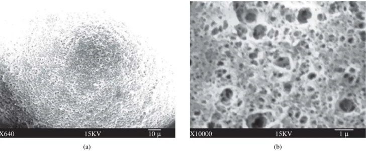
SEM results show that the HPMA copolymer beads (Fig. 1) obtained using the PVA suspending agent were a white fine powder of very small particles (10-20 µm). In contrast, the HEMA copolymer beads (Fig. 2), produced using the Mg(OH)2 suspending agent, appeared as larger white spheres (200 µm). The presence of mesopores (20-500 Å diameter) was verified for the HPMA copoly-mer beads while macropores (> 500 Å diameter) were found in the HEMA copolymer beads.
differ-ences in the porous structure of the beads were certainly related to the method of preparation of these particles to-gether with the presence of a cross-linker and a porogen.
NSA and MIP results (Table 1) showed greater specific surface area (SSA, m2/g) and total pore volume (TPV, 10 -3mL/g) for the HPMA copolymer beads than the HEMA copolymer beads. An average pore radius (APR) of about ten fold is verified for the HEMA copolymer beads. The results shown in Table 1 are consistent with the SEM results for pore diameter ranges. Both types of beads appear to be porous.
The interactions between the cells and the beads after
Figure 1. SEM micrograph of copolymerized HPMA/EDMA (1:1) beads with 41.25% addition of porogen solvent, DOD, before simple extraction (a, 1.250 ×, scale bar 10 µm) and after Soxhlet extraction (b, 10.000 ×, scale bar 1 µm).
Figure 2. SEM micrograph of HEMA/MMA (1:1) and 10% EDMA copolymerized beads with 35% addition of porogen solvent, DOD, before (a, 640 ×, scale bar 10 µm) and after (b, 10.000 ×, scale bar 1 µm) Soxhlet extraction.
Table 1. NSA and MIP results (specific surface area, SSA, m2g-1,
total pore volume, TPV, average pore size, APR) for HPMA and HEMA copolymer beads, respectively.
Polymer SSA TPV APR
(m2/g) (10-3mL/g) (Å)
HPMA copolymer beads 142 351 49 HEMA copolymer beads 31 74 441 (a)
X1250 15KV 10 µ
(b)
X10000 15KV 1 µ
(a)
X640 15KV 10 µ
(b)
mer beads. As shown in Fig. 3b, the proliferation of cells revealed that they were able to attach and grow in the pres-ence of the beads and indeed surrounded these particles with
Figure 3. HPMA copolymer beads control (a, 200 ×) and their interaction with the COS-7 cells (b) seeded in regular medium and incubated at 37 °C for 72 h (200 ×). Note the cell attachment to the beads and cell growth in the background.
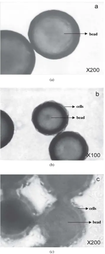
Figure 4. HEMA copolymer beads control (a, 200 ×) and their interaction with the COS-7 cells (b, 100 ×) and (c, 200 ×) seeded in regular medium and incubated at 37 °C for 72 h. Note the cell attachment on the beads in the form of a coating layer and the cell growth in the background (c).
(a)
(b)
(c) (b)
(a)
bead
mately related to the porosity of the material and greatly influence cell growth. Porosity is an important structural parameter to assess cell growth and attachment. These ma-terials may be used as a cell substrate or for other biomedi-cal applications by selecting proper bead and pore size. Furthermore, toxicity in the form of cell death was not de-tected in the described conditions, hence, residual solvent or bead synthesis by-products seem to be absent.
Acknowledgments
Financial support for this work granted by Temple Uni-versity and the fellowship to C.D. Vianna-Soares from Capes/Brasilia/Brazil are gratefully acknowledged. The authors thank Dr. Jennifer Su, Ph.D. for her assistance with the cell experiments.
References
1. Kiremitci, M.; Gürhan, I.; Piskin, E. Biotech. Appl. Biochem., v. 14, p. 170-182, 1991.
2. Kaufmann, P.M.; Heimrath, S.; Kim, B.S.; Mooney, D.J.
Cell Transplant., v. 6, p. 463-468, 1997.
3. Ma, T.; Li, Y.; Yang, S.-T.; Kniss, D.A. Biotechnol. Prog.,
v. 15, p. 715-724, 1999.
4. Koyano, T.; Minoura, N.; Nagura, M.; Kobayashi, K. J. Biomed. Mater. Res., v. 39, p. 486-490, 1998.
5. Higuchi, A.; Tamiya, S.; Tsubomura, T.; Katoh, A.; Cho, C.-S.; Akaike, T.; Hara, M. J. Biomater. Sci. Pol. Ed.,
v. 11, p. 149-168, 2000.
6. Vianna-Soares, C.D.; Kim, C.J.; Borenstein M.R. J. Po-rous Mater., v. 9, p. 67-75. 2002.
7. Vianna-Soares, C.D.; Kim, C.J.; Borenstein, M.R. J. Po-rous Mater., v. 10, p. 123-130, 2003
8. Peluso, G.; Petillo, O.; Anderson, J.M.; Ambrosio, L.; Nicolais, L.; Melone, M.A.B.; Eschbach, F.O.; Huang, S.J. J. Biomed. Mater. Res., v. 34, p. 327-336, 1997.
9. Jayakrishnan, A.; Chithambara Thanoo, B. J. Biomed. Mater. Res., v. 24, p. 913-927, 1990.
a particular agglomerated, confluent pattern. This pattern, nevertheless, was not observed with the large size HEMA copolymer beads (Fig. 4a, control). It is clear from Fig. 4b that the cell-bead association is different from the HPMA copolymer beads. At a higher magnification, as depicted in Fig. 4c, a cell layer coating the bead surface can be visual-ized. In this case, the cell background density for the HEMA copolymer beads was less when compared to the HPMA copolymer beads (Fig. 3b). This can be explained not only by their larger particle size, but also, by the greater porous texture (larger pore size). The superior adhesive character-istics of the cell onto HEMA copolymer beads may be at-tributed to the lower degree of hydrophilicity of the poly-mer beads used, i.e., the additional hydrophobic properties of the chemical structure of the beads, due to the presence of the pore size controller, MMA. Therefore, not only the chemical structure but also the surface roughness affected cell growth, likewise Higuchi et al. have reported5.
Kiremitci, Gürhan, and Piskin1 have observed cell cul-ture in a copolymer using HEMA as a basic monomer, ex-cept that it was on a swellable gel based matrix. They showed that cell attachment and growth can be controlled by chang-ing the degree of hydrophilicity, by the addition of MMA, as well as the degree of charge. Jayakrishnan and Chithambara Thanoo9 have reported the manufacture of beads made with the hydrophilic monomer (HEMA) only and cross-linked with EDMA. They reported that the control of porosity is accom-plished by the addition of a polymeric diluent in the dispersed phase. In fact, HEMA monomer has been proven to be non-toxic to human cells and affords preparation of beads in a wide size range, which is crucial for many biomedical appli-cations. Nevertheless, the same authors did not report on any cell-bead interactions.