SOCIEDADE BRASILEIRA DE ORTOPEDIA E TRAUMATOLOGIA
w w w . r b o . o r g . b r
Original
article
Reconstruction
of
soft-tissue
lesions
of
the
foot
with
the
use
of
the
medial
plantar
flap
夽
Jefferson
Lessa
Soares
de
Macedo
a,b,∗,
Simone
Corrêa
Rosa
a,
Altino
Vieira
de
Rezende
Filho
Neto
a,
Adilson
Alves
da
Silva
a,
Alex
Corcino
Silva
de
Amorim
aaHospitalRegionaldaAsaNorte,Brasília,DF,Brazil
bEscolaSuperiordeCiênciasdaSaúde,CursodeMedicina,Brasília,DF,Brazil
a
r
t
i
c
l
e
i
n
f
o
Articlehistory:
Received15August2016 Accepted4October2016 Availableonline19October2017
Keywords:
Heel
Reconstruction Woundsandinjuries Footinjuries
a
b
s
t
r
a
c
t
Objective:Tostudyuseofthemedialplantarflapforreconstructionoftheheelandfoot.
Method:Theauthorssharetheirclinicalexperiencewiththeuseofthemedialplantarartery
flapforcoverageoftissuedefectsaroundthefootandheelaftertrauma.Twelvecasesof medialplantararteryflapperformedfromJanuary2001toDecember2013wereincluded.
Results:Ofthe12patients,tenweremaleandtwowerefemale.Theindicationswere
trau-maticlossoftheheelpadintencasesandthedorsalfootintwocases.Alltheflapshealed uneventfullywithoutmajorcomplications,exceptonecasewithpartialflaploss.Thedonor sitewascoveredwithasplit-thicknessskingraft.Theflapshadslightlyinferiorprotective sensationcomparedwiththenormalside.
Conclusion: Fromtheseresults,theauthorssuggestthatthemedialplantararteryflapisa
goodadditiontotheexistingarmamentariumforcoverageofthefootandheel.Itisversatile flapthatcancoverdefectsontheheel,overtheAchillestendonandplantarsurface,aswell asthedorsalfoot.Itprovidestissuetotheplantarskinwithasimilartextureandintact protectivesensation.
©2016SociedadeBrasileiradeOrtopediaeTraumatologia.PublishedbyElsevierEditora Ltda.ThisisanopenaccessarticleundertheCCBY-NC-NDlicense(http:// creativecommons.org/licenses/by-nc-nd/4.0/).
Reconstruc¸ão
de
lesões
de
partes
moles
de
pé
com
o
uso
de
retalho
plantar
medial
Palavras-chave:
Calcanhar Reconstruc¸ão Ferimentoselesões Traumatismosdopé
r
e
s
u
m
o
Objetivo:Estudarcasosderetalhoplantarmedialnareconstruc¸ãodocalcanharedopé.
Método:Osautoresapresentamsuaexperiênciacomousodoretalhobaseadonaartéria
plantarmedialparacoberturadedefeitosteciduaisnopé,especialmentedocalcanhar.Doze retalhosdaartériaplantarmedial,feitosentrejaneirode2001edezembrode2013,foram incluídos.
夽
StudyconductedatHospitalRegionaldaAsaNorte,Brasília,DF,Brazil.
∗ Correspondingauthor.
E-mail:[email protected](J.L.Macedo).
http://dx.doi.org/10.1016/j.rboe.2017.10.009
Resultados:Dos12pacientes,dezeramhomenseduasmulheres.Asindicac¸õesforamperda traumáticadocoximdocalcanharemdezpacientesedorsodopéemdoiscasos.Todos osretalhoscicatrizaramsemmaiorescomplicac¸ões,excetoumcasocomperdaparcial. Aáreadoadorafoicobertacomenxertodepeleparcial.Osretalhosapresentaramuma sensibilidadeprotetoralevementeinferioraoladonormal.
Conclusão: Deacordocomosresultados,oretalhoplantarmedialéumaboaopc¸ãopara
coberturadopé,especialmentedaregiãodocalcanhar.Aversatilidadedoretalhopermite a coberturadedefeitosnocalcanhar,sobreotendãodeAquileseapoioplantar,assim comoodorsodopé.Esseretalhoconferepararegiãoplantarumapeledetexturasimilare sensibilidadeprotetoraintacta.
©2016SociedadeBrasileiradeOrtopediaeTraumatologia.PublicadoporElsevier EditoraLtda.Este ´eumartigoOpenAccesssobumalicenc¸aCCBY-NC-ND(http:// creativecommons.org/licenses/by-nc-nd/4.0/).
Introduction
Thereconstruction ofthedistalthird ofthe legremains a challengeforreconstructivesurgeons.Anatomical character-istics,suchasthescarcityofsofttissuesandthinskin,leadto greatdifficultiesinthetreatmentofsofttissuelesionsatthis location.
Theuseoffasciocutaneousflapsinthereconstructionof lesionsofthelowerthirdofthelowerlimb,especiallyofthe foot,iswellindicatedduetothesimilaritieswiththetissues oftheregion. Amongtheseflaps, the medialislandflap is noteworthy.1,2
ThemedialplantarflapwasinitiallydescribedbyHarrison andMorgan.3Itisbasedonthemedialplantararteryand
con-sistsofafasciocutaneousflapthatusesskinfromtheplantar archofthefoot,anidealtissuetocoverdefectsoftheheeland otherregionsofthefoot,duetothestructuralsimilarity.The innervationofthisflapispreserved,givingitsensation,which isaprotectivefactor.4
Thisstudyaimedatpresentingaseriesofcasesinwhich themedialplantarflap wasusedforthe treatmentoffoot injuries,especiallyoftheheel,from2001to2013.
Methods
Thisisaretrospectivestudyofallpatientsadmittedto hos-pitalduringthestudyperiodwhounderwentreconstruction ofthelowerlimbsduetolossofcutaneouscoveragewiththe useofthemedialplantarflap.Thefollowingvariableswere assessed:gender,age,traumaetiology,presenceandlocation ofthefracture,characteristics ofthelossofsubstance,and presenceofboneexposure.
Theinclusioncriteriawerepatientstreatedwithlowerlimb traumain thestudy periodwho underwent reconstruction withamedialplantarflap.Dopplerassessmentofthearterial systemofthefootwasperformedinallpatients.Thedorsal arteryofthefootandtheposteriortibialarterywerepatentin allpatients.
The exclusion criteria were hemodynamically unstable patients,tibialnervelesions,orlesionsintheplantardonor area.
The defect was only measured after preparationof the receptorsite,andthentransferredtothedonorsite.Theflap mustbeslightlylargerthanthereceivingarea.
ThestudywasapprovedbytheResearchEthicsCommittee undertheCAAE(CertificateofPresentationforEthical Consid-eration)number:47391715.6.0000.5553,RecommendationNo.: 1.167.841.
Surgicaltechnique
Thesurgicaltechniquewasasfollows:thelowerlimbisplaced in the supine position, with the hip flexedand externally rotated,kneeflexed,andfootinmaximumsupination.The area ofskin tobe transferred from the plantar cavus was markedoff,accordingtothesizeofthelesiontobecovered, limited bythemargins ofthe footarea that doesnotbear weight.Themidlineoftheplantarsurfaceofthefootandthe prominenceofthenavicularbonedeterminethelateraland medialbordersofthe cutaneousterritory,thatis,10–12cm longand4–6cmwide.Theoriginofthemedialplantarartery (superficialbranch)isidentifiedattheseptumbetweenthe abductorhallucismuscleandtheflexordigitorumbrevis mus-cle and emits several branches through the intermuscular septumtothemedialplantarskin.Thisarterycontinuesalong themedialborderofthefoot,anastomosedwiththefirst plan-tarmetatarsalartery.Themedialplantararteryisgenerally smallerthanthedominantlateralplantarartery.5–7
Themedialplantararteryisattacheddistallytotheflap, andtheproximalstumpissuturedtotheflap.Subfascial dis-sectionoftheflapisthenperformed;theflapiselevatedin adistal-to-proximaldirection.Theabductorhallucismuscle issectionedtoachieveagreaterlengthoftheneurovascular pedicle.Theflapisrotatedcarefullyinordertoavoidbending thepedicle.Thefasciclesofthecutaneousnerveare main-tainedintheflap,andaninterfasciculardissectionismade proximally.Subsequently,apartialskingraftisperformedin thedonorarea,atthesamesurgicaltime.
Results
wereadmittedonanoutpatientbasis,afterclinical/surgical controloftheirwoundsbyother specialties,suchas ortho-pedicsandgeneralsurgery.Themeanageofthepatientsat thetimeofinitialcarewas32years(range:2–53),witha pre-dominanceofthe20–29agegroup.Amalepredominancewas observed,representing83.3%ofthesample.Regardingthe eti-ologyoftrauma,motorcycleaccidents(50%)werenoteworthy, followedbyrun-overinjuries(33.3%),andmotorvehicle acci-dent(16.7%).Regardingthelocationofthelesions,themost frequentwerelossofsofttissueintheplantarsupportregion oftheanteriorheel(58.3%),theposteriorheelovertheAchilles tendon(25%),andthedorsumofthefoot(16.7%)(Figs.1–3). Regardingthepresenceoffracture,83.3%ofthepatientsdid notpresent fractures,while 16.7% presenteda metatarsus fracture.Boneexposurewasobservedin58.3%ofthepatients; theother41.7%presentedsofttissuelosswithoutboneor ten-donexposure.Regardingsurgicaltreatment,in83.3%ofthe casesamedialplantarislandflapwasused.
Inall cases, partialskin graftingwas performedon the donorarea,atthesametime.Surgicalcomplicationsobserved werepartiallossoftheskingraftinonecase(8.3%)and par-tiallossoftheflapinonecase(8.3%).Inthelattercase,the patientlaterunderwentareverseflowsuralfasciocutaneous flap,withoutcomplications.Intheothercases,theuseofthe medialplantarflapwasenoughtocoverthelesion,allowing goodestheticandfunctionalresults.Cutaneoussensationwas preservedinallflaps.Nocasesofdysesthesiaweredetected.
Discussion
Thefirstoptionforthereconstructionofthefootandcalcaneal plantarregionshouldbetheuseoffasciocutaneousflaps;the medialplantarflapisinaprominentpositionforprovidinga resistantskincoveringthatappearsclosetonormal,asitisa regionalflap.5Therefore,itallowsareconstructionofsimilar
tissuewithsimilartissue,thatis,itbringstothe reconstruc-tionregionaglobularskinwithafattycushionandfibrous septafixedtotheskinthatareresistanttosheartraumaand weight-bearing.5,6
Themedialplantarflapisrelativelyeasytoperform,with greatversatility,based onawell-defined vascularanatomy pattern.7 Thisflaphasevenbeenusedinpatientswith
dia-betesmellitus.8
Fromapracticalstandpoint,inthereconstructionofthe softtissueoftheheel,itisimportantthattheheelisdivided into weight-bearing regions (anterior or plantar) and non-weight-bearingregions(posterior, onthe Achillestendon).9
Theskinoftheheelandplantararchhavethesame charac-teristics;therefore,thisisthemainreasonforthepreferential useofmedialplantarflapinlesionsoftheanteriorheel.The factthatthisflapisinnervatedbythe cutaneousbranchof themedialplantarnerveisrelevantasitprovidessensitivity, animportantrequirementforpatientambulation.Theflapis createdalittlelargerthanorthesamesizeasthedefect,as thereisnosignificantprimarycontractionoftheflapdueto itsspecificfibroadiposetissuecharacteristics.
Themedialplantarflaphasalsobeenindicatedforpatients withdiabeticneuropathywhopresentchroniculcersinareas
ofsensoryloss,withalowrateofulcerrecurrenceinthelong term.Sincediabeticpatientsmayhavevascularproblems,this flapmayonlybeindicatedforthosewithgoodvascularflow totheflapregion.8
Locoregional fasciocutaneous flapsare an alternative to freeflapsforlowerlimbreconstruction,especiallyintheheel region.Freeflapswouldbeindicatedformorecomplexcases, whennoneofthelocoregionalflapsareavailable.9
Freeflapsaregood optionsforrebuildinglargelossesof softtissueontheheelandlowerthirdoftheleg.Microsurgery mayrequirelongersurgicaltimethanlocoregionalflaps,and aspecializedteamisneeded.10
Incalcanealreconstructions,thereversesuralflapisalso agoodoption.11,12Thisflapwassuccessfullyusedinonecase
ofpartiallossofthemedialplantarflapinthereconstruction oftheheel.Thedisadvantageofthereversesuralflapislossof sensationinthelateralmalleolus,thelateralsideofthefoot, andonthefifthtoe,duetoligatureofthesuralnerve.
Inthepresentstudy,distalbasedreverseflowmedial plan-tararteryflapswerenotmade.Thistypeofflapisindicated forthereconstructionofdistaldefectsintheplantarregion ofthemetatarsalheads.Theseflapsarebasedonretrograde bloodflowfromthedistalmedialplantararterytothedorsal arteryofthefootthroughthefirstdorsalmetatarsal commu-nicatingbranches.Thedisadvantageofthisflapisitssensory loss.13
Freeflapsbased onthe medialplantararteryare alsoa goodoptionforreconstructionofdistaldefectsofthe plan-tarregion,andtheycanbeflapinnervated.14,15Moreover,the
medialplantarflapcanbemadecrosslegged,withthedonor regionoftheflapbeingthecontralateralfoot.16
Onedisadvantageoftheflapbasedonthemedialplantar arteryisthelossofafootartery.However,themain irriga-tionplantararchofthefootisthedeepone,whichisformed mainlybythelateralplantarartery,allowingtheformationof ananastomoticnetworkbetweenthetwomainarteriesofthe foot(dorsalarteryofthefootandlateralplantarartery).
Thedeepplantararchalsoformsfourplantarmetatarsal arteriesandsomeperforatingarteries.Thecontributionofthe medialplantararterytothedeepplantararchissmallandis limitedtothelateralbranchofitsdeepbranch.17
Another disadvantage of the medial plantar flap is its limitationinsizeandoncoverageofdeepandextensive cav-itydefects.Therefore,largermuscleorfasciocutaneousflaps shouldbeusedtocoversuchdefects.
The options for reconstruction in complex lesions are numerous;thechoiceofanadequatesurgicalplanningbased onthepatient’sage,gender,andoccupation, aswellasthe sizeandlocationoftissueloss,isparamount.Furthermore, thepresenceoftraumaandassociatedinjuriesmustalways beconsidered,especiallyintraumacausedbytheimpactof highenergy.Theconcernwiththedonorareaandthequality oftheresultsintherecipientareahasbeenincreasing.
Fig.1–(A)53-Year-oldpatient,victimofamotorcycleaccident,withlossofsubstanceintheAchillestendontopography.(B)
Flapdonorarea.(C)MedialplantarflapcoveringtheAchillestendonthreemonthspostoperatively.
Fig.2–(A)5-Year-oldchild,victimofarun-overinjury,withlossofsubstanceinthedorsumofthefootwithcompound
metatarsalfracturesassociatedwithtoeamputation.(B)Dissectedmedialplantarislandflap,withitsneurovascular
pedicle.(C)Flappositionedintherecipientarea,onthedorsumofthefoot.(D)Donorareaofthemedialplantarflapwith
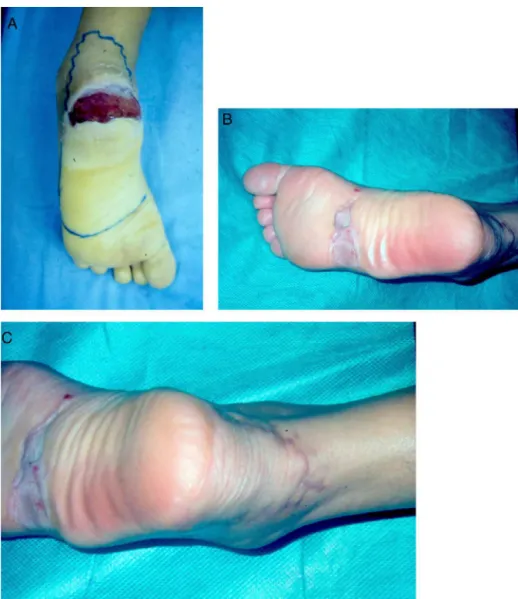
Fig.3–(A)26-Year-oldpatient,victimofamotorcycleaccidentwithlossofcalcanealsubstance.(B)Thedonorareaofthe
skingraftflap,fivemonthspostoperatively.(C)Advancingflapofthemedialplantarregionandtheposteriorregionofthe
heelcoveringthelossofsubstanceintheheel.
Conclusion
Themedialplantarflaphasbeenshowntobeagood treat-mentoptionforinjuriesoftheheelanddorsumofthefoot, withahighsuccessrateandeasyreproducibility.Themain advantagesoftheflaparethepresenceofsensationit pro-videsandthefactthatitbringsspecializedplantartissueto thereconstructed recipientarea,withlowmorbidityinthe donorarea.
Conflicts
of
interest
Theauthorsdeclarenoconflictsofinterest.
r
e
f
e
r
e
n
c
e
s
1. MartinsGB,MoreiraAA,VianaFO.Reconstruc¸ãodelesõesde partesmolesdocalcanharcomusoderetalhos
faciocutâneos.RevBrasCirPlast.2009;24(10):104–9.
2.Benito-RuizJ,YoonT,Guisantes-PintosE,MonnerJ, Serra-RenomJM.Reconstructionofsoft-tissuedefectsofthe heelwithlocalfasciocutaneousflaps.AnnPlastSurg. 2004;52(4):380–4.
3.HarrisonDH,MorganDG.Theinstepislandflaptoresurface plantardefects.BrJPlastSurg.1981;34(3):315–8.
4.MourougayanV.Medialplantarartery(instepflap)flap.Ann PlastSurg.2006;56(2):160–3.
5.MacchiV,TiengoC,PorzionatoA.Correlationbetweenthe courseofthemedialplantararteryandthemorphologyof theabductorhallucismuscle.ClinAnat.2005;18(8):580–8.
6.DrakeRL.TerminologiaanatômicaInternationalAnatomical Terminology.Stuttgart:Thieme;2011.
7.Rodriguez-VegasM.Medialispedisflapinthereconstruction ofpalmarskindefectsofthedigitis:clarifyingtheanatomyof themedialplantarartery.AnnPlastSurg.2014;72(5):542–52.
8.SchwarzRJ,NegriniJF.Medialplantararteryislandflapfor heelreconstruction.AnnPlastSurg.2006;57(6):658–61.
9.BarreiroGC,BatistaRR,BusnardoF,OlivanM,FereiraMC. Reconstruc¸ãodeplantadepédeacordocomoconceitodas subunidadesanatômicas.RevBrasCirPlast.2010;25Supl:81.
11.GarciaAM.Retalhosuralreversoparareconstruc¸ãodistalda perna,tornozelo,calcanharedopé.RevBrasCirPlast. 2009;24(1):96–103.
12.Al-QattanMM.Thereversesuralfasciomusculocutaneous mega-highflap:astudyof20consecutiveflapsforlower-limb reconstruction.AnnPlastSurg.2007;58(5):513–6.
13.CoruhA.Distallybasedperforatormedialplantarflap:anew flapforreconstructionofplantarforefootdefects.AnnPlast Surg.2004;53(4):404–8.
14.ZelkenJA,LinCH.Analgorithmforforefootreconstruction withtheinnervatedfreemedialplantarflap.AnnPlastSurg. 2016;76(2):221–6.
15.LykoudisEG,SeretisK,LykissasMG.Freesensatemedial plantarflapforcontralateralplantarforefootreconstruction withflapreinnervationusingend-to-sideneurorrhaphy:a casereportandliteraturereview.Microsurgery.
2013;33(3):227–31.
16.AcikelC,CelikozB,YukselF,ErgunO.Variousapplicationsof themedialplantarflaptocoverthedefectsofplantarfoot, posteriorheel,andankle.AnnPlastSurg.2003;50(3):498–503.