ABSTRACT
http://dx.doi.org/10.1590/1678-775720130691
Antimicrobial and cytotoxic effects of phosphoric
acid solution compared to other root canal irrigants
Maíra PRADO, Emmanuel João Nogueira Leal da SILVA, Thais Mageste DUQUE, Alexandre Augusto ZAIA, Caio Cezar Randi FERRAZ, José Flávio Affonso de ALMEIDA, Brenda Paula Figueiredo de Almeida GOMES
Department of Restorative Dentistry, Endodontics Division, Piracicaba Dental School, State University of Campinas - UNICAMP, Piracicaba, SP, Brazil.
Corresponding address: Brenda P.F.A. Gomes - Faculdade de Odontologia de Piracicaba - UNICAMP - Departamento de Endodontia - Av. Limeira, 901 - Areião - Piracicaba - São Paulo - Brazil - 13414-903 - e-mail: [email protected] - Phone: (55) 19 2106 5343 - Fax: (55) 19 2106 5218
Submitted: January 8, 2014 - Modiication: July 20, 2014 - Accepted: July 29, 2014
P
hosphoric acid has been suggested as an irrigant due to its effectiveness in removing the smear layer. Objectives: The purpose of this study was to compare the antimicrobial and cytotoxic effects of a 37% phosphoric acid solution to other irrigants commonly used in endodontics. Material and Methods: The substances 37% phosphoric acid, 17% EDTA, 10% citric acid, 2% chlorhexidine (solution and gel), and 5.25% NaOCl were evaluated. The antimicrobial activity was tested against Candida albicans, Staphylococcus aureus, Enterococcus faecalis, Escherichia coli, Actinomyces meyeri, Parvimonas micra, Porphyromonas gingivalis, and Prevotella nigrescens according to the agar diffusion method. The cytotoxicity of the irrigants was determined by using the MTT assay. Results: Phosphoric acid presented higher antimicrobial activity compared to the other tested irrigants. With regard to the cell viability, this solution showed results similar to those with 5.25% NaOCl and 2% chlorhexidine (gel and solution), whereas 17% EDTA and 10% citric acid showed higher cell viability compared to other irrigants. Conclusion: Phosphoric acid demonstrated higher antimicrobial activity and cytotoxicity similar to that of 5.25% NaOCl and 2% chlorhexidine (gel and solution).Keywords: Phosphoric acids. EDTA. Citric acid. Chlorhexidine. Products with antimicrobial action. Toxicity test.
INTRODUCTION
Mechanical instrumentation during endodontic therapy produces an amorphous irregular layer known as smear layer. The smear layer consists of remnants of ground dentin, odontoblastic processes, pulp tissue, and microorganisms in the case of infected teeth10. It forms a barrier between
illing material and sound dentin, which inhibits the penetration of irrigants into dentinal tubules, increases the microleakage of commonly used sealers, and decreases the bond strength of resin-based materials12,13,19.
S o m e c h e m i c a l a g e n t s s u c h a s E D TA (ethylenediaminetetraacetic acid) solutions at concentrations ranging from 15 to 17%, citric acid (5-50%), and phosphoric acid (5-37%) are used to remove this layer1,12,20,22.
Phosphoric acid at 37% is a substance commonly used in restorative dentistry for the conditioning of
both enamel and dentin in the coronal surface. Its use in endodontics for smear layer removal showed favorable results. In a 3-minute application in the apical third, this solution was more effective than 17% EDTA and 10% citric acid12. Additionally, it was
observed that when this solution was associated with NaOCl, the bond strength of gutta-percha/AH Plus was improved as well as when associated with CHX for Resilon/Real Seal SE systems13. Although
this solution has shown favorable results, assessing other characteristics such as its cytotoxic and antimicrobial effects is clinically required in the endodontic practice.
MATERIAL AND METHODS
Root canal irrigants
The substances evaluated in the present study were 37% phosphoric acid, 17% EDTA, 10% citric acid, 5.25% NaOCl, and 2% chlorhexidine (solution and gel). The same drugstore provided all these substances (Drogal, Piracicaba, SP, Brazil).
Antimicrobial assay
Microorganisms
The species of microorganisms Candida albicans (ATCC 10556), Staphylococcus aureus (ATCC 25923), Enterococcus faecalis (ATCC 29212), and Escherichia coli (ATCC 25922) were grown on 5% brain heart infusion agar plates (BHI, Lab M, Bury, UK), whereas Actinomyces meyeri (ATCC 33972), Parvimonas micra (ATCC 33270), Porphyromonas gingivalis (ATCC 49417) and Prevotella nigrescens (ATCC 33563) were grown on fastidious anaerobe agar plates (FAA, Lab M, Bury, UK). In both media, 5% deibrinated sheep blood was added.
Agar diffusion method
The methodology used was adapted from Gomes, et al.7 (2002). All microorganisms were previously
subcultured in appropriate culture media and under gaseous conditions to conirm their purity.
The aerobic (C. albicans) and facultative anaerobic strains (E. coli, S. aureus, E. faecalis) were individually inoculated into tubes containing 5 mL of sterile saline solution. The suspension was adjusted spectrophotometrically at 800 nm (Optical Density 800) to match the turbidity of 1.5x10⁸ CFU/
mL (equivalent to 0.5 McFarland standard). Five-hundred mL of each test microorganism suspension were used to inoculate glass bottles containing 50 mL of BHI Agar (Lab M, Bury, UK) at 46°C before being mixed and poured onto 150 mm plates containing a previously set layer of Mueller Hinton agar (Oxoid, Unipath Ltd, Basingstoke, Hampshire, UK).
The strict anaerobic microorganisms (A. meyeri, P. micra, P. gingivalis, P. nigrescens) were suspended spectrophotometrically at 800 nm to match the turbidity of 3.0x10⁸ CFU/mL (equivalent to 1
McFarland standard). Sterile swabs were dipped into the microbial suspension and were used to inoculate pre-reduced 150 mm plates containing 5% sheep blood fastidious anaerobe agar (FAA - Lab-M, Bury, Lancs, UK). The inoculation procedures used were appropriate to provide a semi-conluent growth of the microorganisms being tested6,7.
Three sterilized stainless steel tubes of 8.0x1.0x10.0 mm were added to the surfaces of the media and illed with 0.2 mL of each pure or composite substance. The plates were maintained for 2 hours at room temperature and appropriate gaseous conditions to allow for diffusion of the agents
through agar and then incubated at 37°C again under appropriate gaseous conditions for the following periods of time: aerobes, 24 hours; facultative anaerobes, 24-48 hours in a CO2 incubator (Jouan SA, Thermo Fisher Scientiic, Saint Herblain, Loire-Atlantique, France) in an atmosphere of 10% CO2; and anaerobes in anaerobic workstation (Don Whitley Scientiic, Bradford, West Yorkshire, UK) in an atmosphere of 10% H2, 10% CO2, 80% N2 for 7 days.
Zones of inhibition of microbial growth around the cylinder containing the tested substances were measured and recorded after the incubation period. The inhibitory zone was considered to be the shortest distance (mm) between the outer margin of the cylinder and the initial point of the microbial growth. Three replicates were made for each microorganism.
Cytotoxicity assay
Balb/c 3T3 cells (American Tissue Type Collection, Manassas, VA, USA) were cultured in Dulbecco modified Eagle medium (DMEM) (Gibco, Grand Island, NY, USA), supplemented with 10% fetal bovine serum (Sigma Chemical Co, St Louis, MO, USA), 100 mg/mL streptomycin, and 100 mg/mL penicillin at 37°C in a humidiied incubator in an atmosphere containing 5% CO2. Conluent cells were detached with 0.25% trypsin and 0.05% EDTA for 5 minutes, with aliquots of separated cells being subcultured. Cells were seeded in 96-well plates (1x10⁴ cells/well). After overnight attachment, cells
were treated with various irrigants (200 µL/well) for 30 minutes, which was representative of the clinical situation. Each irrigant was tested in a 1/1, 1/10, 1/100 and 1/1000 dilution with DMEM.
Statistical analysis
The results were submitted to the Kolmogorov-Smirnov’s test to evaluate the normal distribution. Antimicrobial data were analyzed with Kruskal-Wallis’ and Mann-Whitney’s tests (p<0.05). Cytotoxicity data were analyzed with one-way analysis of variance (ANOVA) and Tukey’s test (p<0.05).
RESULTS
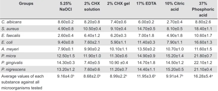
Table 1 shows the average and standard deviation values of growth inhibition zones (in mm) produced by the substances against each tested
microorganism. The in vitro antimicrobial effects of the substances were ranked from strongest to weakest as follows: 37% phosphoric acid, 17% EDTA, 10% citric acid, 5.25% NaOCl, 2% CHX gel, 2% CHX solution. Statistical analyses showed that phosphoric acid presented higher antimicrobial effect than the other irrigants being tested (Figure 1).
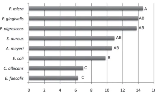
The susceptibility of individual microorganisms to the substances was varied. Strict anaerobic microorganisms such as P. micra, P. gingivalis and P. nigrescens were more susceptible to the substances, while E. faecalis and C. albicans (facultative microorganisms) were more resistant (Figure 2).
Groups 5.25%
NaOCl
2% CHX solution
2% CHX gel 17% EDTA 10% Citric acid
37% Phosphoric
acid
C. albicans 8.60±0.2 8.20±0.8 7.40±0.6 6.00±0.2 2.70±0.4 8.80±2.6
S. aureus 4.90±0.8 10.50±0.4 9.10±0.4 14.70±0.5 8.10±0.5 18.40±1.1
E. faecalis 2.60±0.4 6.40±1.2 6.20±0.3 7.00±1.8 4.90±1.8 10.60±1.7
E. coli 9.40±0.8 7.60±2.1 5.90±1.1 11.40±0.3 7.90±1.1 16.60±1.3
A. meyeri 7.90±0.1 9.90±0.2 10.10±1.1 13.50±0.2 10.70±1.0 11.60±1.5
P. micra 12.50±1.5 11.90±1.0 11.30±0.6 14.90±0.9 15.20±1.4 21.80±0.7
P. gingivalis 14.30±0.3 7.40±0.5 10.90 ±0.4 14.70±1.8 14.50±1.2 22.10±1.2
P. nigrescens 13.20±1.2 7.60±0.6 11.20±0.7 14.40±1.1 15.20±0.5 21.10±0.4
Average values of each substance against all microorganisms tested
9.16±4.0b 8.68±2.0b 8.99±2.2b 11.95±3.6b 9.91±4.7b 16.28±5.4a
Different letters (a, b) indicate statistically signiicant values (p<0.05)
Table 1- Average (±Standard Deviation) area of the zones of microbial growth inhibition in mm provided by the tested substances against all microorganisms
Figure 1- Values of growth inhibition zones (mm) for the eight tested microorganisms
Figure 3 shows the cell viability of the different irrigants. Statistically, 37% phosphoric acid showed cell viability similar to that of 5.25% NaOCl, 2% chlorhexidine (gel and solution), whereas 17% EDTA and 10% citric acid showed higher cell viability than the other irrigants.
DISCUSSION
The need for removing smear layer seems to be a consensus in the literature4,13,19 for several reasons,
including: a) it contains bacteria and necrotic tissues; b) it forms a barrier between the illing material and
sound dentin that limits the penetration of irrigants/ medicaments and sealers into dentinal tubules; c) it increases microleakage with commonly used sealers; d) it decreases the bond strength of resin based materials.
The solution of 37% phosphoric acid has been found to be effective as a inal rinse substance to remove the smear layer from the instrumented surface of root canals and to prevent the formation of a chemical smear layer when used as intermediate irrigant between sodium hypochlorite and chlorhexidine13,14. Additionally, it can increase
the resin-based sealer bond strength to dentin13.
Figure 3- Cell viability to the tested irrigants
Note: Different letters indicate statistically signiicant values (p<0.05)
Figure 2- Average (in mm) of the zones of inhibition of each microorganism against all irrigants tested
However, apart from its chemical properties, it should also have strong antimicrobial properties and minimal cytotoxic effects on the periradicular tissues. The present work compared the antimicrobial and cytotoxic effects of a 37% phosphoric acid solution to other irrigants commonly used in endodontics, including 5.25% NaOCl, 2% chlorhexidine (solution and gel), 17% EDTA, and 10% citric acid.
The agar diffusion test, used in the present study, is a worldwide accepted procedure for determining the in vitro sensitivity under routine laboratory conditions. This method is simple, standardized, and reproducible, being used in several studies, not only to test the antimicrobial properties of irrigants5,18,21,
but also of intracanal medicaments7 and sealers17.
In the present study, E. faecalis and C. albicans were the microorganisms showing the smallest inhibition zone against all tested irrigants, while strict anaerobes such as the Gram-positive P. micra and the Gram-negative bacteria such as P. gingivalis, and P. nigrescens showed the largest inhibition zones. It could be extrapolated that even though the anaerobic bacteria, such as Parvimonas, Prevotella or Porphyromonas spp. are associated with painful infectious exacerbations6,21,they are
more susceptible to the tested irrigants. On the other hand, facultative Gram-positive bacteria such as E. faecalis and C. albicans, which have been associated with asymptomatic root canals6,21, seem
to be more resistant to root canal therapy and are therefore related to endodontic failures. Our indings are in agreement with the literature6,21. Further
investigations using a bioilm model and confocal laser scanning microscopy (CLSM) in order to detect and quantify the bacterial viability after the use of these tested irrigants are required to conirm these preliminary results.
Additionally, all irrigants had an antimicrobial effect on the eight tested strains. However, phosphoric acid showed the highest antimicrobial activity compared to the other irrigants evaluated. The action mechanism of phosphoric acid has not been fully elucidated. One possible explanation is that it may act by increasing the hydrogen-ion concentration in the microorganism, which cannot maintain their vitality under these conditions2,15.
With regard to this aspect, it was observed that the bactericidal activity of 5% phosphoric acid and 10% citric acid was similar, with the former requiring less time than the latter to exert its antimicrobial activity2.
Regarding the values found for 5.25% NaOCl and 2% CHX, they are similar to those reported by Ferraz, et al.5 (2007), but differ from Siqueira, et al.18 (1998).
The cytotoxicity of the irrigants to 3T3 ibroblast cells was evaluated here. Studies on established cell lines are used because of the reproducibility of the results, besides the fact that they multiply rapidly and have unlimited life span3,8,16. Our results
revealed that the cytotoxicity of the 37% phosphoric acid solution was higher than that of other solutions employed for removal of smear layer, such as 17% EDTA and 10% citric acid. However, EDTA and citric acid showed similar cytotoxic effects, which is in disagreement with other studies that observed a lower cytotoxic effect for the citric acid1,9.
In addition, phosphoric acid showed a similar cytotoxicity compared to 5.25% NaOCl and 2% chlorhexidine (solution and gel), in accordance with a previous study4 that compared NaOCl and
chlorhexidine. However, our indings differ from others that observed a lower cytotoxic effect for 2% chlorhexidine11. No study was found in the literature
comparing the cytotoxicity of phosphoric acid with other endodontic irrigants.
CONCLUSION
The solution of 37% phosphoric acid exhibited antimicrobial effect against the tested endodontics pathogens and cytotoxicity similar to that of 5.25% NaOCl and 2% chlorhexidine (solution and gel).
ACKNOWLEDGEMENTS
This study was supported by the Brazilian funding agencies: FAPESP - São Paulo Research Foundation (protocol numbers 2009/53976-0, 2010/50817-5 and 2011/09047-4), CNPq - National Council for Scientiic and Technological Development (protocol number 302575/2009-0) and CAPES - Coordination for the Improvement of Higher Education Personnel.
REFERENCES
1- Amaral KF, Rogero MM, Fock RA, Borelli P, Gavini G. Cytotoxicity analysis of EDTA and citric acid applied on murine resident macrophages culture. Int Endod J. 2007;40:338-43.
2- Arias-Moliz MT, Ferrer-Luque CM, Espigares-Rodríguez E, Liébana-Ureña J, Espigares-García M. Bactericidal activity of phosphoric acid, citric acid, and EDTA solutions against
Enterococcus faecalis. Oral Surg Oral Med Oral Pathol Oral Radiol Endod. 2008;106:e84-9.
3- Bouillaguet S, Wataha JC, Tay FR, Brackett MG, Lockwood PE. Initial in vitro biological response to contemporary endodontic sealers. J Endod. 2006;32:989-92.
4- Chang YC, Huang FM, Tai KW, Chou MY. The effect of sodium hypochlorite and chlorhexidine on cultured human periodontal ligament cells. Oral Surg Oral Med Oral Pathol Oral Radiol Endod. 2001;92:446-50.
5- Ferraz CC, Gomes BP, Zaia AA, Teixeira FB, Souza-Filho FJ. Comparative study of the antimicrobial eficacy of chlorhexidine gel, chlorhexidine solution and sodium hypochlorite as endodontic irrigants. Braz Dent J. 2007;18:294-8.
6- Gomes BP, Ferraz CC, Garrido FD, Rosalen PL, Zaia AA, Teixeira FB, et al. Microbial susceptibility to calcium hydroxide pastes and their vehicles. J Endod. 2002;28:758-61.
7- Koo H, Gomes BP, Rosalen PL, Ambrosano GM, Park YK, Cury JA. In vitro antimicrobial activity of propolis and Arnica montana
8- Loushine BA, Bryan TE, Looney SW, Gillen BM, Loushine RJ, Weller RN, et al. Setting properties and cytotoxicity evaluation of a premixed bioceramic root canal sealer. J Endod. 2011;37:673-7. 9- Malheiros CF, Marques MM, Gavini G. In vitro evaluation of the cytotoxic effects of acid solutions used as canal irrigants. J Endod. 2005;31:746-8.
10- McComb D, Smith DC. A preliminary scanning electron microscopic study of root canals after endodontic procedures. J Endod. 1975;1:238-42.
11- Onçağ O, Hoşgör M, Hilmioğlu S, Zekioğlu O, Eronat C, Burhanoğlu D. Comparison of antibacterial and toxic effects of various root canal irrigants. Int Endod J. 2003;36:423-32. 12- Prado M, Gusman H, Gomes BP, Simão RA. Scanning electron microscopic investigation of the effectiveness of phosphoric acid in smear layer removal when compared with EDTA and citric acid. J Endod. 2011;37:255-8.
13- Prado M, Simão RA, Gomes BP. Effect of different irrigation protocols on resin sealer bond strength to dentin. J Endod. 2013;39:689-92.
14- Prado M, Simão RA, Gomes BP. Evaluation of different irrigation protocols concerning the formation of chemical smear layer. Microsc Res Tech. 2013;76:196-200.
15- Russel AD, Hugo WB, Ayliffe GA. Principles and practice of disinfection, preservation and sterilization. 3rd ed. Oxford:
Blackwell Science Ltd; 1999. p. 124-48.
16- Schwarze T, Leyhausen G, Geurtsen W. Long-term cytocompatibility of various endodontic sealers using a new root canal model. J Endod. 2002;28:749-53.
17- Silva EJ, Herrera DR, Rosa TP, Duque TM, Jacinto RC, Gomes BP, et al. Evaluation of cytotoxicity, antimicrobial activity and physicochemical properties of a calcium aluminate-based endodontic material. J Appl Oral Sci. 2014;22:61-7.
18- Siqueira JF Jr, Batista MM, Fraga RC, de Uzeda M. Antibacterial effects of endodontic irrigants on black-pigmented gram-negative anaerobes and facultative bacteria. J Endod. 1998;24:414-6. 19- Torabinejad M, Handysides R, Khademi AA, Bakland LK. Clinical implications of the smear layer in endodontics: a review. Oral Surg Oral Med Oral Pathol Oral Radiol Endod. 2002;94:658-66. 20- Torabinejad M, Shabahang S, Aprecio RM, Kettering JD. The antimicrobial effect of MTAD: an in vitro investigation. J Endod. 2003;29:400-3.
21- Vianna ME, Gomes BP, Sena NT, Zaia AA, Ferraz CC, Souza Filho FJ. In vitro evaluation of the susceptibility of endodontic pathogens to calcium hydroxide combined with different vehicles. Braz Dent J. 2005;16:175-80.