ABSTRACT
http://dx.doi.org/10.1590/1678-775720140491
Effect of prosthodontic planning on lateral occlusion
scheme: a comparison between conventional and
digital planning
Jaafar ABDUO1,2, Mohammed BENNAMOUN3, Marc TENNANT2, John McGEACHIE2
1- Melbourne Dental School, Melbourne University, Melbourne, Australia.
2- School of Anatomy, Physiology and Human Biology, University of Western Australia, Crawley, Australia. 3- School of Computer Science and Software Engineering, University of Western Australia, Crawley, Australia.
Corresponding address: Jaafar Abduo - Melbourne Dental School - Melbourne University - 720 Swanston Street - Melbourne - Australia - Phone: 0061 3 9035 8998 - Fax: 0061 3 9341 1599 - e-mail: [email protected]
Submitted: December 11, 2014 - Modiication: February 6, 2015 - Accepted: February 25, 2015
R
ecently, digital wax-up is proposed as a tool to aid prosthodontic planning. However, there are no data about the effect of prosthodontic planning on lateral occlusion scheme. Objective: This study aims to evaluate the impact of conventional and digital prosthodontic planning on lateral occlusion scheme. Material and Methods: Dental models of 10 patients were collected. All models had Angle Class I occlusion and were undergoing prosthodontictreatment that would inluence the lateral occlusion scheme. Each set of models had received
both conventional wax-up and digital wax-up. In relation to the lateral occlusion scheme, the following variables were evaluated: the prevalence of the different lateral occlusion scheme, number of contacting teeth and percentage of each contacting tooth. Four excursive positions on the working side were included: 0.5, 1.0, 2.0 and 3.0 mm from the maximal intercuspation position. Results: The lateral occlusion scheme of the two wax-up models was subjected to alterations following excursion. There was a tendency for the prevalence of canine-guided occlusion to increase and for the prevalence of group function occlusion to decrease with increasing excursion. The number of contacting teeth was decreasing with the increasing magnitude of excursion. For the 0.5 mm and 1.0 mm positions, the
two wax-ups had signiicantly greater contacts than the pre-treatment models, while at
the 2.0 mm and 3.0 mm positions, all the models were similar. For all models, canines were the most commonly contacting teeth, followed by the teeth adjacent to them. No difference was observed between the two wax-ups in relation to the number of contacting
teeth. Conclusion: Although the prosthodontic planning had inluenced the pattern of the
lateral occlusion scheme and contacts, there was no difference between the conventional and digital prosthodontic planning.
Keywords: Diagnostic techniques. Wax. Dental occlusion. Dental models.
INTRODUCTION
The ideal lateral occlusion scheme has been a topic of debate for many years. It has been postulated that the lateral occlusion scheme impacts masticatory function, comfort and aesthetics. Several authors discussed the rationale of each lateral occlusion scheme19,23. It is common
throughout prosthodontic treatment that the lateral occlusion scheme be altered by controlling morphologies, alignments and orientations of teeth.
The two commonly applied schemes are canine-guided occlusion and group function occlusion. The canine-guided occlusion is a mutually protected occlusion where the vertical and horizontal overlap of the canine teeth causes disengagement of the posterior teeth in the lateral movement of the mandible9. The group function occlusion is
based on multiple contacts between the maxillary and mandibular teeth in lateral movement on the working side9. It has been speculated that
teeth laterally because of the canines’ strategic location, anatomy and proprioceptive properties19.
On the other hand, group function occlusion might contribute to a wide distribution of occlusal forces on several teeth instead of a single tooth; thus, the occlusion can be more comfortable and functional23.
However, true clinical evidence supporting either scheme is still lacking, therefore, both schemes are deemed acceptable4,24.
More recently, there has been a discussion about
the limitations of deining each lateral occlusion
scheme, as the occlusal presentation is naturally more complex 17. For example, with different
degrees of excursion, the lateral occlusion scheme might differ. Further, any functional occlusion is subjected to changes with time, yet without manifestation of physiological abnormalities2.
It is also acknowledged that most patients are comfortable with their existing dentition and occlusion. Thus, the occlusion scheme can be
considered physiological even if it does not it into any speciic category.
Fixed prosthodontic treatment is indicated to alter the tooth morphology, which can eventually alter the lateral occlusion scheme. To date, the authors are not aware of any study that evaluated
the impact of ixed prosthodontic treatment on
the lateral occlusal scheme. The purpose of this observational study is to evaluate the effect of two
forms of ixed prosthodontic planning (conventional
and digital diagnostic wax-ups) on lateral occlusion scheme. Further the frequency of each tooth
contact will be quantiied. The null hypotheses are
that the prosthodontic planning will change the lateral occlusion scheme and the frequency of teeth contacts, and there will be a difference between the two forms of prosthodontic planning.
MATERIAL AND METHODS
Models of 10 patients who required fixed
prosthodontic treatment (crowns or ixed partial
dentures) were collected for this study. Figure 1 summarizes the inclusion criteria. The treatment indications were managements of heavily restored dentition, tooth wear, aesthetic problems and short-span edentulous area. For all models, the diagnostic
wax-up had an inluence on the lateral occlusion
scheme. The patients received the treatment at the Oral Health Centre of Western Australia and a human research ethics approval was obtained from the Human Research Ethics Committee of the
University of Western Australia (RA/44/1/5079). An irreversible hydrocolloid impression (Alginate,
GC America, IL, USA) was taken for each arch and an occlusal relation record was obtained by intra-oral
polyvinyl siloxane registration material (GC Exabite,
GC America, IL, USA) according to the centric relation position. All the impressions were poured by
type III dental stone (Buff Stone, Adelaide Moulding
& Casting Supplies, South Australia, Australia). These casts comprised the pre-treatment models. All the models were duplicated twice by reversible
hydrocolloid duplicating materials (Magafeel, MKM
System, Haanova, Slovakia). On one model, the conventional wax-up was executed and the other
model was used for the digital wax-up (Figure 2).
Conventional wax-up
On a semi-adjustable articulator (Whip Mix, Louiseville, KY, USA), one set of duplicate
models were articulated according to maximal intercuspation to represent the pre-treatment articulation. The other sets were used for the conventional wax-up and were articulated according to centric relation position with the aid of the intra-oral record. Therefore, after the wax-up, there was be a coincidence between centric relation and maximal intercuspation positions4. Silicone material putty (Dental Speedex Putty, Coltene/Whaledent
AG, Altstatten, Switzerland) was applied on to the buccal aspects of the posterior teeth of the mounted models. This silicone index was used for the digital articulation of the pre-treatment models.
The conventional wax-up procedure involved inlay wax addition on the external tooth surface with the aim of rectifying the defective tooth structure, obtaining natural and aesthetic tooth morphology, achieving symmetry between the two sides, and obtaining even bilateral occlusal contacts. For some patients, an increase of the vertical dimension of occlusion was necessary. All the conventional wax-ups were completed by an experienced dental technician. The dental technician was advised to
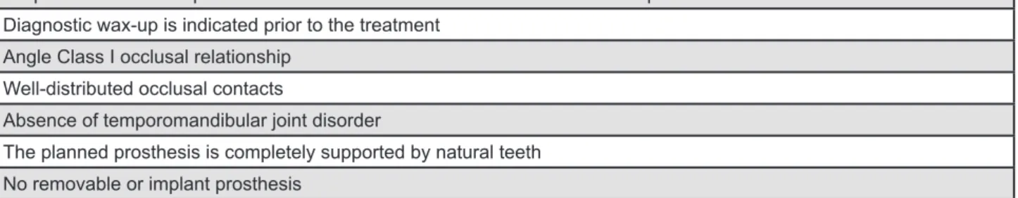
Requirements of ixed prosthodontic treatment in the form of crowns or ixed partial dentures in at least one arch
Diagnostic wax-up is indicated prior to the treatment
Angle Class I occlusal relationship
Well-distributed occlusal contacts
Absence of temporomandibular joint disorder
The planned prosthesis is completely supported by natural teeth
No removable or implant prosthesis
produce a group function lateral occlusion scheme for the initial excursions with no steep occlusion guidance. On the completed wax-up models, silicone indices were formed on the buccal aspect of the posterior teeth.
A micro-CT scanner (SkyScan, Bruker micro CT, Kontich, Belgium) was used to scan the
pre-treatment, conventional wax-up models and the associated silicone indices. The generated Digital Imaging and Communication Medicine
(DICOM) images were used to construct virtual 3D Stereolithography (STL) images of the maxillary and
mandibular models by a DICOM viewing program
(CTvox, Bruker microCT, Kontich, Belgium). The
virtual image of the conventional wax-up was used
for the subsequent analysis (Figure 2B).
In order to digitally articulate the models, the image registration process by a 3D rendering
software package (Geomagic Studio, Raindrop Geomagic Inc., Research Triangle Park, NC, USA) was
applied. Initially, corresponding points on common surfaces between the model and the silicone indices were selected to approximate the positions of the images. This was followed by implementing the
Iterative Closest Point Algorithm, which re-orients the models according to the best it principles. The
same process was repeated for the other silicone index and the opposing arch. Eventually, the silicone indices were deleted digitally, and the two models were digitally articulated.
Digital wax-up
The digital wax-up has been discussed in an earlier report1. In summary, the pre-treatment
models and the associated silicone indices were scanned and converted to virtual images. The virtual pre-treatment models were articulated according to the centric relation position. This was achieved by superimposing the virtual pre-treatment models on the virtually articulated conventional wax-up models. The unaltered tissues were used for the
registration. A 3D rendering software (Geomagic
Studio) was used to execute the digital wax-up. The pre-treatment models were articulated by the process of Image Registration. Virtual physiological
teeth (Phonares Teeth, Ivoclar Vivadent AG,
Schaan, Liechtenstein) were used to alter the teeth morphologies. The virtual tooth alignment involved size alteration, rotation and translation.
Each virtual tooth was itted manually on the model
with the aim of obtaining ideal teeth arrangement,
emergence proile, symmetry and aesthetics. After
the completion of the wax-up, the scanned model and the virtual teeth were merged to generate
a single model (Figure 2C), which enhanced the
computation speed of the subsequent analysis.
Virtual simulation of lateral movement
For each set of articulated models, the occlusion scheme was evaluated for each working side separately. From the maximal intercuspation
position (Figure 3A), four horizontal excursive
positions were considered: 0.5 mm, 1.0 mm, 2.0
mm and 3.0 mm (Figure 3B to Figure 3E). The
lateral movement was simulated virtually by moving the mandibular arch in the working side horizontally
for each speciied location. This was followed by
gradually moving the mandible vertically away from the maxilla by 0.05 mm increments. Once all the contacts on the working side disappeared,
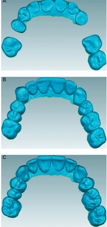
Figure 2- Example of the evaluated virtual models. (A) Pre-treatment model; (B) Conventional wax-up model; (C) Digital wax-up model
A
B
the mandible was moved by 0.05 mm vertically towards the maxilla7. This process detected the
existing working side contacts that dictate the lateral occlusion scheme. Due to the limitation of this process in detecting non-working side contacts, only the working side contacts were considered. Depending on the detected contacts, three occlusion
schemes were considered: (1) canine-guided occlusion, (2) group function occlusion, and (3)
single tooth-guided occlusion. Canine-occlusion was recorded if the lateral contact occurred on a canine tooth. The occlusion is considered group function occlusion if more than two teeth in one arch were contacting laterally. In situations where the lateral contact occurred on a single tooth other than the canine, the lateral occlusion scheme was considered to be a single tooth-guided occlusion.
Analysis
For each position, three variables were evaluated:
(1) the prevalence of each lateral occlusion scheme (2) the average number of contacting teeth and (3)
the percentage of each contacting tooth. All the variables were blotted in bar diagrams. For the last two variables, the maxillary and mandibular teeth contacts were distinguished. In addition, for the
average number of contacting teeth, the
Kruskall-Wallis test was used to determine the presence of a statistical difference between the different
positions (p=0.05). When a signiicant difference
was observed, the Mann-Whitney test was used for post-hoc analysis. Further, the difference in the number of teeth in contact between the pre-treatment models and each of the two wax-up models, and between the two wax-up models was
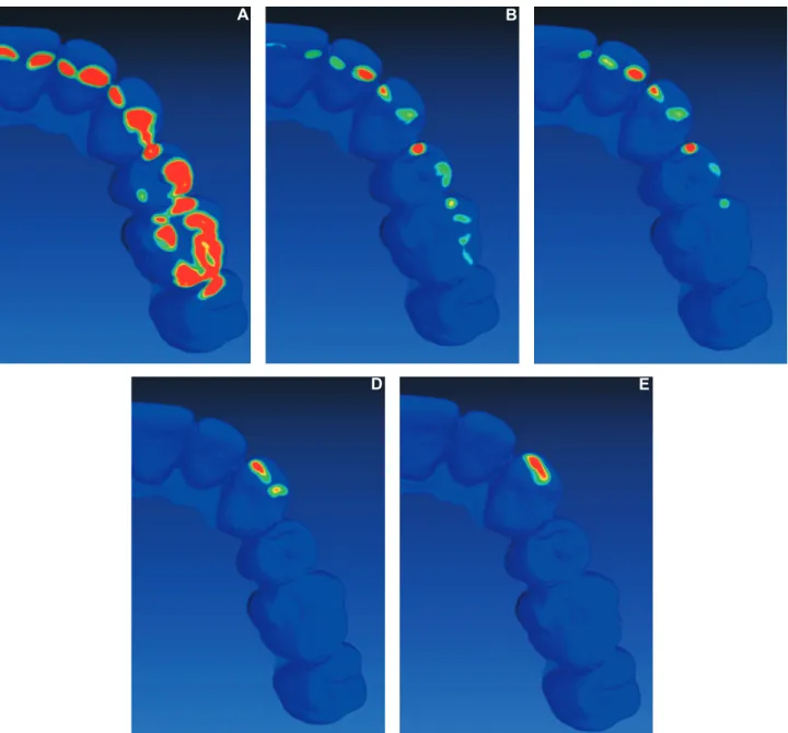
Figure 3- Example of virtual simulation of lateral movement. (A) Maximal intercuspation; (B) 0.5 mm excursion; (C) 1.0 mm excursion; (D) 2.0 mm excursion; (E) 3.0 mm excursion; red colour indicates the existing contacts
A
B
evaluated by the Mann-Whitney test (p=0.05). The
same test was applied to evaluate the difference between maxillary and mandibular arches for each position.
RESULTS
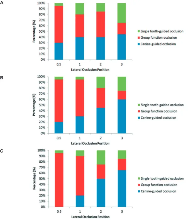
Prevalence of lateral occlusion scheme
As the excursion increases, the lateral occlusion scheme changes for all the evaluated models
(Figure 4). Overall, the models exhibited similar
patterns of lateral occlusion alterations. For the
pre-treatment models (Figure 4A), the prevalence
of canine-guided occlusion had increased minimally
through the excursion (from 30% to 45%). There
was a tendency for the group function occlusion
to reduce with increased excursion (from 65% to 20%). The single tooth occlusion had a tendency to
increase with increased lateral excursion.
For the two wax-up models (Figure 4B and Figure
4C), there was a consistent and gradual increase
of canine-guided occlusion (from 20% to 60% for conventional wax-up, and from 0% to 65% for
digital wax-up) and a reduction of group function
occlusion (from 75% to 15% for conventional wax-up, and from 95% to 29% for digital wax-up) with
increased excursion. It appears that the single tooth occlusion had minimally increased as the excursion increased. The overall patterns for the two wax-ups were similar, except that the conventional wax-up
Figure 4- Proportion of each lateral occlusion scheme in each excursive position. (A) Pre-treatment models; (B)
Conventional wax-up models; (C) Digital wax-up models
A
had a slightly more even pattern of alteration, while the digital wax-up exhibited steeper occlusion alterations.
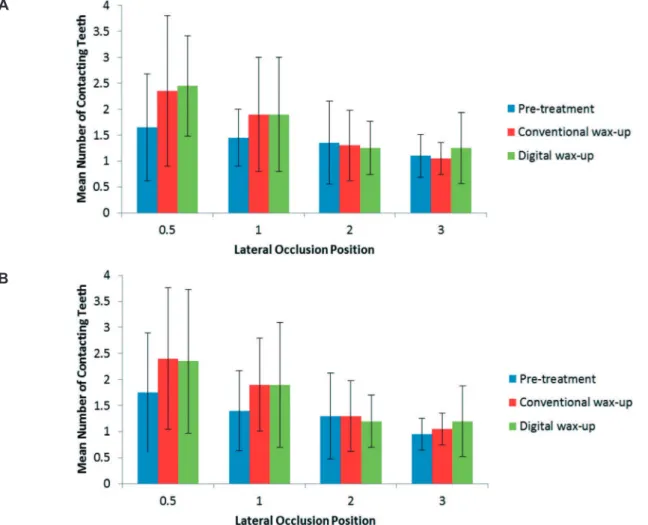
Number of contacting teeth
Regardless of the evaluated model, there was a clear pattern of reduction of the average number of teeth in contact by increasing the degree of
excursion (Figure 5). For the pre-treatment models,
there was a slight reduction in the number of teeth in contact by increasing the degree of excursion, yet there was a statistical difference between all the positions, except between 1.0 mm and 2.0 mm. For the two wax-ups, there was a clear nominal reduction in the number of teeth in contact by
increasing the excursion. A statistically signiicant
difference was observed between all the positions, except between 2.0 and 3.0 mm, for the digital
wax-up. Between the maxillary and mandibular arches, no statistically signiicant difference was
observed in any position for pre-treatment and the two wax-up models.
At the 0.5 mm and 1.0 mm excursion positions,
the pre-treatment models had signiicantly less
teeth contacting than the two wax-up models.
However, at the 2.0 mm and 3.0 mm positions, there was no difference between the pre-treatment and the two wax-up models. At all the positions, there was no statistical difference between the conventional and digital wax-ups.
Percentage of each contacting tooth
For all the arches of all the models, and almost at all positions, the canine had the tendency to be the dominant contacting tooth. Further, it appears that the contacts decreased gradually from canines to the more anterior teeth and from the canines to the posterior teeth. However, there were some differences in the frequency and patterns of
contacts (Figure 6).
For the pre-treatment models (Figure 6A and Figure 6B), the maxillary arch had frequent canine
contacts at all the positions. The lateral incisors,
premolars and the irst molars had consistent
contact at all the positions. In the mandible, there was a reduction in the percentage of canine contacts. Overall, there was similarity in contact frequency between the canines and premolars.
For the conventional wax-up (Figure 6C and Figure 6D), there was a general similarity between
Figure 5- The mean number of contacting teeth for all the models in each excursive position. (A) Maxillary arch; (B)
Mandibular arch
A
the two arches. The canines maintained the frequency of contacts between the positions. The
irst premolar was the second tooth in the frequency
of contacts. For all the remaining teeth, it was clear that the frequency of contacts had a tendency to decrease with increasing the excursion position. The mandibular canines were distinguished in the slight reduction of frequency with increased excursion.
In relation to the digital wax-up (Figure 6E and Figure 6F), the two arches showed similar patterns
and frequency. The frequency of the canines contacts had increased with excursion, while the remaining teeth contact frequency had decreased
signiicantly with excursion.
DISCUSSION
This study indicates that conventionally and
digitally planned prosthodontic treatment inluence
the lateral occlusion in relation to the prevalence of each scheme at different positions, and the contacting teeth type and quantity. Therefore, the hypothesis that the prosthodontic planning will impact the dynamic occlusion was accepted. Such
a inding supports that the lateral occlusion scheme
should be carefully considered before and after the treatment.
In this study, multiple lateral locations were considered in order to provide an insight about the possible contact pattern from the immediate
excursion to the maximal excursion (about 3 mm)17.
This was found to be more clinically relevant than only evaluating the lateral occlusion at maximal excursion22,27. Guiding contacts at the 3 mm
position might occur primarily during parafunctional activity and bruxism in the edge-to-edge position. However, occlusal guiding during mastication and physiological movement tends to occur within 0.5 mm from the maximal intercuspation15,16. Therefore, the range evaluated in this study covers functional and parafunctional jaw movement.
In relation to the prevalence of each lateral occlusion scheme, it is clear that with greater excursion, the prevalence of canine-guided occlusion tends to increase. This was in accordance with the studies that evaluated the prevalence of each lateral occlusion scheme at different positions for the natural dentitions of young participants3,28. Yaffe and Ehlrich28 (1987) evaluated the prevalence
of the different lateral occlusion schemes at 1.0,
Figure 6- Percentage of the contacting teeth in each excursive position for all the models. (A) Pre-treatment maxillary arch; (B) Pre-treatment mandibular arch; (C) Conventional wax-up maxillary arch; (D) Conventional wax-up mandibular arch; (E) Digital wax-up maxillary arch; (F) Digital wax-up mandibular arch
A
C
E
B
D
2.0 and 3.0 mm excursions. They found that the prevalence of canine-guided occlusion had
increased from 16.1% to 48.6%, and the group function occlusion had reduced from 83.9% to 51.5%. A similar inding was observed by Al-Nimri,
et al.3 (2010), when they evaluated the prevalence
of the lateral occlusion scheme at 0.5 and 3 mm positions. They found that the prevalence of
canine-guided occlusion had increased from 21.9% to 59.6%, and the prevalence of group function had reduced from 45.3% to 23.9%. Although
the present study evaluated the lateral occlusion
scheme of restored dentitions, overall, the igures
supported the outcome of the earlier studies. The differences in documenting the prevalence of each occlusion scheme could be related to the different
classiication and recording methods of the lateral
occlusion scheme2,17. In addition, it is important
to emphasize that a single lateral occlusion scheme did not exist for any model for the entire excursive path. Therefore, in accordance with all the published clinical studies, true canine-guided occlusion or group function occlusion seldom exists clinically2,4,24,27.
The dynamic nature of the lateral occlusion scheme at the different arch positions is attributed to teeth morphological factors. In the initial phase of excursion, the cusps are articulated against wider fossa surfaces21. As excursion progresses,
the total contact area is reduced, which means less teeth will be in contact. This observation supports the concepts of “progressive occlusion”, in which many teeth initially control the occlusion, followed primarily by the canines during the maximal excursion8,10. This occlusion scheme appears to be
more physiologically relevant than a single occlusion scheme that controls the lateral movement through the entire excursion. This complex relationship might have a protective role in tolerating lateral forces28. Physiologically, it is thought that such an
arrangement is advantageous in facilitating smooth multidirectional movement of the mandibular arch, which might reduce the risk of patient discomfort21.
Further, broad occlusal contact areas were found to be helpful in mitigating excessive occlusal forces on teeth11, which might contribute to the
dissipation of sudden lateral forces on the teeth. In addition, the greater prevalence of group function occlusion means that greater contacts at less lateral movement can enhance the occlusal phase of chewing25.
The pattern of lateral occlusion scheme alteration with different excursion was observed for the pre-treatment and up models. However, the wax-up models had more consistent gradual occlusion scheme changes. This might be due to the pre-treatment dentitions suffering from morphological abnormalities, such as tooth wear or failed
restorations. Subsequently, a wider contact area between worn down teeth might be evident and will maintain a greater number of contacts, even after maximal excursion5. Whereas the dentitions
of the wax-up models had restored natural dental morphology and less prominent wear facets, which can produce steeper articulation and cause a consistent reduction of the total number of tooth contacts during excursion. This was evident in the
initial phases of excursion (0.5-1.0 mm), in which the contacts quantity was signiicantly greater for
the two wax-up models than at the later stages of excursion. In addition, it was clear that the proportion of canine-guided occlusion at later stages of excursion was greater for the two wax-ups than for the pre-treatment models. This was also likely to be related to restoring the cuspal morphology of the canines to the original canine morphology. The studies that evaluated the implication of individual age on excursion scheme confirmed that the younger the individual, the greater the prevalence of canine-guided occlusion2,18. This was attributed
to the prominent dental morphology and less cuspal wear of the young dentitions2,18. Therefore, it could
be speculated that the planned prosthodontic treatment can return the dentition occlusion scheme
closer to its original state. Between the two
wax-ups, some differences were observed, but not to a
signiicant level. Thus, it is dificult to assume that
a different wax-up protocol will produce a different
outcome on the inal treatment. Therefore, the
hypothesis that there will be a difference between the two prosthodontic planning procedures was rejected.
It is clear that at all positions, the canines had
the greatest number of contacts. The signiicant
contribution of canines was observed even after partial excursion, where group function occlusion
was dominating. This relects the importance of
canines for lateral occlusion scheme development. This finding corroborates the observation by several earlier investigations12,13,17,28. Ogawa, et
al.17 (1998), on natural and young dentition, found that the canines were contacting in about 70% of
the excursive positions, which was similar to the outcome of our pre-treatment models in the present
study. Such a inding supports the protective role
of canines during the lateral occlusal movements. The dominance of canines is related to two factors: the canine’s morphology and position. The canines are innately long teeth, which tend to have more prominent cuspal morphology, which controls the articulation against the opposing teeth19. As
side condyle slides, the working side canine is positioned in the corner, where interferences control occlusal guidance6. The importance of the
location in the arch explains the reason why the teeth immediately adjacent to the canine tend to have frequent occlusal contacts. Likewise, as the teeth are more distant from the canines, they are less likely to be in contact on the working side6,17.
Although some investigators had found that group function was more common, for a great portion of their participants the group function occlusion was composed of canine and premolar occlusion, which reinforces the importance of the location in the arch12,13,26. The strategic importance of canines
is further supported by being the most vulnerable teeth for attrition19,26. Therefore, regardless of
the observed lateral occlusion scheme, it could
be stated that the canines are signiicant teeth to
control the lateral occlusion19.
Following the prosthodontic planning, it was clear that the frequency of the canine contact was
increased, followed primarily by the irst premolars.
As stated earlier, this was attributed to restoring the original canine cuspal morphology, while the canines of pre-treatment models suffered from greater tooth wear5. Therefore, after the prosthodontic
planning, a greater protective role of canines was more evident. The two wax-ups were very similar in the prevalence of the lateral occlusion scheme and the pattern of occlusal contacts frequency. However, for the digital wax-up, there appeared to be an exaggerated role of the canine teeth from the greater frequency of canine contacts. The
most likely explanation is the well-deined dental
morphologies that can be attained digitally by the software14. On the other hand, this means that
the digital wax-up can produce a steeper lateral occlusion than the conventional wax-up, which can result in more restricted lateral movement. Although this may have clinical implications, like patient discomfort21, such assumption should be conirmed by additional studies.
Despite the lack of signiicant statistical difference
between the two wax-up protocols applied in this study, it is important to reinforce that the digital wax-up is still in its early phase of application and requires additional investigation. For example, for the digital wax-up to be applicable clinically, the models have to be produced by computer-aided
manufacturing, which will inevitably inluence the
accuracy of the lateral occlusal contacts. Further, the methodology of this study is limited by not considering the non-working side contacts, which could influence the observed lateral occlusion scheme. The omission of observing the non-working side contacts was necessary, as virtual modelling of the condylar movement is very imprecise20.
However, as the digital wax-up is associated
with steeper occlusal surfaces, the likelihood of developing non-working side contacts is higher.
CONCLUSION
Given the limitations of the present study, the following can be concluded:
The prosthodontic planning influenced the pattern of the lateral occlusion scheme. The influence of the alteration tends to be more prominent at the initial stages of excursion.
For all the models, the initial phase of excursion involved a greater number of contacting teeth and higher prevalence of group function occlusion than maximal excursion. Canine-guided occlusion tends to be more prevalent at the later stage of excursion.
Overall, the difference in the number and pattern of contacts is very minimal between the two wax-ups.
ACKNOWLEDGEMENTS
This research is supported by the Research Development Award from the University of Western
Australia, and the Australian Prosthodontic Society.
The authors acknowledge the facilities, the
scientiic and technical assistance of the National
Imaging Facility at the Centre for Microscopy, Characterization & Analysis, University of Western Australia; a facility funded by the University, State and Commonwealth Governments. The authors would like to thank the staff at the technical facility of the Oral Health Centre of Western Australia.
REFERENCES
1- Abduo J, Bennamoun M, Tennant M, McGeachie J. Precision of virtual prosthodontic planning for oral rehabilitation. Br J Appl Sci Technol. 2014;4(27):3915-29.
2- Abduo J, Tennant M, McGeachie J. Lateral occlusion schemes in natural and minimally restored permanent dentition: a systematic
review. J Oral Rehabil. 2013;40(10):788-802.
3- Al-Nimri KS, Bataineh AB, Abo-Farha S. Functional occlusal
patterns and their relationship to static occlusion. Angle Orthod.
2010;80(1):65-71.
4- Becker CM, Kaiser DA, Schwalm C. Mandibular centricity: centric relation. J Prosthet Dent. 2000;83(2):158-60.
5- Beyron HL. Occlusal changes in adult dentition. J Am Dent Assoc. 1954;48(6):674-86.
6- D'Amico A. Functional occlusion of the natural teeth of man. J Prosthet Dent. 1961;11(5):899-915.
7- DeLong R, Heinzen M, Hodges JS, Ko CC, Douglas WH. Accuracy
of a system for creating 3D computer models of dental arches. J
Dent Res. 2003;82(6):438-42.
8- DiPietro GJ. A study of occlusion as related to the Frankfort-mandibular plane angle. J Prosthet Dent. 1977;38(4):452-8. 9- The glossary of prosthodontic terms. J Prosthet Dent. 2005;94(1):10-92.
10- Goldstein GR. The relationship of canine-protected occlusion
to a periodontal index. J Prosthet Dent. 1979;41(3):277-83. 11- Hidaka O, Iwasaki M, Saito M, Morimoto T. Inluence of
clenching intensity on bite force balance, occlusal contact area,
12- Ingervall B. Tooth contacts on the functional and nonfunctional side in children and young adults. Arch Oral Biol.
1972;17(1):191-200.
13- Ingervall B, Hähner R, Kessi S. Pattern of tooth contacts in eccentric mandibular positions in young adults. J Prosthet Dent. 1991;66(2):169-76.
14- Mehl A, Blanz V, Hickel R. A new mathematical process for the calculation of average forms of teeth. J Prosthet Dent. 2005;94(6):561-6.
15- Ogawa T, Koyano K, Suetsugu T. The relationship between inclination of the occlusal plane and jaw closing path. J Prosthet Dent. 1996;76(6):576-80.
16- Ogawa T, Koyano K, Suetsugu T. Characteristics of masticatory
movement in relation to inclination of occlusal plane. J Oral Rehabil.
1997;24(9):652-7.
17- Ogawa T, Ogimoto T, Koyano K. Pattern of occlusal contacts in
lateral positions: canine protection and group function validity in
classifying guidance patterns. J Prosthet Dent. 1998;80(1):67-74. 18- Panek H, Matthews-Brzozowska T, Nowakowska D, Panek B, Bielicki G, Makacewicz S, et al. Dynamic occlusions in natural permanent dentition. Quintessence Int. 2008;39(4):337-42. 19- Rinchuse DJ, Kandasamy S, Sciote J. A contemporary and
evidence-based view of canine protected occlusion. Am J Orthod
Dentofacial Orthop. 2007;132(1):90-102.
20- Schierz O, Klinger N, Schön G, Reissmann DR. The reliability
of computerized condylar path angle assessment. Int J Comput
Dent. 2014;17(1):35-51.
21- Schuyler CH. The function and importance of incisal guidance
in oral rehabilitation. 1963. J Prosthet Dent. 2001;86(3):219-32. 22- Suit SR, Gibbs CH, Benz ST. Study of gliding tooth contacts during mastication. J Periodontol. 1976;47(6):331-4.
23- Thornton LJ. Anterior guidance: group function/canine guidance. A literature review. J Prosthet Dent. 1990;64(4):479-82.
24- Turp JC, Greene CS, Strub JR. Dental occlusion: a critical
relection on past, present and future concepts. J Oral Rehabil. 2008;35(6):446-53.
25- Wang M, Mehta N. A possible biomechanical role of occlusal
cusp-fossa contact relationships. J Oral Rehabil.
2013;40(1):69-79.
26- Weinberg LA. A cinematic study of centric and eccentric occlusions. J Prosthet Dent. 1964;14(2):290-3.
27- Woda A, Vigneron P, Kay D. Nonfunctional and functional occlusal contacts: a review of the literature. J Prosthet Dent. 1979;42(3):335-41.