313
Bispo IGA et al. Brain metastasis as a presentation of lung adenocarcinoma
Radiol Bras. 2013 Set/Out;46(5):313–316
Brain metastasis as initial presentation of papillary
adenocarcinoma of the lung: case report
*
Metástase cerebral como apresentação inicial de adenocarcinoma papilífero de pulmão: relato de caso
Irving Gabriel Araújo Bispo1, Diego Teixeira Nascimento2, Karina Oliveira Ferreira3, Ricardo Fakhouri4, Atilano Salvador Godinho5, Thiago de Oliveira Ferrão6
The authors describe the case of a 33-year-old patient with history of seizures alone without any previous symptom, being diagnosed with brain metastases from primary papillary adenocarcinoma of the lung. Emphasis is given to the diagnostic investigation for brain metastasis and prognostic evaluation of papillary adenocarcinoma of the lung, and a brief literature review on such diseases is performed.
Keywords: Neoplastic metastasis; Papillary adenocarcinoma; Immunohistochemistry.
Os autores descrevem um caso de paciente de 33 anos de idade com história de crises convulsivas isoladas sem qual-quer antecedente, sendo diagnosticadas metástases cerebrais tendo como sítio primário um adenocarcinoma papilífero de pulmão. É enfatizada a investigação diagnóstica para metástase cerebral e avaliação prognóstica do adenocarcinoma papilífero de pulmão, além de realizar breve revisão sobre essas doenças.
Unitermos: Metástase neoplásica; Adenocarcinoma papilar; Imuno-histoquímica.
Abstract
Resumo
* Study developed at Hospital Universitário – Universidade Federal de Sergipe (HU-UFS), Aracaju, SE, Brazil.
1. MD, Resident of Medical Practice, Hospital Universitário – Universidade Federal de Sergipe (HU-UFS), Aracaju, SE, Bra-zil.
2. MD, Resident of Radiology and Imaging Diagnosis, Hos-pital Universitário – Universidade Federal de Sergipe (HU-UFS), Aracaju, SE, Brazil.
3. MD, Clinical Oncologist, Hospital Universitário – Univer-sidade Federal de Sergipe (HU-UFS), Aracaju, SE, Brazil.
4. Associate Professor of General Pathology and Patholog-ical Anatomy, Universidade Federal de Sergipe (UFS), Aracaju, SE, Brazil.
5. MD, Radiologist, Residency Coordinator, Department of Radiology and Imaging Diagnosis, Hospital Universitário – Uni-versidade Federal de Sergipe (HU-UFS), Aracaju, SE, Brazil.
6. Titular Member of Colégio Brasileiro de Radiologia e Diag-nóstico por Imagem (CBR), Fellow PhD Degree and Residency Preceptor, Department of Radiology and Imaging Diagnosis, Hospital Universitário – Universidade Federal de Sergipe (HU-UFS), Aracaju, SE, Brazil.
Mailing Address: Dr. Irving Gabriel Araújo Bispo. Avenida Augusto Franco, 3553, Condomínio Recanto dos Pássaros, Bloco I, ap. 504, Bairro Ponto Novo. Aracaju, SE, Brazil, 49047-040. E-mail: [email protected].
Received February 10, 2012. Accepted after revision April 1st, 2013.
Bispo IGA, Nascimento DT, Ferreira KO, Fakhouri R, Godinho AS, Ferrão TO. Brain metastasis as initial presentation of papillary adeno-carcinoma of the lung: case report. Radiol Bras. 2013 Set/Out;46(5):313–316.
0100-3984 © Colégio Brasileiro de Radiologia e Diagnóstico por Imagem CASE REPORT
be evaluated. Poorly differentiated carcino-mas arising from the lungs are hardly clas-sified as some specific characteristics may be absent. Thus immunohistochemistry becomes imprescindible to define the type of lung cancer(5).
In the present report, the authors de-scribe the case of a previously healthy, 33-year-old female patient presenting with sei-zures who was diagnosed with brain me-tastasis secondary to papillary adenocarci-noma of the lung.
CASE REPORT
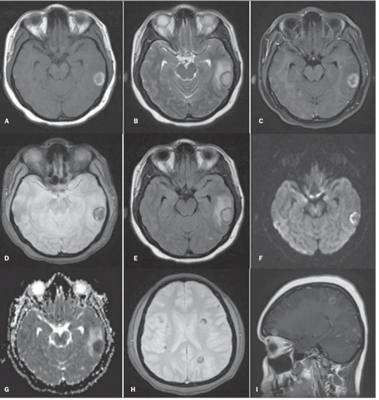
A female, 33-year-old patient was ad-mitted complaining of tonic-clonic seizure without any other previous seizure and was referred to the emergency hospital where she remained for 20 days, being submitted to MRI. Such study demonstrated the pres-ence of several expansile cortical and sub-cortical masses in both brain hemispheres (Figure 1).
The patient was referred to the Univer-sity Hospital for diagnosis elucidation. At admission, the patient did not show any sig-nificant alteration. Serologic tests for para-sitic infection were negative and total ab-dominal and chest CT did not reveal any raphy (CT) is the initial imaging method in
the approach to brain diseases, but MRI is the best method to diagnose brain metasta-sis. The utilization of contrast agents in-creases the MRI sensitivity to identify pa-tients with suspicion of brain metastasis, differentiating it from other lesions origi-nating from the central nervous system (CNS)(3,4). Imaging findings that may be
useful in the differential diagnosis of brain metastasis with other diseases include pres-ence of multiple lesions; lesion location at the white-grey matter junction; circum-scribed margins; great accumulation of vasogenic edema as compared with the le-sion size(2). Currently, with the use of
ad-vanced techniques, MRI can provide more than just lesions location and anatomic details. Among such techniques, spectros-copy, diffusion and perfusion are high-lighted for providing information on the physiology and chemical composition of CNS tumors(3,4). Even so in some cases the
imaging features do not allow the distinc-tion between brain metastasis and primary malignant tumors. In this setting, biopsy is required to define the primary lesion site. In patients submitted to brain biopsy with-out identification of a primary tumor, the pulmonary site should be the first focus to
INTRODUCTION
Brain metastases represent the most fre-quent intracranial tumors in adults(1). The
tomog-314
Bispo IGA et al. Brain metastasis as a presentation of lung adenocarcinoma
Radiol Bras. 2013 Set/Out;46(5):313–316
alteration. In the face of nonspecific find-ings, and considering the non-availability of perfusion and/or spectroscopy studies, brain biopsy was performed. The histo-pathological findings were suggestive of
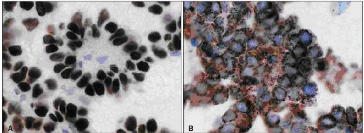
metastasis from papillary carcinoma. The thyroid was the probable site. At immuno-histochemistry, the expression of TTF-1 and napsin-A markers (Figure 2), besides the negativity for thyroglobulin, identified
the pulmonary tissue as the primary site – brain metastasis from papillary adenocar-cinoma of the lung.
The patient was referred for radio-therapy. After the third radiotherapy
ses-Figure 1. Cranial MRI. A: Axial, T1-weighted image – cortical-subcortical, expansile, nodular lesion in the left temporal lobe with a central isointense component and hypersignal halo. B: Axial, T2-weighted image – mild central hypersignal and peripheral hyposignal. C: Axial, contrast-enhanced T1-weighted image with fat-sat – subtle peripheral gadolinium enhancement. D: Axial, T2-weighted image – marked and slightly heterogeneous hyposignal. E: Axial, FLAIR image – mild hypersignal in the central component and hyposignal halo, with perilesional edema. F: Axial, diffusion-weighted image. G: Axial, ADC mapping. H: Axial, T2-weighted gradient image – multiple lesions with peripheral hyposignal. I: Sagittal, contrast-enhanced T1-weighted image – multiple lesions with ring-shaped uptake.
A B C
D E F
315
Bispo IGA et al. Brain metastasis as a presentation of lung adenocarcinoma
Radiol Bras. 2013 Set/Out;46(5):313–316
sion, she presented erythematous papular lesions on her face, besides papular lesions on her chest. The hypothesis of Steven-Johnson syndrome was raised and the pa-tient was transferred to the intensive care unit. After 45 days, the patient was dis-charged with scheduled continuation of radiotherapy sessions.
DISCUSSION
Brain metastases represent the most common intracranial tumors in adults. The most frequent primary carcinoma sites in-clude lung, breast, kidney and colorectal. The clinical signs of brain metastasis are variable. Seizures in previously healthy pa-tients are observed in 20% of cases(1,6). In
the present case, the patient did not present any previous history of seizures. Besides general clinical tests, imaging investigation was indicated to rule out any disease. CT is the initial imaging method to be utilized in the suspicion of intracerebral neoplasm (ei-ther primary or secondary), mostly because of the easy access and swiftness of the ex-amination. However, because of the lower anatomical resolution and/or the presence of small lesions with no hemorrhagic com-ponents, CT results may be dubious or even negative, so MRI utilizing contrast agents and advanced techniques as necessary is the most appropriate imaging method for the diagnosis of brain metastases(7). CT
pre-sents good sensitivity in cases of brain le-sions in association with hemorrhage, even if contrast agents are not utilized. In the present case, MRI demonstrated the pres-ence of multiple, cortical and subcortical nodular lesions, bilaterally, with varied sizes and spontaneously hyperintense on
T1-weighted sequences, possibly as a result from bleeding. Additionally, some lesions presented a halo corresponding to circum-jacent vasogenic edema (Figure 1). In cases of brain metastases, the images are round-shaped, with iso- or hyposignal on T1-weighted sequences, and iso-or hypersignal on T2-weighted sequences, besides a marked perilesional edema. Cystic degen-eration and necrosis can also be detected by MRI, manifesting as areas of signal inten-sity similar to that of the cerebrospinal fluid(4). The spontaneous hypersignal
pat-tern on T1-weighted images may also oc-cur in cases of secondary hemorrhagic im-plantation or melanomas. Primary tumors most frequently associated with hemor-rhagic metastases include renal tumors, thyroid tumors, melanomas and choriocar-cinomas. In many cases, metastases from adenocarcinoma present hyposignal on T2-weighted sequences. In cases where solid lesions are not associated with hemorrhagic components, the enhancement is usually peripheral on both methods(4). It is
impor-tant to observe that in most cases MRI can-not define the primary origin of the me-tastasis(8). The patterns observed in cases of
secondary brain neoplasms by means of ad-vanced MRI techniques include increased amounts of lactate, choline and lipid and decreased N-acetylaspartate at spectros-copy. Depending on the histological grade of the metastatic lesion, the diffusion may be either restricted or facilitated. In cases of hemorrhage, a false hypersignal is ob-served on diffusion-weighted images, as a result of an artifact produced by the pres-ence of blood. Such data are useful in the differential diagnosis with inflammatory and infectious processes, since most of
times such processes restrict the diffusion and the perfusion is said to be “cold” (low capillary density). Several authors have demonstrated lower values for relative ce-rebral blood volume (rCBV) in metastases as compared with multiform glioblastomas (primary tumors), except for hypervascular metastases such as renal carcinomas and melanomas which present higher rCBV values as compared with high grade glial tumors and other secondary lesions. Me-tastases present well-defined limits, with displaced and non infiltrated brain paren-chyma; and this seems to be the main ex-planation for the rCBV efficacy in the dif-ferentiation between metastases and brain tumors(4). A study developed by Itagiba et
al. on the role of diffusion tensor MRI in the assessment of white matter involvement in 44 patients with brain tumors, have dem-onstrated four patterns of involvement, namely, edematous, displaced, infiltrated and disrupted. In such series, the edematous and displaced patterns were found in pa-tients with metastasis, but the second one is nonspecific for such a condition(9). As
regards primary neoplasms, the assessment of the peritumoral region is quite useful in the differentiation with metastases(10).
Another point to be highlighted is role played by immunohistochemistry in the definition of the primary lesion site(5). The
thyroid transcription factor-1 (TTF-1) is the main immunohistochemical marker for pulmonary site. Napsin-A is abundant in the cytoplasm of pulmonary cells of lung adenocarcinomas(11). Stoll et al. have
dem-onstrated that TTF-1 and napsin-A can be markers for adenocarcinomas of probable pulmonary origin. Such authors have also demonstrated that napsin-A was more
sen-Figure 2. Image showing immunohistochemistry test results with positive markers for TTF-1 (A) and napsin-A
316
Bispo IGA et al. Brain metastasis as a presentation of lung adenocarcinoma
Radiol Bras. 2013 Set/Out;46(5):313–316
sitive, as compared with TTF-1, to differ-entiate the papillary subtype of adenocar-cinoma (96% versus 78%, respectively)(11)
Histopathological analysis demonstrated metastatic papillary carcinoma. Imaging studies did not detect any type of suspicious lesion, so immunohistochemistry, due to its broad spectrum, was fundamental to deter-mine the disease site. Several factors play a significant role in the determination of brain metastases prognosis according to a combined classification system called re-cursive partitioning analysis (RPA)(6).
As regards lung adenocarcinoma, Rus-sell et al. have correlated survival with sub-types. In 26 cases of predominantly papil-lary adenocarcinoma, the five-year survival reached 71%. As a comparison is made with other invasive adenocarcinoma subtypes, for example, it is observed that in 14 cases of the predominantly micropapillary sub-type, the five-year survival reached 38%(12).
Recent studies have correlated radiological findings with pathology results. In large cen-ters, with the recent developments in imag-ing methods, it is startimag-ing to be possible to suspect the histological pattern of lung ad-enocarcinomas with basis on the tomo-graphic pattern of the lesion(13). So there is
an opportunity for radiologists, pulmonolo-gists and surgeons to get an early notion on the patients’ prognosis, gaining time in the definition of the approach to be adopted(14).
Finally, the present report describes a case of metastatic, hemorrhagic nodular lesions as initial manifestation of papillary adenocarcinoma of the lung, and empha-sizes the relevance of MRI in the assess-ment of patients with seizures of probable secondary etiology, as well as the determin-ing role of immunohistochemistry in the case definition.
REFERENCES
1. Sperduto PW, Chao ST, Sneed PK, et al. Diagno-sis-specific prognostic factors, indexes, and treat-ment outcomes for patients with newly diagnosed brain metastases: a multi-institutional analysis of 4,259 patients. Int J Radiat Oncol Biol Phys. 2010;77:655–61.
2. Mehta MP. Current management of patients with metastatic brain tumors. Medscape Education Oncology. [acessado em 7 de junho de 2011]. Disponível em: http://www.medscape.org/ viewarticle/742360
3. Lacerda S, Law M. Magnetic resonance perfusion and permeability imaging in brain tumors. Neuro-imaging Clin N Am. 2009;19:527–57. 4. Otaduy MCG, Toyama C, Nagae LM, et al.
Téc-nicas de obtenção das imagens em neurorradio-logia. In: Leite CC, editor. Neurorradiologia – diagnósticos por imagens das alterações encefá-licas. 1ª ed. Rio de Janeiro: Guanabara-Koogan; 2008. p. 1–47.
5. Capelozzi VL. Papel da imuno-histoquímica no diagnóstico do câncer de pulmão. J Bras Pneumol. 2009;35:375–82.
6. Caroli M, Di Cristofori A, Lucarella F, et al. Sur-gical brain metastases: management and outcome related to prognostic indexes: a critical review of a ten-year series. ISRN Surg. 2011;2011:207103. 7. Travis WD, Brambilla E, Noguchi M, et al.
Inter-national association for the study of lung cancer/ american thoracic society/european respiratory society international multidisciplinary classifica-tion of lung adenocarcinoma. J Thorac Oncol. 2011;6:244–85.
8. Kieffer SA, Brace JR. Neoplasias intracranianas. In: Haaga JR, Dogra VS, Forsting M, et al, edito-res. TC e RM – uma abordagem do corpo humano completo. 5ª ed. Rio de Janeiro: Elsevier; 2010. p. 49–146.
9. Itagiba VGA, Borges R, Cruz Jr LCH, et al. Uso do tensor de difusão na avaliação dos padrões de acometimento da substância branca em pacien-tes com tumores cerebrais: é uma ferramenta útil para o diagnóstico diferencial? Radiol Bras. 2010; 43:362–8.
10. Al-Okaili RN, Krejza J, Wang S, et al. Advanced MR imaging techniques in the diagnosis of intraaxial brain tumors in adults. Radiographics. 2006;26:S173–89.
11. Stoll LM, Johnson MW, Gabrielson E, et al. The utility of napsin-A in the identification of primary and metastatic lung adenocarcinoma among cy-tologically poorly differentiated carcinomas. Can-cer Cytopathol. 2010;118:441–9.
12. Russell PA, Wainer Z, Wright GM, et al. Does lung adenocarcinoma subtype predict patient sur-vival?: A clinicopathologic study based on the new International Association for the Study of Lung Cancer/American Thoracic Society/Euro-pean Respiratory Society international multi-disciplinary lung adenocarcinoma classification. J Thorac Oncol. 2011;6:1496–504.
13. Maia Junior ACM, Lucas Junior A, Rocha AJ. Neoplasias parenquimatosas infratentoriais. In: Silva CIS, D’Ippolito G, Rocha AJ, editores. En-céfalo. 1ª ed. Rio de Janeiro: Elsevier; 2012. p. 323–53.