STRUCTURAL AND FUNCTIONAL ANALYSES OF THE
OCCIPITAL CORTEX IN VISUAL IM PAIRED PATIENTS
WITH VISUAL LOSS BEFORE 14 YEARS OLD
Elcio Juliat o Piovesan
1, M arcos Crist iano Lange
1, Pedro André Kow acs
1, Hudson Famelli
1,
Lineu Cesar Werneck
1, Airt on Yamada
2, Guilbert o M inguet t i
3ABSTRACT - Single phot on emission t omography (SPECT) perfusion images of t he brain of individuals w it h complet e visual loss before t he age of 14 w ere carried out and compared t o t hose of visually normal subject s, in order t o assess hypot het ical differences in brain st ruct ural and met abolism bet w een t he t w o groups. St udy group w as comprised by 2 females and 3 males, aged 30 ± 10 years. Cont rols w ere composed by 6 females and 5 males aged 41.5 ± 3.8 years. All individuals w ere submit t ed t o physical and neurological examinat ions, and t o M RI and t o SPECT. Blind subject s present ed larger perfusion measurement s bilat erally in t heir medial t emporal lobes (p= 0.030, right side; p= 0.01, left side), but smaller perfusion measurement s in t heir left front ot emporal area t han cont rols (p= 0.026). Int ragroup analysis of t he st udy group disclosed asymmet ric perfusion, lesser in t he left t emporal and pariet al areas (p= 0.026 and p< 0.0001, respect ively) compared t o t he right side. In t he healt hy cont rols, reduced perfusion w as also not ed at t he left pariet al areas w hen compared t o t he right side (p= 0.035). The st udy revealed t hat complet ely blind pat ient s t hat became visually impaired before t he age of 14 in spit e of not having M RI det ect able changes in t heir brain’s anat omy do present increases in perfusion of t heir left and right medial t emporal lobes, and a reduct ion in t he perfusion of t he left front ot emporal area, as compared t o normal cont rols. While t he increases in blood flow may reflect compensat ory mechanisms for visual deprivat ion, t he significance of t he diminished perfusion in t he left front ot emporal area remains elusive.
KEY WORDS:blindness, cort ical perfusion, neuroplast icit y, visual deprivat ion, visual loss.
Análise estrutural e funcional do córtex visual em pacientes cegos com perda visual antes dos 14 anos de idade
RESUM O - O est udo analisou o comport ament o do córt ex occipit al em indivíduos com perda visual t ot al ant es dos 14 anos de idade. Foram est udados cinco pacient es, 2 femininos e 3 masculinos, com idade média de 30 anos (± 10) const it uindo grupo cegos. Para o grupo cont role foram est udados 11 volunt ários com visão normal, 5 masculinos e 6 femininos com idade média de 41,53 anos (± 3,81). Todos os pacient es foram submet idos a exame físico, neurológico e a exames de ressonância magnét ica encefálica (RM E) e t omografia por emissão de fót on único (SPECT). Os volunt ários apresent aram RM E e SPECT normais. A imagem de RM E não demonst rou alt erações no córt ex occipit al dos cegos. Ent ret ant o, est es apresent avam fluxo sanguíneo maior nas regiões t emporal medial direit a (TM D) (p= 0,030) e t emporal medial esquerda (TM E) (p= 0,010) e menor fluxo sanguíneo na região front o-t emporal esquerda (FTE) (p= 0,026). Comparando-se os lados direit o e esquerdo, os cegos apresent avam redução de fluxo sanguíneo na região t emporal esquerda (p= 0,026) e pariet al esquerda (p< 0,0001). Os volunt ários apresent avam fluxo sanguíneo reduzido na região pariet al esquerda (p = 0,035). O est udo most ra que pacient es deficient es visuais t ot ais, com perda visual adquirida ant es dos 14 anos, não apresent am alt erações est rut urais na RM E quando comparados com o grupo de volunt ários. Observa-se um aument o do fluxo sanguíneo nas regiões TM D e TM E e redução na região FTE. PALAVRAS-CHAVE: cegos, met abolismo cort ical, neuroplast icidade, perda visual, privação visual.
1Unidade de Cefaléia, Especialidade de Neurologia do Depart ament o de Clínica M édica do Hospit al de Clínicas da Universidade Federal
do Paraná (UFPR), Curit iba PR, Brazil; 2Cent ro de Radio Imunoensaio (CERM EN); 3Cent ro de Tomografia Axial Comput adorizada (CETAC).
This project w as financed part ially by t he PIBIC/CNPq, CETAC and CERM EN.
Received 31 January 2002, received in final from 22 M ay 2002. Accept ed 4 June 2002.
Dr. Elcio Juliat o Piovesan – Serviço de Neurologia, Hospit al de Clínicas da UFPR – Rua General Carneiro 181/ 120 andar – 80060-900
Curit iba PR- Brasil. E-mail: [email protected]
The out er layer of t he cerebral cort ex is divided int o several areas specialised in det ect ing and pro-cessing sensory signals from t he eyes and ears and from recept ors for t ouch and nocicept ors. Everyday experience illust rat es t hat , in spit e of dealing w it h dif f erent m odalit ies of inf orm at ion, t he sensorial
regions of t he cort ex must cooperat e w it h each ot her t o int egrat e st imuli received from out side w orld1.
occi-w it h complet e visual loss before t he age of 14, by using magnet ic resonance imaging (M RI) and single phot on emission t omography (SPECT) .
M ETHOD
Selection criteria consisted of: presence of Visual Defici-ency Class 52; installation of the visual loss before the age of 14; a retrobulbar location of the aetiology; lack of active systemic or neurological disease; older than 18; ability for reading in Braille and completion of the informed consent.
Pat ient s.The St udy Group (BLINDS) w as composed ini-t ially of seven individuals, w iini-t h visual loss before ini-t he age of 14, living in t he cit y of Curit iba, Sout h Brazil. Pat ient s w ere recruit ed t hrough phone calls, and t w o of t hem refu-sed t o cont inue in t he prot ocol. Of t he five remaining pat i-ent s, t hree w ere male, and t w o female. M ean age w as 30 ± 10 years. Tw o of t hem w ere blind since birt h, one became blind at t he age of t w o, and anot her at t he age of five. M easles caused blindness in one of t he pat ient s, glaucoma in other, cataract in a other and a retinoblastoma in anot her. In t he fift h pat ient aet iology of t he blindness w as unknow n.
Controls. For the Control Group (CONTROLS) 11 healthy volunt eers w ere st udied. Five of t hem w ere male and six female. M ean age w as 41.53 ± 3.81 years.
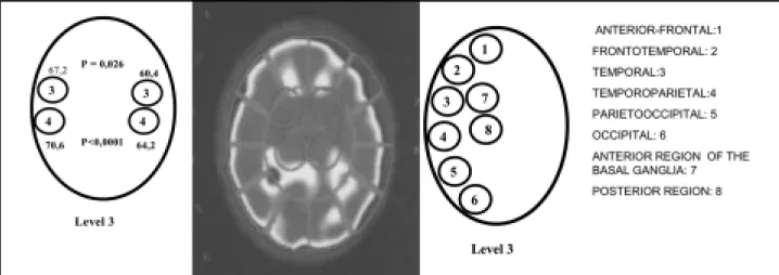
Imaging and image processing t echniques. A 1.5 Tesla Philips Gyroscan ACS – NT w as used t o obt ain t he M RI images, w eighted in T1, T2 and FLAIR protocols for sagittal, coronal and axial slices. SPECT images w ere obt ained w it h a Cardial-Elscint equipment , w it h t w o fixed rot at ing det ec-t ors in 90o, and high spacial resolut ion collimat ors. As 740 M Bq (20mCi) of et ylcyst einat e dimer (ECD) marked w it h Tecnecium-99mm w ere used as radiot racer. All M RI and SPECT exams w ere int erpret ed by t he same professionals, respect ively. SPECT images w ere analyzed at six levels, st ar-t ing caudally ar-t ow ards ar-t he ar-t op of ar-t he head: Level 1: fronar-t
o-pixels as level 3, w it hout subcort ical o-pixels. Values obt ained at t he cerebellar placed pixels w ere used as reference for t he analysis of subsequent findings.
Et hics. The st udy w as approved by t he Et hics Commi-t Commi-t ee on Human Research of HospiCommi-t al de Clínicas – UFPR. All t he included individuals w ere asked t o read and sign an inf orm ed consent bef ore being included. A Braille w rit t en consent form w as given t o t he blind volunt eers.
Data analysis and statistics. The St udent t -t est w as used for t he analysis of t he independent samples, t hrough t he “ Primer of Biost at ist ics” soft w are. The level of significance required w as 5% (0.05).
RESULTS
Control group MRI Imaging and SPECT results.All
the CONTROLS presented normal M R images of the brain and normal SPECT perfusion images of the brain.
St udy group M RI imaging. None of t he f ive
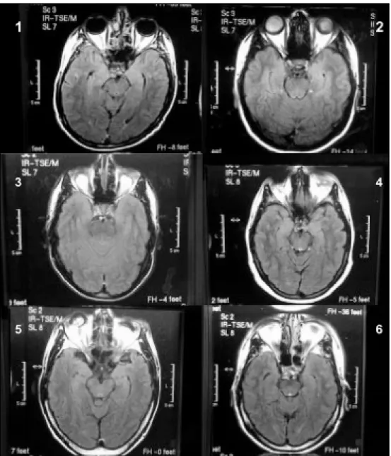
individuals of t he BLINDS group present ed at rophy of t he occipit al cort ex or of ot her regions. Brain ima-ges w ere normal in 4; how ever, in Pat ient 2 diffuse w hit e mat t er abnormalit ies suggest ive of microan-giopat hy w ere observed. Regarding t he opt ical pat h-w ays, t hree pat ient s lacked t he eyeballs, one pat ient present ed signal abnormalit ies in bot h eyeballs and one pat ient present ed absence of t he cryst alline. Bi-lat eral at rophy of t he opt ic nerve w as seen in one pat ient , and unilat eral at rophy of t he left opt ic nerve in ot her (Fig 1).
SPECT comparat ive analysis bet w een groups
(BLINDS versus CONTROLS). At Level 2, BLINDS pre-sented higher perfusion values at the right (70.4± 4.2 right versus 61.1± 5.1 left ) and left medial t emporal
regions (70± 4.6 right versus 60.4± 3.8 left ), as
com-pared t o CONTROLS (p= 0.03 and p= 0.026, respec-t ively). Arespec-t Level 3, BLINDS presenrespec-t ed low er perfusion values at t he lef t f ront ot em poral pixel (59.8± 4.1
versus 69± 4.8, p= 0.026). Borderline dim inished
perfusion w as also observed at t he left t emporal and left t emporopariet al areas (p= 0.066 and p= 0.055, respect ively) in t he BLINDS (Figs 2 and 3).
SPECT int ragroup analysis
Study group. Significant asymmet ries w ere found
in t he brain of t he BLINDS individuals: at Level 3, low er values w ere observed at t he left t emporal
re-Table. Demographic charact erist ics of t he st udy group.
Pat ient Sex Age Cause of t he Age of visual loss visual loss
1 M 20 Ignored rising
2 M 41 M easles 2 years
3 M 40 Glaucoma rising
4 F 30 Ret inoblast oma 5 years
5 F 21 Cat aract 10 years
gion (67.2± 4.1 right versus 60.4± 3.8 left , p= 0.026)
and at t he left t emporopariet al region (70.6± 2.1 right versus 64.2± 1.6 left , p< 0.0001) as compared
t o t heir right count erpart s. Borderline result s w ere ob served in Level 3 at t he lef t f ront ot em p oral (p= 0.080) and in Level 5 at t he lef t f ront al pixel (p= 0.095).
Controls. Significant perfusion asymmet ries w ere
observed in t he brains of t he CONTROLS in t he Level 4, low er at t he left t emporal pixel (68.3± 4.1 right
versus 65.2± 2 left , p= 0.035).
DISCUSSION
The loss of visual input s from eye disease or ret ro-bulbar disorders results in a disconnection of the
occi-pit al cort ex from ext ernal st imuli in complet ely blind individuals. How ever, previous M RI st udies have fai-led t o reveal st ruct ural development al abnormalities in those patients such as occipital lobe atrophy3-10.
There is how ever, experiment al evidence show ing t hat precocious visual privat ion in non human pri-mat es result s in microscopic changes in t his area4.
Positron emission tomography (PET) studies, func-t ional M RI (f-M RI) and func-t ranscranial magnefunc-t ic sfunc-t imu-lat ion (TM S) st udies in blind individuals w it h early visual loss have demonst rat ed a recruit ment of t he occipit al cort ex for ot her neuronal st imuli, such as t act ile and audit ory8,11-13. These cross-modal act
iva-t ion refleciva-t s coriva-t ical plasiva-t iciiva-t y and depends on iva-t he frequency and t ime of use of t he area analyzed. It has been proposed t hat t he plast icit y of t he occipit al
cort ex in blind individuals can be relevant for t he increased abilit ies in t act ile percept ion disclosed by t hem9,11,14-16.
St udies comparing t he met abolism of glucose, t he consumpt ion of oxygen, t he cerebral blood flow and t he elect ric act ivit y of t he primary visual cort ex of blind subject s w it h t hat of healt hy cont rols have failed t o show significant differences6,7,10. Cohen and
collaborators, by comparing subjects w ith congenital or early visual loss w it h individuals w it h lat e visual loss, have demonst rat ed t hat occipit al cort ex plas-t iciplas-t y occurs only in individuals plas-t haplas-t became blind before t he age of 145.
Visual experience is an import ant fact or for t he early organizat ion of t he visual cort ex. Cort ical plas-t iciplas-t y of plas-t he occipiplas-t al corplas-t ex w ill be direcplas-t ly relaplas-t ed t o t he t iming of visual loss17,18, being int errupt ed at
t he age of 145. This upper bracket limit coincides
w it h pubert y and is associat ed w it h a reduct ion of supernumerary synapses inside t he visual cort ex19,20.
Our finding of lack of anat omical differences bet -w een t he BLINDS and t he CONTROLS is similar t o previous st udies t hat have analyzed t he st ruct ure of t he cerebral cort ex of visually impaired subject s3-5.
The abnormalit ies found in t he opt ic pat hw ays in 40% of our cases echoed t he findings of Breit enseher and col., t hat have observed hypoplasia or at rophies in t he opt ical nerve, t ract or opt ic radiat ions in 58% of t heir pat ient s, w it hout ot her abnormalit ies in ano-t her paano-t ienano-t3. How ever, it is reasonable t o assume
t hat t he abnormalit ies found in t he opt ical pat hw ays of t he BLINDS w ere secondary t o t he cause of t he blindness rat her t han being a consequence of t he lack of visual st imuli.
The lack of differences in occipital cortex perfusion values bet w een BLINDS and CONTROLS observed in our st udy suggest ed similar met abolic act ivit y rat es in t he occipit al cort ical st ruct ures of bot h groups. This finding is similar t o t hat obt ained by Ishikaw a and col., that compared through SPECT the perfusion of t he primary visual cort ex of four visually impaired
subject s w it h t hat of normal cont rols7. How ever, in
our st udy, dif f erences w ere observed, such as an bilateral increase in the blood flow in the medial tem-poral voxels and a reduced perfusion in t he left fron-t ofron-t emporal areas. If our findings are nofron-t confron-t ami-nat ed by a bet a t ype error, t hen t hey may reflect f unct ional changes induced by t he loss of visual input . Along t he same line of t hough, it is reasonable t o conceive t hat t he normal blood flow found in t he occipit al cort ex of t he BLINDS reflect s t he effect s of neuroplast icit y-induced funct ional changes.
Neuroplast icit y is a process t hat happens t hanks t o t he int egrat ion of several sensorial channels, such as visual, t act ile and nocicept ive, processed t hrough mult imodal neurons and at cort ical “ cross modal” associat ion areas21. Cort ical plast icit y does not occur
only in t he occipit al cort ex but also in ot her cort ical areas such as m ot or, sensory and t act ile, being dependent of t he frequency and using t ime of t he analyzed area9,15. Being so, t he increased blood flow
in t he t emporal voxels of t he BLINDS could perhaps reflect increase hearing aw areness. Cross modal plas-t iciplas-t y has been hypoplas-t hesized plas-t o help plas-t he plas-t acplas-t ile per-ception abilities of visual impaired subjects11,16,22.
Plas-t iciPlas-t y involves changes and an increase in Plas-t he cor-t ical represencor-t acor-t ion of cor-t he analyzed area, due cor-t o an enhanced peripheral input secondary t o an adapt a-t ive phenomenon of cora-t ical represena-t aa-t ion15,23.
This crossmodal effect arises even w hen the tactile cues t asked are irrelevant and do not predict t he locat ion of t he visual t arget s, suggest ing an exoge-nous (st im ulus driven) at t ent ional m echanism20.
Recent st udies t hat analyzed t he effect ive connec-tivity have suggested that tactile input to the somato-sensory cort ex may influence t he visual cort ex via back project ion t hrough associat ion areas in t he pa-riet al lobe24. The exist ing relat ionships bet w een t he
primary and secondary visual cort ical areas and t he t act ile sensory cort ex is explained by t he bimodal neurons and by t he crossmodal connect ions. The loss of t he visual input s t o t he occipit al cort ex also influ-ences t he t rigeminal nocicept ive discriminat ion25. To
conclude, t he visual cort ex neurons are not t ight ly bound t o visual funct ion, but also able t o process ot her t asks such as Braille reading, and t act ile and t rigeminal nocicept ive discriminat ion13, 25,26.
Finally, our dat a has reinforced t he previous fin-dings of t he lack of st ruct ural changes in t he visual cort ex in pat ient s w it h early visual loss due t o disease of t he opt ical pat hw ays. How ever, t he f inding of significant changes in cerebral cort ical perfusion
sug-gest t he exist ence of act ive neuroplast icit y mecha-nisms operat ing on t he pat ient ´ s adapt at ion.
REFERENCES
1. Gelder B. More to seeing than meets the eye. Science 2000;289:1148-1149. 2. OMS. Classificação estatística internacional de doenças e problemas relacionados a saúde CID10 . 4.Ed, São Paulo: EDUSP, 1997;1:442-443. 3. Breitenseher M, Uhl F, Prayer Wimberger D, Deecke L, Trattnig S, Kramer J. Morphological dissociation between visual pathways and cortex: MRI of visually-deprived patients with congenital peripheral blindness. Neuroradiology 1998;40:424-427 .
4. Buchel C, Price C, Frackow iak RSJ, Friston K. Different activation patterns in the visual cortex of late and congenitally blind subjects. Brain 1998;121:409-419.
5. Cohen LG, Weeks RA, Sadato N, Celnik P, Ishii K, Hallet M. Period of susceptibility for cross-modal plasticity in the blind. A nn Neurol 1999;45:451-460.
6. De Volder AG, Bol A, Blin J, et al. Brain energy metabolism in early blind subjects: neural activ ity in the v isual co rtex. Brain Res 1997;750:235-244.
7. Ishikawa N, Nishijo K, Satou M, Takeda T, Itai Y. Study on the primary cortex of visually impaired subjects by means of 123 I-IMP SPECT and MRI. Ann Nucl Med 1995;9:105-108.
8. Kujala T, Huotilainen M, Sinkkonen J. et al. Visual cortex activation in blind humans during sound discrimination. Neurosci Lett 1995;183:143-146. 9. Pascual-Leone A, Wassermann EM, Sadato N, Hallet M. The role of
reading activity on the modulation of motor cortical outputs to the reading hand in Braille readers. Ann Neurol 1995;38:910-915. 10. Wanet-Defalque MC, Veraart C, De Volder A, et al. High metabolic
activity in the visual cortex of early blind human subjects. Brain Res 1988;446:369-373.
11. Cohen LG, Celnik P, Pascual-Leone A, et al. Functional relevance of cross-modal plasticity in blind humans. Nature 1997;389:180-183. 12. Sadato N, Hallet M. fMRI occipital activation by tactile stimulation in
a blind man. Neurology 1999;52:423.
13. Sadato N, Pascual-Leone A, Grafman J, Deiber MP, Ibañez V, Hallet M. Neural networks for Braille reading by the blind. Brain 1998;121:1213-1229. 14. Pascual-Leone A , Cammarota A , Wassermann EM, Brasil-Neto JP,
Cohen LG, Hallet M. Modulation of motor cortical outputs to the reading hand of Braille readers. Ann Neurol 1993;34:33-37. 15. Sterr A, Muller MM, Elbert T, Rockstroh B, Pantev C, Taub E. Perceptual
correlates of changes in cortical representation of fingers in blind multifinger Braille readers. J Neurosci 1998;18:4417-4423.
16. Sunanto J, Nakata H. Indirect tactual discrimination of heights by blind and blindfolded sighted subjects. Percept Mot Skills 1998;86:383-386. 17. Hubel DH, Wiesel TN. Ferrier Lecture. Functional architecture of
macaque monkey visual cortex. Proc R Soc Lond Biol Sci 1977;198:1-59. 18. Price DJ, Ferrer JM, Blakemore C, Kato N. Postnatal development and plasticity of corticocortical projections from area 17 to area 18 in the cat’s visual cortex. J Neurosci 1994;14:2747-2762.
19. Bourgeois JP, Rakic P. Changes of synaptic density in the primary visual cortex of the macaque monkey from fetal to adult stage. J Neurosci 1993;13:2801-2820.
20. Veraart C, De Volder A G, Wanet-Defalque MC, Bol A , Michel C, Goffinet AM. Glucose utilization in human visual cortex is abnormally elevated in blindness of early onset but decreased in blindness of late onset. Brain Res 1990;510:115-121.
21. Pio v esan EJ, Lange MC, Ko w acs PA , Pacheco C, W erneck LC. Evaluation of headache intensity in migrainous patients with visual handicap through the tactile analogical scale (TAS). Arq Neuropsiquiatr 2001;59:702–707.
22. Deibert E, Kraut M, Kremen S, Hart J Jr. Neural pathways in tactile object recognition. Neurology 1999;52:1413-1417.
23. Werring DJ, Bullmore ET, Toosy A, et al. Recovery from optic neuritis is associated with a change in the distribution of cerebral response to visual stimulation: a functional magnetic resonance imaging study. J Neurol Neurosurg Psychiatry 2000;68:441-449.
24. Macaluso E, Frith CD, Driver J. Modulation of human visual cortex by crossmodal spatial attention. Science 2000;289:1206-1208.
25. Piovesan EJ, Lange MC, Pacheco CG, Fameli H, Kowacs PA, Werneck LC. Influence of visual loss on the threshold of pain perception. Neurology 2001;56(Suppl 3):A65.