*Correspondence: Surjyanarayan Mandal. Research Scholar. School of Phar-maceutical Sciences, Siksha ‘O’ Anusandhan University - Khandagiri Square - Bhubaneswar, Orissa, India. E-mail: [email protected]
http://dx.doi.org/10.1590/S1984-82502015000300024
Mucoadhesive microemulsion of ibuprofen: design and evaluation
for brain targeting efficiency through intranasal route
Surjyanarayan Mandal
1,*, Snigdha Das Mandal
2, Krishna Chuttani
3, Bharat Bhushan Subudhi
11School of Pharmaceutical Sciences, Siksha ‘O’ Anusandhan University, Khandagiri Square, Bhubaneswar, Orissa, India, 2Department of Pharmacology, Parul Institute of Pharmacy and Research, Vadodara, Gujarat, India, 3Division of Cyclotron &
Radiopharmaceutical Sciences, Institute of Nuclear Medicine and Allied Sciences (INMAS), DRDO, Delhi, India
This study aimed at designing mucoadhesive microemulsion gel to enhance the brain uptake of Ibuprofen through intranasal route. Ibuprofen loaded mucoadhesive microemulsion (MMEI) was developed by incorporating polycarbophil as mucoadhesive polymer into Capmul MCM based optimal microemulsion (MEI) and was subjected to characterization, stability, mucoadhesion and naso-ciliotoxicity study. Brain uptake of ibuprofen via nasal route was studied by performing biodistribution study in Swiss albino rats. MEI was found to be transparent, stable and non ciliotoxic with 66.29 ± 4.15 nm, -20.9 ± 3.98 mV and 98.66 ± 1.01% as average globule size, zeta potential and drug content respectively. Transmission Electron Microscopy (TEM) study revealed the narrow globule size distribution of MEI. Following single intranasal administration of MMEI and MEI at a dose of 2.86 mg/kg, uptake of ibuprofen in the olfactory bulb was around 3.0 and 1.7 folds compared with intravenous injection of ibuprofen solution (IDS). The ratios of AUC in brain tissues to that in plasma obtained after nasal administration of MMEI were signiicantly higher than those after intravenous administration of IDS. Findings of the present investigation revealed that the developed mucoadhesive microemulsion gel could be a promising approach for brain targeting of ibuprofen through intranasal route.
Uniterms: Ibuprofen/brain-targeting. Ibuprofen/intranasal administration. Ibuprofen/mucoadhesion. Mucoadhesive microemulsion gel. Drugs/design. Transmission electron microscopy.
O objetivo deste trabalho foi planejar microemulsão/mucoaesiva em gel a im de melhorar a captação cerebral de ibuprofeno por via intranasal. A microemulsão para mucoadesão com ibuprofeno (MMEI) foi desenvolvida pela incorporação de policarboil como polímero mucoadesivo em microemulsão otimizada (MEI) com base em Capmul (MCM) e foi submetida à caracterização, estabilidade, mucoadesão e naso-ciliotoxicidade. A captação cerebral de ibuprofeno pela via nasal foi estudada por meio de estudo de biodistribuição em ratos albinos suíços. MEI se mostrou transparente, estável e não ciliotóxica, com 66,29 ± 4,15 nm, -20,9 ± 3,98 mV e 98,66 ± 1,01%, respectivamente, de tamanho médio dos glóbulos, potencial zeta e conteúdo do fármaco. O estudo revelou o estreita distribuição do tamanho dos glóbulos de MEI. Após administração intranasal única de MMEI e MEI, em dose de 2,86 mg/kg, a captação de ibuprofeno no bulbo olfativo foi em torno de 3,0 e 1,7 vezes maior, comparativamente, à injeção endovenosa de ibuprofeno (IDS). As taxas de ASC em tecido cerebral em relação ao plasma, obtidas após administração da MMEI nasal, foram, signiicativamente, mais elevadas do que aquelas observadas após a administração intravenosa de IDS. Os resultados do presente estudo mostraram que a microemulsão/mucoadesiva em gel poderia ser uma abordagem promissora para o direcionamento cerebral de ibuprofeno por via intranasal.
Unitermos: Ibuprofeno/captação cerebral. Ibuprofeno/administração intranasal. Ibuprofeno/mucoadesão.
INTRODUCTION
Recently, nonsteroidal anti-inflammatory drugs (NSAIDs) appear to be protective in animal models of Parkinson’s disease (PD) and regular use of NSAIDs may reduce the risk of PD in humans (Lang, Lozano, 1998, Tansey, Goldberg, 2010). Some other findings showed that NSAIDs may delay or prevent the onset of PD. Ibuprofen (2-(4-(2-methylpropyl) phenyl)propanoic acid), a NSAID used for relief of symptoms of arthritis and fever. However, the clinical usefulness of ibuprofen
is limited by its high irst-pass metabolism in liver, which
leads to low oral bioavailability and poor brain entry due to over expression of p-glycoprotein (Chen et al., 2008). Therefore, looking for alternative routes of administration
to improve therapeutic efects is necessary.
In recent years, drug delivery through nasal route
has received a lot of attention, because it ofers several
advantages including rapid absorption of drug, avoidance of hepatic first-pass metabolism, and drug delivery preferentially to brain via the olfactory region (Woensel
et al., 2013). Therefore, the nasal route to the brain may
provide a better alternative to oral administration of Ibuprofen. For nasal delivery of Ibuprofen, a challenge existing in formulation development is the solubilization of poorly water-soluble Ibuprofen. Secondly effective
nasal delivery volume in human is ≤400μl (200μl per
nostril), hence normal solution did not provide satisfactory solubility for nasal delivery of Ibuprofen.
M i c r o e m u l s i o n s ( M E s ) a r e i s o t r o p i c , thermodynamically stable transparent systems of oil, water and surfactant, frequently in combination with a cosurfactant with a droplet size usually in the range of 20-200 nm (Shah et al., 2014). These systems are currently of
great scientiic interest to the researchers because of their
potential to incorporate a wide range of drug molecules (hydrophilic and hydrophobic) due to the presence of both lipophilic and hydrophilic domains (Pathak et al., 2014). Moreover, these versatile delivery systems provide protection against oxidation, enzymatic hydrolysis and improve the solubilization of lipophilic drugs and hence enhance their bioavailability (Talegaonkar et al., 2008). In addition, they are amenable for sustained and targeted delivery through oral, ophthalmic, pulmonary, vaginal, intravenous and topical routes (Ghosh, Murthy, 2006, Abdelrahim, Abdelbary, Ghorab, 2011). MEs, by virtue of their lipophilic nature and low globule size, are widely explored as a delivery system to enhance CNS uptake across nasal mucosa. Addition of mucoadhesive agent such as a polyelectrolyte polymer helps in retention of formulation in nasal cavity that further facilitates the absorption. The
retention is due to the electronic interaction between mucin of nasal mucosa and the polymer (Rhee et al., 2007). Evidences of intranasal drug delivery systems formulated
using mucoadhesive agent and its beneits in enhancing
nose-to-brain drug transport have been reported by various scientists in the literature (Porecha et al., 2009).
Hence the objective of this investigation was to prepare, characterize and to perform comparative biodistribution study of the developed formulations in rat, and to also assess direct nose-to-brain transport. It was hypothesized that MEI / MMEI will result in effective nose-to-brain transport, enhanced rate and extent of drug transport, and greater distribution of Ibuprofen within and into the brain. This can help to maximize the therapeutic
index of the drug, reduce its side efects, reduce the dose
and frequency of dosing, and perhaps result into cost
efectiveness (Ponchel et al., 1997; Botner, Sintov, 2011).
MATERIAL AND METHODS
Material
Ibuprofen was obtained as gift sample from Abbott India Ltd., Goa, India. Aceclofenac was procured from Alembic ltd., Baroda, India. Capmul MCM, Captex, Labrafac CC, Cremophor EL, Accenon CC and Transcutol P were received as gifts from ABITEC, Mumbai, India. Polycarbophil (AA-1, pharmagrade, molecular weight approximately 3.5 million) was procured as gift sample from Lubrizol Advanced Material India Pvt Ltd. (Mumbai, India). Isopropyl myristate (IPM), Oleic Acid (OA), polyethylene glycol 400 (PEG 400), PEG 600, Tween 60, Tween 80, glycerol, isopropyl alcohol, isobutyl alcohol, sodium deoxycholate, citric acid (monohydrate), disodium phosphate (dihydrate) and propranolol were purchased from Baroda Chemicals, Vadodara, India. HPLC grade sodium dihydrogen phosphate (NaH2PO4), disodium hydrogen phosphate Na2HPO4), n-hexane, diethyl ether, p-phenylphenol and acetonitrile procured from Merck, India. Double distilled water was prepared by using
Millipore Milli Q plus puriication system.
METHODS
Analytical methodology
using Waters 515 Series pumps combined with a Waters PDA 2998 series photo diode array detector (DAD). The column used was Agilent C18 column (150 mm×4.6 mm, particle size 5-micron Agilent, USA). Analysis was
isocratic at 0.8 ml/min low rate with ACN: Bufer (0.025
M potassium dihydrogen ortho phosphate) pH 4.5 at 11:9 v/v as mobile phase. The mobile phase was developed by
mixing and iltered through a 0.2 µm nylon membrane ilter followed by degassing by sonication before use. The
elution of drug was detected at 230nm.
Selection of formulation components
Selection of the oil was mainly based on the solubility
of the drug. Diferent oils like isopropyl myristate, Capmul MCM, Labrafac CC, Oleic acid, Labrail M 1944CS and
Soyabean oil were screened for solubility study. Selection criteria for surfactant were its HLB value, drug solubility and non-toxic nature. Several surfactants like Tween-60, Tween-80, Captex-355, Accenon CC, and Cremophor EL having HLB value ranging in between 12 to 18, were screened. Screening of co-surfactants was based on their ability to form stable and clear microemulsion at a minimum concentration and several co-surfactants like PEG 400, PEG 600, propylene glycol, glycerol, isobutyl alcohol, isopropyl alcohol and Transcutol P were screened. Excess of Ibuprofen was added to each cap vial containing 5 ml of each of the selected vehicle. After sealing, mixtures were shaken with shaker at 37 oC for 48 h. After reaching equilibrium, each vial was centrifuged at 8000 rpm for 10 min and excess insoluble Ibuprofen was separated by
iltration using Whatman ilter (0.45μ). Both free drug as well as solubilized drug concentration was quantiied
by UV-VIS Spectrophotometer (Shimadzu UV1800) and mass balance was done (Vogt, Kunath, Dressman et al., 2008; Patel et al., 2010).
Pseudoternary phase diagram study
The pseudoternary phase diagrams of oil, surfactant: co-surfactant (Smix), and water were developed using distilled water titration method. The mixture of oil and at
diferent ratio of Smix (1:0.5, 1:1, 1.5:1, 2:1, 2.5:1 and 3:1)
at certain volume ratio were titrated with distilled water in drop wise manner. Using Chemix software, microemulsion regions were determined and Smix showing maximum microemulsion region was used for the development of microemulsion (Moghimipour, Anayatollah, Fatemeh, 2012; Subudhi, Mandal, 2013).
Preparation of formulations
Ibuprofen loaded microemulsion (MEI) was prepared by water titration method. Predetermined amount
of Ibuprofen was dissolved in the required quantity of selected oil. Surfactant and cosurfactant concentration (Smix) from the result of pseudo-ternary phase diagrams was added to the above drug oil mixture. Finally this mixture of oil and Smix was titrated dropwise by distilled water with mild but continuous stirring until a transparent and homogenous microemulsion was produced (Patel, Mandal, Rajesh, 2012; Khan et al., 2009). Then microemulsion was formulated into microemulsion based gel (MMEI) using polycarbophil as mucoadhesive agent (Sharma et al., 2009). Ibuprofen solution (IDS, 20 mg/mL) was prepared by addition of Ibuprofen (200 mg) to 8 ml of distilled water and ethanol (8:2) with continuous stirring. The pH was adjusted to 6.8 ± 0.25 using glacial acetic acid (approximately 0.06 ml/ml). The mixture was stirred for 10 min and a clear solution was obtained. Final volume was made up to 10 ml using distilled water (Piao et al.,
2010). pH of the inal formulation was also checked.
Characterization
Characterization of microemulsion formulations
All measurements were performed in triplicates. Optimized MEI was characterized for % Transmittance, globule size, zeta potential, pH and drug content. % Transmittance and drug content were measured by UV-VIS Spectrophotometer (Shimadzu UV1800) at 650 nm (Mandal et al., 2010) and 222 nm (Joshi et al., 2011) respectively. Globule size and zeta potential were determined by Zetasizer (Nano ZS, Malvern Instruments, UK) at a temperature of 25 ± 2 oC.
For determination of drug content, about 0.5 g of
MMEI was weighed in a 100 ml volumetric lask and dissolved in 50 mL of phosphate bufer of pH 7.4. The volumetric lask was kept for 2 h and shaken well in a
shaker to mix it properly. It was diluted appropriately and analyzed on a UV-VIS Spectrophotometer (Shimadzu UV 1800) at 222 nm.
For spreadability of the gel, 0.5 g of MMEI was placed on a glass plate within a premarked circle of 1 cm diameter. Over which a second glass plate was placed and a weight of 500 g was allowed to rest on the upper glass plate (Joshi, Patravale, 2006). Spreading of the gel with respect to the increase in the diameter after 1 min was noted.
MEI and MMEI pH were determined by digital pH meter (Systronics, India). For pH of the gel, 1.0 g of gel was dissolved in 100 mL of distilled water, stored for 2 h and then the pH was measured in triplicate by calibrated digital pH meter (Dhawan, Medhi, Chopra, 2009).
Viscometer (Model RVT). The sample (30 g) was placed in a beaker and was allowed to equilibrate for 5 min before measuring the dial reading using T-C spindle (spindle no 64 ) at 10 rpm (Joshi, Patravale, 2006; Fanun, 2010).
A drop of diluted MMEI was placed onto the surface of a copper grid coated with carbon. Upon drying, the grids with mesh size of 300 were stained with 2%(w/v) phosphotungstic acid for 120 s and dried at room temperature and probed with Transmission Electron Microscopy (TEM). The process was performed at room
temperature. Images were captured at the magniication of
×48,000 to ×1,50,000 (Chudasama et al., 2011).
Nasal ciliotoxicity
Nasal ciliotoxicity studies were carried out using excised sheep nasal mucosa. In brief, the excise nasal mucosa of sheep was exposed and treated with 1 g of test MMEI containing Ibuprofen at 2.86 mg/mL for 1 h, then rinsed with saline. The mucocilia was then examined with an optical microscope (Nikon Fx-35A, Japan). Saline and propranolol (a serious nasal mucociliary toxicity agent, 1%(w/v) solution) were used as a negative and positive control, respectively (Mandal, Mandal 2010, Mandal et al., 2010).
Stability study
Stability of the developed MMEI was carried according to ICH guideline for 6 months (Bachhav, Patravale, 2009). Optimized microemulsion was kept at cold temp (4-8 °C), room temperature and at elevated temperature (50 ± 2 °C). After every 2 month for 6 months, MMEI was analyzed for phase separation, % transmittance, globule size and % assay.
Animal experiment
A l l a n i m a l e x p e r i m e n t s w e r e a p p r o v e d b y Committee for the Purpose of Control and Supervision of Experiments on Animals (CPCSEA), Ministry of Social Justice and Empowerment, Government of India, New Delhi, India. Male Albino rats (250 ± 20 g) were used for the comparative in-vivo permeation study. The animals were maintained at temperature (25 ± 2 °C), and humidity (60 ± 5 %) and were supplied with food and water ad libitum. Male Albino rats weighing 250 ± 20 g were anesthetized with an intraperitoneal pentobarbital injection (40 mg/kg) and kept on a heating pad to maintain
the body temperature (Mandal, 2011). 35-40 μL of
the developed nasal formulation containing Ibuprofen
equivalent to 2.86 mg/kg were administered intranasally
with the help of a micropipette (200 μL) attached to low
density polyethylene tubing with internal diameter 0.1 mm at the delivery site. For i.v. administration, 0.5 mL of IDS was delivered (dose equivalent to 2.86 mg/kg) through the femoral vein (Elshafeey, Bendas, Mohamed, 2009). At 2, 5, 10, 15, 30, 60, 120, 240 and 360 min after
the dose, blood was collected by cardiac puncture (Hof,
2000; Hamza et al., 2014). Then the skull was cut open and the olfactory bulb (OB), olfactory tract (OT), cerebrum (CER) and cerebellum (CEB) were carefully excised. Each brain tissue was quickly rinsed with saline and bottled up
with ilter paper to get rid of blood-taint and macroscopic
blood vessels as much as possible. After weighing, the brain tissue samples were homogenized with 5 ml of saline in a tissue homogenizer (BD-144 Tissue Homogenizer, India). Blood samples were anticoagulated with heparin and centrifuged at 6000 rpm for 10 min to obtain plasma. All samples, i.e. aliquots of plasma, and brain tissues homogenates, were stored for up to 48 h in a deep freezer (-20 oC) until HPLC analysis. Measurements were made using 3 rats at each time point.
Analytical procedures
Ibuprofen in plasma and brain tissue was assayed
according to a modiied HPLC method (Thomas, Savage,
1985; Choudhary, Arora, Sharma, 2012). Ibuprofen is extracted from plasma treated with 1.0 Mphosphoric acid
for acidiication using hexane and using p-phenylphenol
as an internal standard. To 200 μL plasma samples
internal standard was added and was vortexing for 2 mins. Chromatographic separation achieved isocratically on a C18 column (Inertsil, 150 mm x 4.6 mm) utilizing a mobile
phase of acetonitrile/phosphate bufer/water (60:30:10, v/v, pH 7.0) at a low rate of 1.2 ml/min with UV detection at 230 nm. 80-400 μL brain tissue homogenates, 30 μg/
mL of aceclofenac as internal standard was added and was vortexing for 2 min. After centrifugation at 10,000 rpm for 10 min, the drug was extracted from the supernatant by adding 0.7 mL of n-hexane and diethylether (1:1). The separated organic phases were then evaporated to dryness at 40 0C. The residue was reconstituted in 100 μL mobile
phase and then 20 μL was injected onto an HPLC system
consisting of a LC-20AT VP solvent delivery system and SPD-20A UV detector (Shimadzu, Japan). In order to analyze brain tissue samples, same mobile phase i.e., 60%
acetonitrile, 30% phosphate bufer and 10% water (v/v, pH 7.0) at a low rate of 1.2 mL/min was used and analyzed
20-1000 ng/g for plasma, and brain tissue respectively. Extraction recoveries of ibuprofen from plasma and tissue homogenates were more than 86% and 77% respectively.
Statistical analysis
All data are reported as mean ± SD and the diference
between the groups were tested using Student’s t-test at the level of P < 0.05. More than two groups were compared
using ANOVA and diferences greater at P < 0.05 were considered signiicant.
RESULTS AND DISCUSSION
Preparation and characterization of MMEI
Since Capmul MCM is a biocompatible vehicle and has got maximum drug solubilizing capacity compared to the screened oils i.e., 62.54 ± 1.23 mg/mL, hence it was selected as oil phase. Accenon CC has got less drug solubilizing capacity with HLB value equals to 15.6 was selected as surfactant with the intension to provide sustained release. Results of pseudo-ternary phase diagrams were also depicted that Accenon CC as surfactant and Transcutol P as cosurfactant with Smix (3:1) were suitable for the development of microemulsion.
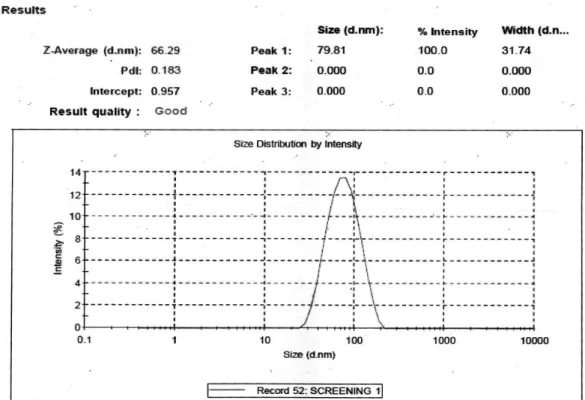
% Transmittance of MEI was found to 99.7 ± 0.12 % indicating the transparency of the system. The average globule size was 66.29 ± 4.15 nm with PdI equals to
0.183 ± 0.19. Moreover results of % Transmittance and average globule size as shown in Figure 1, indicates the nano size range of the formulation. PdI value indicated about monodisperse property of the formulation (Rao, Mcclements, 2011).
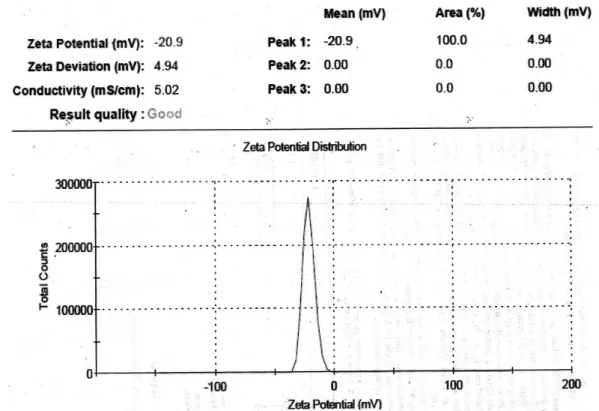
Average zeta potential of IME was found to be -20.9 ± 3.98 mV as shown in Figure 2 indicates the stability of the formulation. This may be because moderate negative surface charges resulted neither into strong aggregation nor repulsion of the globules (Mandal, 2011). Ibuprofen content was found to be 99.38 ± 0.17 %, 99.22 ± 0.23 % and 98.99 ± 0.27 % for MEI, MMEI and IDS respectively. pH of the developed formulations were around 6.4-6.8 indicating the suitability for nasal
administration. No signiicant deviations were observed
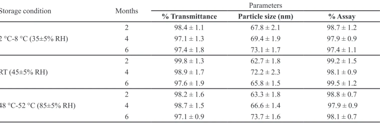
as shown in Table 1, in terms of phase separation, % transmittance, globule size and % assay indicating that developed formulations were physical stable for 6 months at all three storage conditions and hence the formulation may be stored at room temperature. The altered storage
conditions were found to have no adverse efect on the
stability of the optimal formulation. TEM result as shown in Figure 3 revealed the narrow particle size distribution.
Optical microscopic results showed that there was no epithelial damage of cilia on the edge of mucosa that was treated with developed microemulsion formulations for 1 h. This result as shown in Figure 4 [A] revealed that the developed MMEI was non-ciliotoxic and was suitable
for nasal application and as in Figure 4 [C] treated with saline. Formulation treated with 1% v/v propranolol showed severe alteration of mucosal layer as shown in Figure 4 [B].
The spreading diameter in mm of the prepared MMEI was found to be 56 ± 5 mm after 1 min. This spreadability result indicated that the prepared MMEI was easily spreadable by small amount of shear and hence showed excellent spreadability (Misal, Dixit, Gulkari, 2012). The drug content of the MMEI was found to be 97.44 ± 0.71% and pH was found to be 6.29 ± 0.18, which are within the acceptable limits and suitable for FIGURE 2 - Average zeta potential result of the optimized MEI indicating the stability of the formulation.
FIGURE 3 - TEM result of the optimized MEI indicating the narrow particle size distribution.
FIGURE 4 - Result of nasocilio toxicity study (A)Indicating the nontoxic property of developed MMEI on the nasal cilia. (B)Result
of nasocilio toxicity study treated with propranolol showing destruction of cilia. (C)Result of nasocilio toxicity study treated with
intranasal application. Viscosity of the gel was found to be 32,540 ± 7.63 cPs indicating the easy applicability of the formulation into the nasal cavity.
Brain uptake study
Plasma and brain tissue concentration-time proiles
of ibuprofen after nasal administration of MMEI at 2.86 mg/kg dose in rats and intravenous administration of IDS was estimated. The non-compartmental pharmacokinetic parameters were given in Table II.
Sustained absorption of Ibuprofen from MMEI into systemic circulation following intranasal delivery was observed and the maximum plasma concentration was achieved at 1 h. The plasma levels remained constant for 3 h. Following intravenous administration of IDS, brain Ibuprofen concentrations reached peak level at 10 min and declined fast with time. Following intranasal administration of developed MMEI, Ibuprofen level in brain displayed that maximum concentration achieved after about 30 min in OB and 60-120 min in other
regions of the brain. Ibuprofen content was also difered
considerably in different brain regions. The highest
concentration was observed in the OB (the peak drug level was 1090.2 ± 332.4 ng/g), followed by the olfactory tract,
then the cerebellum and cerebrum. These indings support
the existence of a nose–brain direct pathway following the intranasal administration. As shown in Figure 5, Ibuprofen was particularly concentrated in the OB after nasally administered MMEI, the AUC0→360 value was 2.98 times (74569 ± 3617 ng.min/g versus 24992 ± 1622 ng.min/g) that obtained after i.v. injection of IDS.
However, the uptakes of Ibuprofen into other brain regions after nasal dosing of MMEI were lower than those after i.v. injection as shown in Table II. This may be due to the following reason, in contrast to cerebrum and cerebellum, the mass of the OB is small. The distribution of Ibuprofen from OB through olfactory pathway into other brain regions could be counteracted by their large masses, thus leading to the insignificant increase in Ibuprofen content in other brain tissues (Bechgaard, Gizurarson, Hjortkjaer, 1997). In order to evaluate the brain targeting through nasal delivery of MMEI, the brain-to-plasma Ibuprofen AUC ratios at 10, 60, 360 min following both i.v. and i.n. routes were calculated. Results showed that the ratio of AUC in brain tissues to
TABLE I - Results of stability study
Storage condition Months Parameters
% Transmittance Particle size (nm) % Assay
2 °C-8 °C (35±5% RH)
2 98.4 ± 1.1 67.8 ± 2.1 98.7 ± 1.2
4 97.1 ± 1.3 69.4 ± 1.9 97.9 ± 0.9
6 97.4 ± 1.8 73.1 ± 1.7 97.4 ± 1.1
RT (45±5% RH)
2 99.8 ± 1.3 62.7 ± 1.8 99.2 ± 1.5
4 98.9 ± 1.7 72.2 ± 2.3 98.1 ± 0.9
6 97.6 ± 1.9 65.8 ± 1.5 99.5 ± 1.2
48 °C-52 °C (85±5% RH)
2 98.2 ± 1.6 63.3 ± 1.8 98.8 ± 0.7
4 98.7 ± 1.5 66.6 ± 1.4 97.9 ± 0.9
6 97.1 ± 0.9 73.7 ± 1.6 98.1 ± 0.7
(Result = mean ± SD, n =3).
TABLE II - Pharmacokinetic parameters after intravenous and intranasal administration of IDS and MMEI respectively (n=3)
Parameters and routes Plasma OB OT CER CEL
Cmax (ng/mL.g) (intranasal) 73.4±14.4 1090.2 ±452.4 315.2 ±101.6 157.8 ±19.3 7164.4 ±9.8
Cmax (ng/mL.g) (intravenous) 807.6±86.9 2976.2±255.5 3211 ±199.3 3164 ±246.9 3016 ±184.8
AUC0→360 (ng.min/mL.g) Intranasal 23185±1330 74569±3617 9228±1201 6829±4217 5164±958
AUC0→360 (ng.min/mL.g) Intravenous 61655±2348 24992±1622 13612±1016 17292±627 10405±1988
% [AUCi.n. /AUCi.v.] 37.61 298.37 67.79 39.49 49.63
that in plasma after nasal administration of MMEI were
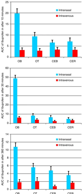
signiicantly higher (P<0.05) than those after i.v. injection of IDS. For example, at 10 min after nasal dosing, the AUC ratio was 6.8 times higher than that after i.v. dosing (19.8 ± 2.2 versus 3.2 ± 0.4) in OB (Figure 6 A). Similarly 17.2 times (48.8 ± 9.2 versus 5.31 ± 0.81) and 9.4 times (10.66 ± 1.01 versus 1.14 ± 0.36) increment of Ibuprofen concentration in OB following intranasal and intravenous delivery at the time points 30 and 360 min as shown in Figure 6 B and Figure 6 C respectively.
Following intranasal administration, about 19-86% of Ibuprofen content at 6 h was transported to the brain via the olfactory pathway (Botner, Sintov, 2011). In short, these results support the existence of an alternative brain entry pathway for Ibuprofen by formulating in o/w mucoadhesive microemulsion because of its protection against enzymatic hydrolysis which may be due to the rapid drug absorption (Yang, Gao, Mitra, 2001; Bitter, Katja, Surber, 2011; Haque et al., 2012) and increasing the quantity of drug to the brain.
CONCLUSION
Results demonstrated that polycarbophil based mucoadhesive microemulsion system comprising Capmul MCM (3% v/v), Accenon CC (26% v/v), Transcutol P (9% v/v) and distilled water (62% v/v) was optimal for intranasal delivery of Ibuprofen. The microemulsion systems are transparent and physical stable at ambient conditions for 6 months. Results of Bioditribution study
conirmed that a fraction of Ibuprofen could be transported
directly into the brain after nasal delivery. Enhanced rate and extent of transport of Ibuprofen following intranasal administration of MMEI may help in decreasing the dose and frequency of dosing and possibly maximize the 0 200 400 600 800 1000 1200
0 50 100 150 200 250 300 350 400
Drug Concentration (ng/ml)
Time (min) OB OT CEB CER Plasma
FIGURE 5 - Ibuprofen concentration in diferent parts of brain
and blood after intranasal delivery.
0 2 4 6 8 10 12 14
OB OT CEB CER
A
UC of Ib
uprof
en in after 360 min
utes Intranasal Intravenous 0 10 20 30 40 50 60
OB OT CEB CER
A
UC of Ib
uprof
en in after 30 min
utes Intranasal Intravenous 0 5 10 15 20 25
OB OT CEB CER
A
UC of Ib
uprof
en in after 10 min
utes Intranasal
Intravenous
FIGURE 6 - AUC of Ibuprofen in plasma and diferent parts of
therapeutic index. However, ratio of clinical beneits to the
risk of the formulation developed in this investigation will decide its appropriateness in the clinical practice.
ACKNOWLEDGEMENTS
The authors are thankful to Abbott India Ltd., Goa, India and Alembic ltd., Baroda, India for providing Ibuprofen and Aceclofenac as gift sample. The authors express their gratitude to ABITECH, Mumbai, India for providing oils and surfactants as gift sample. The authors are also grateful to Mr Anand Pithadia, PIPH, Vadodara, Gujarat for providing assistance for animal study and Mrs. Kavitha Pillai for proof-reading the manuscript for grammatical and spelling errors.
REFERENCES
ABDELRAHIM, M.E.; ABDELBARY, A.; GHORAB, M. Formulation and evaluation of medicated microemulsion
for topical application. Int. J. Nov. Drug. Deliv. Tech., v.1,
n.4, p.208-212, 2011.
ALBERT, K.S.; RAABE, A.; GARRY, M.; ANTAL, E.J.; GILLESPIE, W.R. Determination of ibuprofen in capillary and venous plasma by high-performance liquid
chromatography with ultraviolet detection. J. Pharm. Sci.,
v.73, n.10, p.1487-1489, 2006.
BACHHAV, Y.G.; PATRAVALE, V.B. Microemulsion-based
vaginal gel of clotrimazole: formulation, in vitro evaluation,
and stability studies. AAPS PharmSciTech., v.10, n.2,
p.476-481, 2009.
BECHGAARD, E.; GIZURARSON, S.; HJORTKJAER, R.K. Pharmacokinetic and pharmacodynamic response after
intranasal administration of diazepam to rabbits. J. Pharm.
Pharmacol. v.49, n. 8, p.747-750, 1997.
BITTER, C.; KATJA, S.Z., SURBER, C. Nasal drug delivery
in humans. Curr. Probl. Dermatol., v. 40, p. 20-35, 2011.
BOTNER, S.; SINTOV, A.C. Intranasal delivery of two benzodiazepines, midazolam and diazepam, by a
microemulsion system. Pharmacol. Pharm., v. 2, n.3, p.
180-188, 2011.
C H E N , H . ; Z H A N G , S . M . ; H E R N A N , M . A . ; SCHWARZSCHILD, M.A.; WILLETT, W.C.; COLDITZ, G.A.; SPEIZER, F.E.; ASCHERIO, A. Nonsteroidal anti-inlammatory drugs and the risk of parkinson disease. Arch. Neurol., v.60, n.8, p.1059-1064, 2008.
CHOUDHARY, S.; ARORA, S.; SHARMA, T. Bioanalytical method development and validation of ibuprofen using
RP-HPLC. Am. J. PharmTech Res., v.2, n.6, p.312-321, 2012.
CHUDASAMA, A.; PATEL, V.; NIVSARKAR, M.; VASU, K.; SHISHOO, C. Investigation of microemulsion system
for transdermal delivery of itraconazole. JAPTR, v.2, n.1,
p.30-38, 2011.
DHAWAN, S.; MEDHI, B.; CHOPRA, S. Formulation and evaluation of diltiazem hydrochloride gels for the treatment of anal issures. Sci. Pharm., v. 77, p. 465-482, 2009.
ELSHAFEEY, A.H.; BENDAS, E.R.; MOHAMED, O.H.
Intranasal microemulsion of sildenafil citrate: in vitro
evaluation and in vivo pharmacokinetic study in rabbits.
AAPS PharmSciTech., v.10, n.2, p.361-367, 2009.
FANUN, M. Microemulsions with nonionic surfactants and mint
oil. The Open Coll. Sci. J., v.3, p.9-14, 2010.
GHOSH, P.K.; MURTHY, R.S.R. Microemulsions: A potential
drug delivery system. Curr. Drug. Deliv., v.3, n.2,
p.167-180, 2006.
HAMZA, B.; RIHAB, O.; SAMAR, M.; EL-SHAMY, A.A. Chitosan and cyclodextrin in intranasal microemulsion for improved brain buspirone hydrochloride pharmacokinetics
in rats. Carbohydr. Polym., v.99, p.297-305, 2014.
HAQUE, S.; MD, S.; FAZIL, M.; KUMAR, M.; SAHNI, J.K.; ALI, J. Venlafaxine loaded chitosan NPs for brain targeting: Pharmacokinetic and pharmacodynamic evaluation.
Carbohydr. Polym., v.89, n.1, p.72-79, 2012.
HOFF, J. Methods of blood collection in the mouse. Lab
Animals, v.29, p.47-53, 2000.
JING, Y.; JIAN, P.Z.; QI, N.P.; YUN, L.; LIANG, C. Distribution of nobiletin chitosan-based microemulsions in brain
following i.v. injection in mice. Int. J. Pharm., v.352,
JOSHI, R.S.; PAWAR, N.S.; KATIYAR, S.S.; ZOPE, D.B.; SHINDE, A.T. Development and validation of UV spectrophotometric methods for simultaneous estimation of Paracetamol and Ibuprofen in pure and tablet dosage form.
Pharm. Sin., v.2, n.3, p.164-171, 2011.
JOSHI, M.; PATRAVALE, V. Formulation and evaluation of nanostructured lipid carrier (NLC)-based Gel of Valdecoxib.
Drug Dev. Ind. Pharm., v.32, n.8, p.911-918, 2006.
KHAN, S.; PATIL, K.; YEOLE, P.; GAIKWAD, R. Brain targeting studies on buspirone hydrochloride after intranasal
administration of mucoadhesive formulation in rats. J.
Pharm. Pharmacol., v.61, n.5, p.669-675, 2009.
LANG, A.E.; LOZANO, A.M. Parkinson’s disease: irst of two
parts. N. Engl. J. Med., v.339, n.15, p.1044-1053, 1998.
MANDAL, S.; MANDAL, S.D.; SURTI, N.; PATEL, V.B. Development of microemulsion formulation for the solubility enhancement of lunarizine. Pharm. Lett., v.2, n.3, p.227-236, 2010.
MANDAL, S.; MANDAL, S.D. Design and development of carbamazepine mucoadhesive Microemulsion for intranasal
delivery: an ex-vivo study. Int. J. Pharm. Sci. Rev. Res., v.3,
n.1, p.56-60, 2010.
MANDAL, S. Microemulsion drug delivery system: design and development for oral bioavailability enhancement of
lovastatin. S. Afr. Pharm. J., v.78, n.3, p.44-50, 2011.
MISAL, G.; DIXIT, G.; GULKARI, V. Formulation and
evaluation of herbal gel. Indian J. Nat. Prod. Resour., v.3,
n.4, p.501-505, 2012.
MOGHIMIPOUR, E.; ANAYATOLLAH, S.; FATEMEH, L. Preparation and evaluation of tretinoin microemulsion
based on pseudo-ternary phase diagram. Adv. Pharm. Bull.,
v.2, n.2, p.141-147, 2012.
PATEL, V.; KUKADIYA, H.; MASHRU, R.; SURTI, N.; MANDAL, S. Development of microemulsion for solubility
enhancement of clopidogrel. IJPR, v.9, n.4, p.327-334,
2010.
PATEL, B.M.; MANDAL, S.; RAJESH, K.S. Formulation and kinetic modeling of curcumin loaded intranasal
mucoadhesive microemulsion. J. Pharm. Bioall. Sci., v.4,
p.81-83, 2012. Suppl. 1.
PATHAK, R.; DASH R. P.; MISRA, M.; NIVSARKAR, M. Role of mucoadhesive polymers in enhancing delivery of
nimodipine microemulsion to brain via intranasal route.
Acta Pharm. Sinica B., v.4, n.2, p.151-160, 2014.
PONCHEL, M.; MONTISCI, J.; DEMBRI, A.; DURRER, C.; DUCHENE, D. Mucoadhesion of colloidal particulate
systems in the gastrointestinal tract. Eur. J. Pharm.
Biopharm., v.44, n.1, p.25-31, 1997.
PIAO, H.; BALAKRISHNAN, P.; CHO, H.; KIMB, H.; KIM,Y.; CHUNG, S.; SHIM, C.; KIM, D. Preparation and evaluation
of fexofenadine microemulsions for intranasal delivery. Int.
J. Pharm., v.395, n.1-2, p.309-316, 2010.
PORECHA, S.; SHAH, T.; JOGANI, V.; NAIK, S.; MISRA, A. Microemulsion based intranasal delivery system for
treatment of Insomnia. Drug Deliv., v.16, n.3, p.128-134,
2009.
RAO, J.; MCCLEMENTS, D.J.Formation of flavor oil
microemulsions, nanoemulsions and emulsions: inluence
of composition and preparation method. J. Agric. Food
Chem., v.59, n.9, p.5026-5035, 2011.
RHEE, Y.S.; PARK, C.W.; NAM, T.Y.; SHIN, Y.S.; CHIC, S.C.; PARK, E.S. Formulation of parenteral microemulsion
containing itraconazole. Arch. Pharm. Res., v.30, n.1,
p.114-123, 2007.
SHAH, B.M.; MISRA, M.; SHISHOO, C.J.; PADH, H. Nose to brain microemulsion-based drug delivery system of rivastigmine: formulation and ex-vivo characterization.
Drug Deliv., v.45, p.89-121, 2014.
SHARMA, G.; MISHRA, A.K.; MISHRA, P.; MISRA, A . I n t r a n a s a l c a b e rg o l i n e : p h a r m a c o k i n e t i c a n d
pharmacodynamic studies. AAPS PharmSciTech., v.10,
n.4, p.1321-133, 2009.
SUBUDHI, B.B.; MANDAL, S. Self-microemulsifying drug delivery system: formulation and study intestinal
permeability of ibuprofen in rats. J. Pharm., v.2013, p.1-7,
2013.
TALEGAONKAR, S.; AZEEM, A.; AHMAD, F.J.; KHAR, R.K.; PATHAN, S.A.; KHAN, Z.I. Microemulsions: a novel
approach to enhanced drug delivery. Rec. Pat. Drug Deliv.
TANSEY, M.G.; GOLDBERG, M.S. Neuroinflammation in parkinson’s disease: its role in neuronal death and
implications for therapeutic intervention. Neurobiol. Dis.,
v.37, n.3, p.510-518, 2010.
THOMAS, C.J.T., SAVAGE, S. High-performance liquid chromatographic determination of ibuprofen in bulk drug
and tablets. Drug Dev. Ind. Pharm., v.11, n.5, p.1123-1131,
1985.
VOGT, M.; KUNATH, K.; DRESSMAN, J.B. Dissolution enhancement of fenoibrate by micronization, cogrinding a n d s p r a y - d r y i n g : c o m p a r i s o n w i t h c o m m e r c i a l
preparations. Eur. J. Pharm. Biopharm., v.68, n.2,
p.283-288, 2008.
WOENSEL, M.; WAUTHOZ, N.; ROSIERE, R.; AMIGHI, K . ; M AT H I E U , V. ; F L O R E N C E , L . ; G O O L , S . ; VLEESCHOUWER, S. Formulations for intranasal delivery of pharmacological agents to combat brain disease:
a new opportunity to tackle GBM. Cancer, v.5, n.3,
p.1020-1048, 2013.
XUAN, Z.; XING, L.; TAO, G.; XUN, S.; ZHI-RONG, Z. In
vitro and in vivo investigation of dexibuprofen derivatives
for CNS delivery. Acta Pharm. Sin.. v.33, p.279-288, 2012.
YANG, C.; GAO, H.; MITRA, A.K. Chemical stability, enzymatic hydrolysis, and nasal uptake of amino acid ester
prodrugs of acyclovir. J. Pharm. Sci., v.90, n.5, p.617-624,
2001.
Received for publication on 25th November 2013