1 Received on July 4, 2012.
Accepted for publication on September 4, 2012.
2 Programa de Pós-Graduação em Ciências Veterinárias, Faculdade de
Agronomia, Medicina Veterinária e Zootecnia (Famevz), Universidade Fe-deral de Mato Grosso (UFMT), Av. Fernando Corrêa da Costa 2367, Bairro Boa Esperança, Cuiabá, MT 78068-900, Brazil. *Corresponding author: [email protected]
Experimental osteoarthritis in rabbits: lesion progression
1Wilma N.S. Campos2*, Marcos A. Souza5, Thaís Ruiz2, Thalita P. Peres3, Pedro B. Néspoli5, Alessandro T.C. Marques4, Edson M. Colodel5 and Roberto Lopes de Souza5
ABSTRACT.-Campos W.N.S., Souza M.A., Ruiz T., Peres T.P., Néspoli P.B., Marques A.T.C., Co-lodel E.M. & Souza R.L. 2013. Experimental osteoarthritis in rabbits: lesion progres-sion.Pesquisa Veterinária Brasileira33(3):279-285. Programa de Pós-Graduação em Ciên-cias Veterinárias, Faculdade de Agronomia, Medicina Veterinária e Zootecnia, Universidade Federal de Mato Grosso, Av. Fernando Corrêa da Costa 2367, Bairro Boa Esperança, Cuiabá, MT 78068-900, Brazil. E-mail: [email protected]
The aim of this study was to evaluate the progression of lesions in different stages of osteoarthritis (OA) experimental by radiography (RX), computed tomography (CT), macroscopic and histopathology, linking these different diagnostic methods, helped to provide information that helps the best time for the therapeutic approach. Four experi-mental periods were delineated at 3, 6, 9 and 12 weeks after induction of OA, known as PI, PII, PIII and PIV, respectively, each with six animals. We evaluated the five compart -ments of the femorotibial joint: medial femoral condyle (MFC), lateral femoral condyle (LFC), medial tibial plateau (MTP), lateral tibial plateau (LTP) and femoral trochlea (FT). Therefore we established an index by compartment (IC) and by adding such an index was estimated joint femorotibial (IFT). It was observed that the CFM was the compart-ment with the highest IC also differed significantly (p<0.05) from other compartcompart-ments. Compartments showed no significant difference (p>0.05) between the PI and PII, ho -wever contrary fact occurred between the PII and PIII (p<0.05), PIII and PIV (p<0.01) and between PI and PIV (p<0.001). Similarly the IFT, showed a significant difference in the animals of PIV compared to PI (p<0.001), PII (p<0.001) and PIII (p<0.01), and there was no statistical difference (p> 0.05) between the PI and PII. In the variation of the average interval between periods, there was a higher value between the PIII PIV and for the other intervals of time periods (PI, PII, and PIII-PII). However, these intervals showed no statistically significant difference (p>0.05). Through the RX, CT, macroscopic and histopathological findings, we found similar patterns among individuals within the same period demonstrating a gradual progression of the disease. These results show that between 3 and 6 weeks progression of the lesion is slower and probably also can be reversed in comparison to other ranges where proved further progression between 9 and 12 weeks after induction of trauma OA. These results may provide a better thera-peutic approach aimed at reversing the lesions in early stages of OA. We conclude that the interconnection of the four diagnostic methods individually classified into scores, which were unified in both indices in the evaluation by the femorotibial joint compart -ment and may represent a diagnostic condition closer to the true condition of the injury and its progression.
INDEX TERMS: Osteoarthritis, progression, diagnosis, injury, rabbits, experimental induction.
3 Curso de Graduação em Medicina Veterinária, Famevz-UFMT, Cuiabá,
MT.
4 Departamento de Radiologia, Universidade de Cuiabá (Unic), Avenida
Beira Rio 3100, Bairro Jardim Europa, Cuiabá, MT 78015-480.
5 Departamento de Clínica Médica Veterinária (Climev), Faculdade de
RESUMO.- [Osteoartrite experimental em coelhos: Pro-gressão lesional.] O objetivo deste estudo foi avaliar a pro-gressão das lesões em diferentes períodos da osteoartrite (OA) experimental em coelhos, através da radiografia (RX), tomografia computadorizada (TC), macroscopia e histopa -tologia, interligar estes diferentes métodos diagnósticos, bem como trazer informações que ajudem na decisão do melhor momento para a abordagem terapêutica. Foram delineados quatro períodos experimentais às 3, 6, 9 e 12 semanas após a indução da OA, denominados como PI, PII, PIII e PIV, respectivamente, cada qual com seis animais. Fo-ram avaliados os cinco compartimentos da articulação fe-morotibial: côndilo femoral medial (CFM), côndilo femoral lateral (CFL), platô tibial medial (PTM), platô tibial lateral (PTL) e tróclea femoral (TF). Por conseguinte, estabeleceu--se um índice por compartimento (IC) e através da soma destes foi obtido um índice da articulação femorotibial (IFT). Observou-se que, o CFM foi o compartimento com maior valor de IC, além disso, diferiu significativamente (p<0,05) dos demais compartimentos. Os compartimen -tos não apresentaram diferença significativa (p>0,05) en -tre o PI e o PII, no entanto fato contrário ocorreu en-tre o PII e o PIII (p<0,05), PIII e PIV (p<0,01) e entre o PI e PIV (p<0,001). Similarmente o IFT, apresentou diferença sig -nificativa nos animais do PIV em relação ao PI (p<0,001), ao PII (p<0,001) e ao PIII (p<0,01), e não houve diferen -ça estatística (p>0,05) entre o PI e PII. Na variação média dos intervalos entre os períodos, observou-se um maior valor entre o PIII e o PIV em relação aos outros intervalos de períodos (PI-PII e PII-PIII). No entanto, estes intervalos não apresentaram diferença estatisticamente significativa (p>0.05). Através do RX, TC, macroscopia e histopatológico, verificamos padrões similares entre os indivíduos dentro do mesmo período demonstrando assim uma gradual pro-gressão da doença. Tais resultados evidenciam que entre as 3 e 6 semanas a progressão da lesão é mais lenta e muito provavelmente ainda pode ser reversível em comparação aos outros intervalos onde se comprovou uma maior pro-gressão entre as 9 e 12 semanas após a indução traumá-tica da OA. Estes resultados podem propiciar uma melhor abordagem terapêutica objetivando a reversão das lesões em fases iniciais da OA. Concluímos que, a interligação dos quatro métodos diagnósticos, individualmente classifica -dos em escores e que foram unifica-dos em índices tanto na avaliação por compartimento quanto pela articulação femorotibial pode representar uma condição diagnóstica mais próxima à verdadeira condição da lesão quanto de sua progressão.
TERMOS DE INDEXAÇÃO: Osteoartrite, progressão, diagnóstico, lesão, coelhos, indução experimental.
INTRODUCTION
Osteoarthritis (OA)is one of the most common joint disease in humans and animals and is related to the deterioration of articular cartilage, subchondral bone and/or synovial membrane, leading to failure of the synovial joint (Amiel et al. 2003, Castro et al. 2006). In the early stages of OA, the lesions may be focused on some of the femorotibial joint compartment, among them the most common is the
femo-ral condyles (Kamei, Sumen & Sakaridani 2008). Injuries resulting from trauma may be reversible if diagnosed early or irreversible due to progressive degeneration of articular cartilage, causing pain, swelling, and loss of limb function (Salminen 2002).
The osteoarthritic changes often are diagnosed in an advanced stage of disease, when the cartilage repair pro-cess often has already changed the joint structure. Because of this, new approaches in the treatment of OA, including the development of new drugs, are hampered by the lack of objective and measurable criteria for disease progression (Takahashi et al. 2004). Thus it is essential to know the characteristics of development and progression of cartilage destruction in OA in order to develop and improve diagnos-tic and therapeudiagnos-tic approaches (Sage & Turner 2002).
Among the diagnostic methods currently used to assess OA are non-invasive methods such as radiography (RX), ul-trasound, magnetic resonance imaging and computed to-mography (CT), and invasive procedures such as arthrosco-py, macroscopic and histopathological examination with its advantages and disadvantages (Bruyere et al. 2006). High quality radiographs are accurate in identifying structural changes resulted from OA, but the diagnosis can be challen-ging in the early stages of the disease (Widmer & Blevins 1994, Carrig 1997, Mahaffey 1998). The CT scan allows earlier identification of OA in relation to conventional ra -diography. The programs of cone beam CT, also in the tra-ditional TC, allow multiplanar reconstructionof the volume scanned, for instance, visualization of axial images, coronal and sagittal sections, as well as 3D reconstruction (Reetz et al. 2006). The macroscopic and histological evaluations are used in many studies as a “gold” standard in the evaluation of disease progression. The scoring system of histological OA introduced by Mankin et al. (1971), is currently the most commonly applied scale. This system was developed to describe the hip OA in humans, but was subsequently ap-plied directly or with modifications to other joints and in various animal models (Pearson et al. 2011).
It is in many situations, more than one diagnostic me-thod, is used to determine the presence and progression (Stickle & Hathcock 1993, Torelli et al. 2004). The present study aims to evaluate the evolution of lesions associated with different stages of OA by radiography, computed to-mography, macroscopic and histopathologic evaluation, linking these different diagnostic methods, as well as pro-vide information that will help to decide the best timepoint to approach therapy considering the increasing trend of se-condary lesions in rabbits with experimental OA.
MATERIALS AND METHODS Experimental protocol
We used 24 male New Zealand white, rabbits, aged 5-6 mon-ths and weighing 2.0 to 3.0kg, from the Central Animal House of Federal University of Mato Grosso, Brazil. For the animals inclu-sion criteria for the study bilateral joints femorotibia radiographs were used in craniocaudal and mediolateral projections, for the
verification of the absence of structural changes in the joints.
For the experimental induction of OA, we used the techni-que of cranial cruciate ligament transection (CCLT), proposed
anesthetized intramuscularly with ketamine (S Agener
Keta-mine® 10%, 50mg /kg), midazolam (2mg/kg) and meperidine
(Dolosal®, 10mg/kg) and maintained with isoflurane mask (Iso
-forine®). After trichotomy and antisepsis of the surgical area,
pa-rapatellar incision was made medial in the right knee, followed by arthrotomy and lateral dislocation of the patella, the cranial cruciate ligament (CCL) was then sectioned without injuring the adjacent structures. The joint was irrigated with sterile saline and synthesis of joint capsule was made using polyglactin 910
(Vi-cryl®5-0) in simple continuous pattern, subsequent synthesis of
the skin using nylon (Shalon®,sutures) 3-0 in simple interrupted
pattern. For analgesia and prevention of infection after surgery, animals were treated subcutaneously with cephalothin sodium (12.5mg/kg) every 6 hours for 7 days and morphine (Dimorf
®3mg/kg) every four hours for 4 days. The animals were housed
in appropriate room and housed in individual cages measuring 70cmx70cmx70cm, fed with standard comercial pellets and wa-ter ad libitum.
Subsequently, the animals were devided into four experimen-tal periods referred to as PI, PII, PIII and PIV, each of six animals, which were respectively evaluated at 3, 6, 9 and 12 weeks after induction of OA by X-ray, CT, macroscopically and
histopatholo-gically. These four diagnostic procedures were performed in five
different compartments of the femorotibial joint: medial femo-ral condyle (MFC), latefemo-ral femofemo-ral condyle (LFC), medial tibial plateau(MTP), lateral tibial plateau (LTP) and femoral trochlea (FT). Since each diagnostic method was evaluated by two inde-pendent observers in a blind study, they were unaware of the samples evaluated in relation to their periods.
Image
Radiographs were obtained in craniocaudal and mediolateral projections of thefemorotibial joint using X-ray machine set with
300mA/56kV, screens and films of medium speed and revelation
by an automatic processor Vision® line, LX-2 model. To obtain the
CT images, we used an apparatus for cone beam computed tomo-graphy (i-Cat®, Imaging Sciences International, Hatfield, Pennsyl
-vania, USA), in which the animals were positioned supined with the knee extended right, and the detection limits were determi-ned by na initial cursor proximal to the femorotibial joint and a cursor on the end of the tibial tuberosity. The acquisition time was 20 seconds, using 0.2 at full resolution voxel, 64 kilovolt (kV) and 3.2 milliamps per second (mAs). After its acquisition, the images were downloaded to a computer alocated in adjoining room, whe-re the animals could observed through plumbiferous glass. In a subsequent step was carried out detailed evaluation of axial
ima-ges, coronal, sagittal, and oblique slices with a thickness of 76μm
using a 3D software (Kodak® imaging). Both examinations were
performed with the animals under general anesthesia.
For both methods of diagnostic imaging the evaluation para-meters were: presence and size of osteophytes (grade 0 = absent, 1 = small degree, grade 2 = moderate, grade 3 = severe) and sub-chondral sclerosis (grade 0 = absent, grade 1 =mild, 2 = moderate degree, grade 3 = severe) according to Torelli et al. (2004) with
modifications. Through the sum of these two parameters, a partial
score was assigned to each compartment evaluated and total joint score was obtained by the sum of the parts of each compartment, the joint being scored from 0 to 30, both the RX and CT. At the end of the respective periods and imaging studies, animals were euthanized by intramuscular injection of 50mg/kg ketamine so-dium 10% and after 10 minutes, anesthetized with intravenous
overdose of sodium thiopental (Thiopentax®). This study was
ap-proved by the Ethics Committee on Animal Research at the Fede-ral University of Mato Grosso (CEPA-UFMT), registered under the Protocol No. 23108.035345/10-5.
Macroscopic evaluation
Immediately after animals euthanasia the femorotibiais joints were dissected, examined macroscopically and the compartments
were classified according to criteria established by Laverty et al. (2010), with modifications. To grade 0 (absent) surface was nor
-mal, smooth and without significant alterations, to grade 1 (mild), rough, with minimal fibrillation on the articular surface; to grade
2 (moderate), rough surface, with erosion and/or the presence of
osteophytes, but without exposure of subchondral bone; to grade
3 (severe), there is erosion with exposure of subchondral bone injury and repair in the presence of marginal osteophytes and in-tense bone remodeling. A total joint score was obtained by sum-ming the partial scores of each compartment scored from 0 to 15. The lesions were then documented using a digital câmera (Nikon D7000, Tokyo, Japan).
Histopathologic evaluation
Samples were taken from each compartment joint, fixed in 10% buffered formalin solution for 48 hours, decalcified in a solu -tion of 5% formic acid for 1 week. These were then processed in
gradients of alcohol and xylene, embedded in paraffin blocks and
stained with hematoxylin and eosin (H &E). The histological slides were evaluated by scale according to the criteria established by Mankin et al. (1971). Thus, the partial score of each compartment was obtained by calculating the evaluation system standardized by the Osteoarthritis Research Society International (OARSI) and established by Bao et al. 2009 and Rutgers et al. 2010 where
sco-re= grade x stage, grade (scale, 0–6) was defined as OA depth pro
-gression into cartilage irrespective of its horizontal extent; stage (scale, 0–4) was defined as the horizontal extent of cartilage invol -vement within one side of a joint compartment irrespective of the
underlying grade; score (scale, 0–24) was defined as assessment
of combined OA grade and OA stage. Thus the partial score ranged from 0 to 24 in each compartment, and the total joint score was obtained by summing the partial scores.
Statistical analysis
The data were tabulated in spreadsheets, and established a rate per compartment (IC) using partial scores according to each diagnostic procedure (Rx=x-ray, computerized tomography Tc =, Macro= macroscopic and histopathology =Histo) the following formula:
0> Rx + Tc + Macro + Histo <1;
6 6 3 24
Thus, the IC varied 0-1 and femorotibial índex (IFT) varied
from 0 to 5, obtained by adding the ICs of the five compartments
evaluated. Data were analyzed regarding their normal distribu-tion using the Kolmogorov-Smirnov test. Therefore, the normal data were compared using one-way ANOVA, subjected to analysis
of variance (F test) and in cases of significant F test, means were
compared using the Tukey test. The values were analyzed using the computer program R (2012). For all statistical analyzes was
adopteda significance level of 5%.
RESULTS
no statistically significant difference (p>0.05) between the PI and PII, however different fact occurred between the PII and PIII (p<0.05), PIII and PIV (p<0.01) and between the PI and PIV (p<0.001), as shown in Table 1.
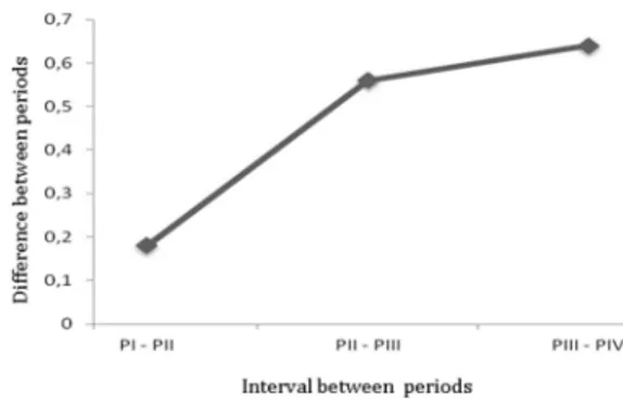
Similarly the IFT, showed a significant difference in the animals of PIV compared to PI (p<0.001), PII (p<0.001) and PIII (p<0.01), and there was no statistical difference betwe -en the PI and PII, as shown in Figure 2. Analyzing the
va-riation of the average interval between periods, there was a greater interval value between the PIII PIV and for the other intervals of time periods (PI, PII, and PIII-PII). Never-theless, these intervals showed no statistically significant difference (p>0.05) as shown in Figure 3. By radiography, CT, macroscopic and histopathological findings, we found similar patterns among individuals within the same pe-riod demonstrating a gradual evolution of the disease. In PI, mild signs of osteolysis was the only obvious change to imaging tests (X-rays and CT) and, in most animals, no signs of OA were features observed in the radiographic exami-nation. Macroscopically, the articular surface proved to be a rough exterior and small fibrillations, only the MFC and the MTP. While the major histopathological changes were hypertrophy, cracks and disorientation of the columns of chondrocytes, as illustrated in Figure 4a.
In PII, the main radiographic changes included mild subchondral sclerosis but the animal five of the period sho -wed moderate level for this parameter. By CT was observed signs of osteolysis and osteophyte formation. The macros-copic changes were observed as surface roughness, atrial, formation of small gaps and erosion (Fig.4b). Disruption of the cartilage surface, hypertrophic chondrocytes, diso-rientation and clusters proliferation of chondrocytes in the column less than 10% evaluated in the entire region, were found in histological changes over time.
In PIII, radiographic and tomographic included, presen-ce of osteophytes, sclerosis and osteolysis, most evident in MFC (Figure 4c). By gross analysis was evident signs of fibrillation, erosion, and fibrocartilage. On histologic eva -luation showed the formation of cysts in the middle zone, hyperplasia and cellular disorganization. In PIV, it was evi-dent the formation of multiple osteophytes, sclerosis and bone remodeling by the intense X-rays and CT scans. Mo-reover, the joints showed thickness articular capsule ero-sion total thickness of bone cartilage, deterioration of the meniscus, soft tissue fibrosis and osteophytes the presence of X-ray (Fig.4d). The macroscopic, there was erosion of the articular surface, wear and presence of osteophytes. For the histopathology of the lesions changes were found ex-tending for more than 50% of the region evaluated, such as cyst formation, fibroplasia and repair.
Fig.1. Schematic representation of the mean variations of the ICs in each compartment articular (CFM: medial femoral
con-dyle; CFL: lateral femoral concon-dyle; PTM: medial tibial plateau:
PTL: lateral tibial plateau and TF: femoral trochlea) to 3 (PI), 6 (PII), 9 (PIII) and 12 (PIV) weeks after induction of OA in rabbits. Notes the highest and lowest value of IC to CFM and TF, respectively, in all periods. Values expressed as mean and standard deviation.
Fig.2. Schematic representation of the mean values of the IFT (femorotibial Index) obtained by X-ray, CT scan, macroscopic
and histopathological findings at different stages of experi -mental OA in rabbits. PI, PII, and PIII PIV 3, 6, 9 and 12 weeks respectively. Values are expressed as mean and standard de-viation. *P<0.05 vs. IP; € *p <0.05 vs PII, *p<0.01 vs PIII
Fig.3. Schematic representation of the means values between pe-riods of 3 (PI), 6 (PII), 9 (PIII) and 12 (PIV) weeks after induc-tion of experimental OA in rabbits. Notes the highest interval between PIII and PIV. However, no statistically difference
(p>0.05) was noted.
Table 1. Average values of the partial indices of joint compartments, obtained by radiography, CT, macroscopic
and histopathologic at different stages of OA*
Compartmentsb PI PII PIII PIV
MFC 0,13 ±0.05Ac 0,19 ±0,09Ac 0,36 ±0,13Ab 0,54 ±0,10Aa
LFC 0,03 ±0,03Cc 0,07 ±0,06Cc 0,16 ±0,10Cb 0,31 ±0,10Ca
MTP 0,09 ±0,07Bc 0,12 ±0,11Bc 0,27 ±0,07Bb 0,40 ±0,12Ba
LTP 0,02 ±0,03Cc 0,05 ±0,04Cc 0,16 ±0,07Cb 0,25 ±0,12Ca
FT 0,01 ±0,01Cc 0,04 ±0,04Cc 0,09±0,05Cb 0,18 ±0,08Ca aAssessments conducted at 3, 6, 9 and 12 weeks after induction of OA (PI,
PII, and PIII PIV, respectively) through the cranial cruciate ligament
tran-section of the rabbits. bMFC: medial femoral condyle; LFC: lateral femoral condyle; MTP: medial tibial plateau; LTP: lateral tibial plateau and FT: fe -moral trochlea. Means followed by same uppercase and lowercase in the
DISCUSSION AND CONCLUSIONS
The initial loss of articular cartilage in early OA is classi-cally considered a focal process that can progressively in-volve all joint compartments, inducing biological changes in the molecular composition of the joint surfaces (Lorenz & Richter 2006). Tiraloche et al. (2005), evaluating the effect of glucosamine on cartilage degradation in rabbits, observed differences in therapeutic responses between the regions analyzed, and no effect on MFC and MTP, indi-cating greater severity in these compartments. Such fact could also be demonstrated in our study, which evaluated the five compartments, the MFC and MTP were the most affected.
Similarly, Buchanan & Kean (2002) and Glasson (2007) report that in humans the medial compartment is the most commonly involved, which should, however, relate this to the presence and functionality of the meniscus, and the medial meniscus is the most injured, precisely because it is more attached and monitor the movements of the tibia at the knee. However, according Sah et al. (1997), this effect depends on the chronicity of traumatic injury. The same au-thors also reported that in acute lesions induced by CCLT,
the lateral meniscus and the LFC are the most adversely affected due to the large rotational forces created.
In chronic lesions, the medial compartments are the most affected as result of repeated episodes of cranial trans-lation of the tibia. Nevertheless, this study showed higher medial compartments gravity in all periods. These discre-pancies can be attributed to the species studied and the mode of exercise trauma. Furthermore, the extent of injury of cartilage appears to be highly dependent on the joint re-gion, which can be explained by different loading conditions in different regions (Lorenz & Richter 2006). This explains the fact that the FT compartment be less commonly affected in all periods in this study is characterized as an area of lo-wer joint friction, and yet the lolo-wer levels of stress.
Another important fact observed in this study is that the highest and lowest values increase from the interval between periods occurred between the PIII and PIV and PI and PII, respectively. It can be observed based on the studies discussed above, there was an increase in the in-tensity of the lesions, which is probably due to an environ-ment created by CCLT instability in a period longer than six weeks. For this same fact has been observed in humans by
Fig.4. (A) Histological appearance, (B) macroscopic, (C) CT, and (D) X-ray, at 3, 6, 9 and 12 weeks respectively after the induction of
experimental rabbits in OA. (A) Observe intense disorientation of the columns of chondrocytes and extracellular matrix deposition
distributed unevenly in the articular cartilage (*) in the CFM. HE, 20x; (B) rough aspect and erosion, (*) CFL - initial lesion (dashed
outline), (**) CFM - advanced lesions (dashed outline); (C) Note areas of sclerosis (*) in the medial portion tibiofemoral joint and
the axial projection of the medial femoral condyle (MFC) observed in B, note bone osteolysis and bone remodeling in CFM (arrow);
(D) regions presenting femorotibial joint sclerosis, osteophytes (arrows) and intense bone remodeling in the medial compartments
O’Connor, Laughlin & Woods (2005) and Richardson et al. (2007) in a similar study.
We believe that this fact gives also the chronic and ir-reversible lesions of articular cartilage in the later periods of OA, because according to Salminen (2002) with the pro-gression of lesions, tissues degenerate more rapidly than can be regenerated. Mainly due to the erosions of cartilage matrix and death of the chondrocytes articular cartilage OA advanced. This erosion is associated with extracellular matrix degradation by proteolytic enzymes, exceeding the amount and speed of production of new matrix. This phe-nomenon has a strong influence of inflammatory cytokines released from fibroblasts and macrophages, which can per -petuate and lead to an irreversible process of degeneration of cartilage (Sandell & Aigner 2001).
In this study, the different diagnostic methods were able to show the progression of OA lesions during the stu-dy periods. The model used CCLT to induce morphological changes allowed the evaluation of this common disease, as described by Biasi et al. (2005), Gonçalves et al. (2008) and Melo et al. (2008). The development and progression of OA occurred due to joint instability, which changed the distri-bution of weight on the joint (Glasson et al. 2007, Herzog & Longino 2007). In PI and PII, the histological findings in this study as the presence of few areas of atrial and rare cluster groups, as observed in the superficial and middle layers, are found also found in normal cartilage under me-chanical induction, not being associated OA (Fernandes et al. 1998). However, Hashimoto et al. (1998) report that in the initial phase of OA degeneration is often observed in articular surface in the form of disruption of the columns of chondrocytes, as disclosed in our study.
However, it should be noted that the rate of disease progression appears to be highly dependent on the species studied. While Graverand Le et al. (2002) found a level of 9.10 using the criteria established by Mankin et al. (1971) as early as 3 weeks after CCLR in rabbits, these levels were observed in dogs after 12 weeks of induction of OA (Lorenz 2005).
Although in our study the values of the interval between periods have not shown statistically significant difference between them, show that between 3 and 6 weeks the lesion progression is slower and most likely can still be reversed in comparison to other intervals which proved further pro-gression between 9 and 12 weeks after induction of trauma OA. The extrapolation of experimental results to the clini-cal routine is not always possible and should be done with caution, because events such as the chronic instability and pre-existing degenerative changes present in clinical cases, can not be disregarded. Moreover, these results are impor-tant because they can provide a better therapeutic approa-ch aimed at reversing the damage still in early stages of OA.
We conclude that the interconnection of the four diag-nostic methods (radiography, tomography, macroscopic and histopathology), individually classified into scores, which were unified in both indices in the evaluation by the femorotibial joint compartment and may represent a diag-nostic condition closer to the true condition of the lesion and its progression.
Acknowledgements.- To Coordenação de Aperfeiçoamento de
Pesso-al de Nível Superior (CAPES) for providing graduate student stipend; to
the trainees Keyla D’Agostin, Dábila Sonego for helping during the
expe-riment; and to. Prof. Dr. Anderson Castro for their assistance in the statis -tical analyzes.
REFERENCES
Amiel D., Toyoguchi T., Kobayashi K., Bowden K., Amiel M.E. & Healey R.M. 2003. Long term effect of sodium hyaluronate (Hyalgan) on osteoarthri-tis progression in a rabbit model. Osteoarth. Cartil. 9:636-643. Bao J.P., Chen W.P., Feng J., Zhao J., Shi Z.L. & Huang K. 2009. Variation
pat-tern of two degradation enzymes systems in articular cartilage in di-fferent stages of osteoarthritis: regulation by dehydroepiandrosterone. Clin. Chim. Acta 408:1-7.
Biasi F., Rahal S.C. & Volpi R.S. 2005. Reconstrução do ligamento cruzado cranial em cães, associado ou não ao sulfato de condroitina. Arq. Bras. Med. Vet. Zootec. 57:442-447.
Bruyere O., Collette J., Kothari M., Zaim S., White D. & Genant H. 2006. Os-teoarthritis, magnetic resonance imaging, and biochemical markers: a one year prospective study. Ann. Rheum. Dis. 65:1050-1054.
Buchanan W.W. & Kean W.F. 2002. Osteoarthritis. III. Radiological and
cli-nical definition. Inflammopharm. 10:53-78.
Carrig C.B. 1997. Diagnostic imaging of osteoarthritis. Veter. Clin. North Am., Small Anim. Pract. 27:777-813.
Castro R.R., Cunha F.Q., Silva F.S. & Rocha F.A.C. 2006. A quantitative appro-ach to measure joint pain in experimental Osteoarthritis - evidence of a role for nitric oxide. Osteoarth. Cartil. 14:769-776.
Fernandes J.C., Martel-Pelletier J., Lascau-Coman V., Moldovan F., Jovanovic D. & Raynauld J.P. 1998. Collagenase-1 and collagenase-3 synthesis in normal and early experimental osteoarthritic canine cartilage: an im-munohistochemical study. J. Rheumatol. 25:1585-1594.
Glasson S.S., Blanchet T.J. & Morris E.A. 2007. The surgical destabilization of the medial meniscus (DMM) model of osteoarthritis in the 129/SvEv mouse. Osteoarth. Cartil. 15:1061-1069.
Gonçalves G., Melo E.G. & Gomes M.G. 2008. Effects of chondroitin sulfate and sodium hyaluronate on chondrocytes and extracellular matrix of ar-ticular cartilage in dogs with joint disease. Arq. Bras. Med. Vet. Zootec. 60:93-102.
Hashimoto S., Ochs R.L., Komiya S. & Lotz M. 1998. Linkage of chondrocyte apoptosis and cartilage degradation in human osteoarthritis. Arthritis Rheum. 41:1632-1638.
Herzog W. & Longino D. 2007. The role of muscles in joint degeneration and osteoarthritis. J. Biomech. 40:54-63.
Kamei G., Sumen Y. & Sakaridani K. 2008. Evaluation of cartilage defect at medial femoral condyle in early osteoarthritis of the knee. Magn. Reson. Imag. 26:567-571.
Laverty S., Girard C.A., Williams J.M., Hunziker E.B. & Pritzker P.H. 2010. The OARSI histopathology initiative e recommendations for histological assessments of osteoarthritis in the rabbit. Osteoarth. Cartil. 18:53-65. Le Graverand M.P., Eggerer J., Vignon E., Otterness I.G., Barclay L. & Hart
D.A. 2002. Assessment of specific mRNA levels in cartilage regions in a
lapine model of osteoarthritis. J. Orthop. Res. 20:535-544.
Lorenz H., Wenz W., Ivancic M., Steck E. & Richter W. 2005. Early and stable upregulation of collagen type II, collagen type I and YKL40 expression levels in cartilage during early experimental osteoarthritis occurs inde-pendent of joint location and histological grading. Arthritis Res. Ther. 7:156-165.
Lorenz H. & Richter W. 2006. Cellular and molecular changes in degenera-ting cartilage. Progr. Histochem. Cytochem. 40:135-163.
Mahaffey M.B. 1998. The stifle and tarsus, p.194-199. In: Thrall D.E. (Ed.),
Textbook of Veterinary Diagnostic Radiology. 3rd ed. Saunders Elsevier,
Philadelphia.
Melo E.G., Nunes V.A. & Rezende C.M.F. 2008. Sulfato de condroitina e hia-luronato de sódio no tratamento da doença articular degenerativa em cães. Estudo histológico da cartilagem articular e membrana sinovial. Arq. Bras. Med. Vet. Zootec. 60:83-92.
O´Connor D.P., Laughlin M.S. & Woods G.W. 2005. Factors related to addi-tional knee injuries after anterior cruciate ligament injury. Arthroscopy 21:431-8.
Pearson R.G., Kurien T., Shu K.S.S. & Scamell B.E. 2011. Histopathology grading systems for characterisation of human knee osteoarthritis e re-producibility, variability, reliability, correlation, and validity. Osteoarth. Cartil. 19:324-331.
Pritzker K.P.H., Gay S., Jimenez S.A., Ostergaard K. & Pelletier J.P. 2006. Osteoarthritis cartilage histopathology: grading and staging. Osteoarth. Cartil. 14:13-29.
R Development Core Team 2012. R: A language and environment for sta-tistical computing. R Foundation for Stasta-tistical Computing, Vienna,
Aus-tria. ISBN 3-900051-07-0, URL <http://www.R-project.org/>
Reetz J.A., Mai W., Muravnick K.B., Goldschmidt M.H. & Schartz T. 2006. Computed tomographic evaluation of anatomic and pathologic varia-tions in the feline nasal septum and paranasal sinuses. Vet. Radiol. Ul-trasound 47:321-327.
Rocha I.D., Moraes T.M.S., Rezende M.U. & Pécora J.R. 2007. Avaliação da evolução de lesões associadas à lesão do ligamento cruzado anterior. Acta Ortop. Bras. 15:105-108.
Rutgers M.. Van Pelt M.J.P., Dhert W.J.A., Creemers L.B. & Saris D.B.F. 2010. Evaluation of histological scoring systems for tissue-engineered, repai-red and osteoarthritic cartilage. Osteoarth. Cartil. 18:12-23.
Sage A.M. & Turner T.A. 2002. Ultrasonography of the soft tissue structu-res of the equine foot. Equine Vet. Educ. 14:212-221.
Sah R.L., Yang A.S., Chen A.C., Hant J.J., Halili R.B. & Yoshioka M. 1997. Phy-sical properties of rabbit articular cartilage after transection of the an-terior cruciate ligament. J. Orthop. Res. 15:197-203.
Salminen H.J., Saamanen A.M., Vankemmelbeke M.N., Auho P.K., Perala M.P. & Vuorio E.I. 2002. Differential expression patterns of matrix metallo-proteinases and their inhibitors during development of osteoarthritis in a transgenic mouse model. Ann. Rheum. Dis. 61:591-597.
Sandell L.J. & Aigner T. 2001. Articular cartilage and changes in arthritis: An introduction on cell biology of osteoarthritis. Arthritis Res. 3:107-113.
Stickle R.L. & Hathcock J.T. 1993. Interpretation of computed tomographic images. Vet. Clin. North Am. 23:417-435.
Takahashi M., Naito K., Abe M., Sawada T. & Nagano A. 2004.Relationship between radiographic grading of osteoarthritis and the biochemical ma-rkers for arthritis in knee osteoarthritis. Arthritis Res. Ther. 16:208-212. Tiraloche G., Girard C., Chouinard L., Sampalis J., Moquin L., Ionescu M.,
Reiner A., Poole R. & Laverty S. 2005. Effect of oral glucosamine on cartilage degradation in a rabbit model of osteoarthritis. Arthritis Res. 52:1118-1128.
Torelli S.R., Rahal S.C., Volpi R.S., Yamashita S., Mamprim M.J. & Crocci A.J. 2004. Radiography, computed tomography and magnetic resonance imaging at 0.5 Tesla of mechanically induced osteoarthritis in rabbit knees. Braz. J. Med. Biol. Res. 37:493-501.
Yoshioka M., Coutts R.D., Amiel D. & Hacker S.A. 1996. Characterization of a model of osteoarthritis in the rabbit knee. Osteoarth. Cartil. 4:87-98. Widmer W.R., Buckwalter K.A., Braunstein E.M., Hill M.A., O’Connor B.L.
& Visco D.M. 1994. Radiographic and magnetic resonance imaging of