Alexandra Furtado Guerreiro
[Nome completo do autor]
[Nome completo do autor]
[Nome completo do autor]
[Nome completo do autor]
[Nome completo do autor]
[Nome completo do autor]
[Nome completo do autor]
Master of Science [Habilitações Académicas] [Habilitações Académicas] [Habilitações Académicas] [Habilitações Académicas] [Habilitações Académicas] [Habilitações Académicas] [Habilitações Académicas] May, 2020
Comparison of the radiosensitisation ability of
metal oxide nanoparticles using clinical
megavoltage X-rays
[Título da Tese]
Thesis submitted in partial fulfilment of the requirements for the degree of Doctor of Philosophy in Radiation Biology and BiophysicsDissertação para obtenção do Grau de Mestre em [Engenharia Informática]
Supervisor: Jon Golding, Senior Lecturer, Open University
Co-supervisors: Nigel Mason, Professor and Head of School, University of Kent Maria Alice Santos Pereira, Assistant Professor, NOVA School
of Sciences and Technology
Examination Committee:
Chairperson: Professor Paulo Manuel Assis Loureiro Limão-Vieira Raporteurs: Professor Frederick Currell
Dr Octávia Gabriela da Silva Viegas Nené Monteiro Gil Members: Professor Malgorzata S"mialek-Telega
Professor Pedro António de Brito Tavares Professor Jon Golding
NOVA Lisbon University
Faculty of Science and Technology
[Nome completo do autor]
[Nome completo do autor]
[Nome completo do autor]
[Nome completo do autor]
[Nome completo do autor]
[Nome completo do autor]
[Nome completo do autor]
Alexandra Furtado Guerreiro
[Nome completo do autor]
[Nome completo do autor]
[Nome completo do autor]
[Nome completo do autor]
[Nome completo do autor]
[Nome completo do autor]
[Nome completo do autor]
Master of Science [Habilitações Académicas] [Habilitações Académicas] [Habilitações Académicas] [Habilitações Académicas] [Habilitações Académicas] [Habilitações Académicas] [Habilitações Académicas]
Comparison of the radiosensitisation ability of
metal oxide nanoparticles using clinical
megavoltage X-rays
[Título da Tese]
May, 2020
Thesis submitted in partial fulfilment of the requirements for the degree of Doctor of Philosophy in
Radiation Biology and Biophysics
Dissertação para obtenção do Grau de Mestre em [Engenharia Informática]
Comparison of the radiosensitisation ability of metal oxide nanoparticles using clinical megavoltage X-rays
Copyright © Alexandra Furtado Guerreiro, Faculty of Sciences and Technology, NOVA University Lisbon.
The Faculty of Sciences and Technology and the NOVA University Lisbon have the right, perpetual and without geographical boundaries, to file and publish this dissertation through printed copies reproduced on paper or on digital form, or by any other means known or that may be invented, and to disseminate through scientific repositories and admit its copying and distribution for non-commercial, educational or research purposes, as long as credit is given to the author and editor.
To my mother, sister and
niece
Acknowledgements
The work presented in this thesis was performed at Open University and at GenesisCare in Milton Keynes and I would like to thank everyone that contributed to make this work possible.
To my supervisor, Dr Jon Golding, I would like to thank his commitment in my work and for always being there when I needed. His guidance and assistance were crucial to make this thesis come true.
To Eleanor Crabb and Nicholas Chatterton I would like to thank for all their input and all the brain storm that helped developed this thesis.
To Genesis Care staff, such as Aquila Sharif and all the radiographers, I would like to thank all their availability and effort to assist me in this thesis and make it possible.
Also, I would like to thank Open University staff such as Dr Igor Kraev, Dr Radka Gromnicova, Brett Keith, George Bryant and Dr Matthew Kershaw for all their technical assistance.
I would like to thank professor Nigel Mason and Sir John Mason Academic Trust for the support provided for the development of this work. To professor Alice S. Pereira for all her support from Portugal. Many thanks to Fundação para a Ciências e Tecnologia (FCT/MCTES), Radiation Biology and Biophysics Doctoral Training Programme (RaBBiT, PD/00193/2012); UID/Multi/04378/2013 (UCIBIO); UID/FIS/00068/2013 (CEFITEC); and grant number PD/ BD/114450/2016 for granting me this PhD fellowship.
I would like to thank all the friends I made at Open University and that made my journey much easier and happier. A special thanks to Marcelle Silva, Sonia Azeggagh, Edu Frias and Radka Gromnicova for all the shared moments and their amazing support as friends that I will keep for life.
Also, I would like to thank my friends Nidia Almeida and Fausto that I met in the beginning of this journey and that made my experience so much better and fun. I hope we share many other journeys together. To Dr Cristina Timóteo for the support and friendship provided whenever I needed.
I would like to thank my closest friends and family for their support and for being present in my life. Thank you, Alexandros Kostopoulos, for all the moments we shared, all your support, encouragement, and companionship. A special thanks to my mother, sister and father for believing in me, and being so patient and so supportive. Mum and sister there are no words to describe how important you are for me and I dedicate this thesis to you. Also, I want to welcome our new member of the family, my niece Alice, which although still very young I hope in the future she will be able to read this thesis and know that my life became a brighter place with her presence. Thank you so much!
Abstract
A wide range of nanoparticles (NPs), composed of different elements and their compounds, are being developed by several groups as possible radiosensitisers, with some already in clinical trials. However, no systematic experimental survey of the clinical X-ray radiosensitising potential of different element NPs has been made. In this thesis, a direct comparison is made through the irradiation-induced (10 Gy of 6 MeV X-ray photon) production of reactive oxygen species (ROS) such as hydroxyl radicals, superoxide anion radicals and singlet oxygen in aqueous solutions of the different metal oxide NPs. Also, cancer cell and DNA damage due to these NPs is studied under irradiation conditions.
The results of this thesis showed that without any X-rays, several NPs produced different radicals. Furthermore, NPs such as vanadium oxide (V2O5) produced a significant amount of radicals compared to water and to the other types of NPs when irradiated with 10 Gy of 6 MeV X-rays and NPs such as copper oxide (CuO) showed significant cell and DNA damage that is not related to the production of ROS.
Beyond identifying promising metal oxide NPs radiosensitisers, these results have shown evidence that the chemical and biological effects of these NPs are also present in the mode of action of these radiosensitisers challenging the pure physical radio-enhancement concept.
Keywords: metal oxide nanoparticles; clinical X-ray; ROS; hydroxyl and superoxide radical; singlet oxygen; radiosensitiser.
Resumo
Uma gama ampla de nanopartículas (NPs), compostas de diferentes elementos e dos seus compostos, encontram-se a ser desenvolvidas por diferentes grupos como possíveis radiossensibilizadores, onde algumas encontram-se em ensaios clínicos. No entanto, nenhum estudo sistemático comparativo do potencial de radiossensibilização por raios-X usados clinicamente dos diferentes elementos de NPs foi realizado. Nesta tese, a comparação directa é realizada através da indução de espécies reactivas de oxigénio (ROS), como os radicais hidroxilo e o anião superóxido, e o oxigénio singleto, por irradiação (10 Gy de 6 MeV de fotões raios-X) em soluções aquosas das NPs dos diferentes óxidos de metais. Para além disso, o dano causado a células cancerígenas e ao DNA devido à acção destas NPs é estudado nestas condições de irradiação.
Os resultados desta tese mostram que, sem a acção dos raios-X, diferentes NPs produziram diferentes radicais. Nanopartículas como o óxido de vanádio (V2O5) produziram uma quantidade significativa de radicais quando comparado à ausência de NPs e também quando comparado aos outros tipos de NPs quando estas foram irradiadas com 10 Gy de 6 MeV de fotões raios-X e NPs de óxido de cobre (CuO) mostraram um dano significativo celular e ao DNA que não se encontra relacionado com a produção de ROS.
Para além de identificarem NPs de óxidos de metais promissores à radiossensibilização, estes resultados mostraram evidência de que os efeitos químicos e biológicos destas NPs estão também presentes no seu modo de acção enquanto radiossensibilizadores, desafiando assim o conceito da acção puramente física da radiossensibilização.
Palavras-chave: nanopartículas de óxidos de metais; raios-X clínicos; espécies reactivas de oxigénio; radicais hidroxilo e o anião superóxido; oxigénio singleto; radiossensibilizador.
Table of Contents
Acknowledgements ... vii
Abstract ... ix
Resumo ... xi
List of Figures ... xvii
List of Tables ... xix
List of Abbreviations and Symbols ... xxi
1 – INTRODUCTION ... 1
1.1 Radiotherapy ... 3
X-ray beam treatment ... 3
Megavoltage X-ray physical properties ... 4
Current methods of improving radiotherapy targeting ... 7
1.2 Importance of Reactive Oxygen Species (ROS) ... 8
1.3 Effects of radiation in cells ... 10
1.4 Tumour environment ... 12
1.5 Radiosensitisers ... 16
1.6 Radioprotectors ... 22
1.7 Gold NPs as radiosensitisers ... 25
1.8 Metal oxides as radiosensitisers ... 31
1.9 Research aims ... 35
2 – MATERIALS AND METHODS ... 39
2.1 Metal oxide nanoparticle-based methods ... 41
Nanoparticle characteristics ... 41
Nanoparticle coating ... 42
Nanoparticle physico-chemical characterization ... 43
2.1.3.1 Transmission Electron Microscopy (TEM) ... 43
2.1.3.2 Dynamic Light Scattering (DLS)/Zeta potential ... 43
2.1.3.3 X-ray diffraction ... 43
2.2 Cell culture-based methods ... 44
Cell culture ... 44
Cytotoxicity of metal oxide nanoparticles ... 44
2.2.2.1 Clonogenic assay ... 44
Cellular uptake of metal oxide nanoparticles ... 44
2.2.3.1 Inductively coupled plasma mass spectrometer (ICP-MS) ... 44
2.3 Reactive oxygen species (ROS) based methods ... 45
ROS probes ... 45
2.3.1.1 Hydroxyl radical probe ... 45
2.3.1.3 Singlet oxygen probe ... 45
2.3.1.4 Superoxide anion probe ... 46
ROS measurement ... 46
ROS calibration measurement ... 46
2.4 Plasmid DNA based methods ... 47
Plasmid DNA extraction ... 47
Plasmid DNA measurement ... 47
2.5 Hole quenching-based methods ... 47
Hole scavenger ... 47
2.5.1.1 Formic acid ... 47
Hole quenching measurement ... 47
2.6 Nanoparticle catalytic ability ... 48
2.7 Hypoxia ... 49
2.8 X-ray irradiation ... 49
2.9 Statistical analysis ... 49
3 COMPARISON OF THE RADIOSENSITISING ABILITY OF PERIOD 4
TRANSITION METAL OXIDE NANOPARTICLES ... 51
3.1 Introduction ... 53
3.2 Experimental design ... 56
Choice of nanoparticle size, shape and concentration ... 56
Choice of nanoparticle coating ... 57
Choice of cell line ... 58
Choice of irradiation parameters and radical probes ... 59
3.3 Results ... 60
Physico-chemical characterization of NPs ... 60
3.3.1.1 Transmission electron microscopy (TEM) and histogram size ... 60
3.3.1.2 Analysis of zeta potentials of NPs ... 64
Catalytic ability ... 67
ROS production ... 68
3.3.3.1 ROS probe fluorescence quenching ... 68
3.3.3.2 Testing coumarin-3-carboxylic acid (3-CCA) for best dose ... 73
3.3.3.3 Evaluation of hydroxyl radicals in normoxia and hypoxia conditions ... 74
3.3.3.4 Evaluation of hydrogen peroxide in normoxia conditions ... 76
3.3.3.5 Quenching results effect on radiosensitisation ... 77
3.3.3.6 Spearman correlation results for fluorescence data ... 79
Nanoparticle cytotoxicity results ... 80
3.3.4.1 Clonogenic assay ... 80
3.3.4.2 Inductively coupled plasma mass spectrometry (ICP-MS) ... 82
3.3.4.3 Irradiation of cancer cells in normoxia and hypoxia ... 84
3.3.4.4 Spearman correlation results for clonogenic data ... 86
Spearman correlation results between fluorescence and clonogenic data ... 86
4 A COMPARISON OF THE RADIOSENSITISATION ABILITY OF 22
DIFFERENT ELEMENT METAL OXIDE NANOPARTICLES USING
CLINICAL MEGAVOLTAGE X
-
RAYS – PUBLISHED PAPER ... 99
4.1 Introduction ... 101 4.2 Paper ... 101 Background ... 102 Results ... 104 4.2.2.1 NP characterisation. ... 104 4.2.2.2 Radical formation ... 105 4.2.2.3 Hole scavenger ... 111
4.2.2.4 DNA damage assay ... 112
Discussion ... 115 Conclusions ... 119 Methods ... 120 4.2.5.1 Nanoparticles ... 120 4.2.5.2 TEM ... 120 4.2.5.3 DLS ... 120 4.2.5.4 XRD ... 121
4.2.5.5 Hydroxyl radical probe ... 121
4.2.5.6 Singlet oxygen probe ... 121
4.2.5.7 Superoxide anion probe ... 121
4.2.5.8 Radical measurement ... 121
4.2.5.9 Radical probe calibration curves ... 122
4.2.5.10 Hole quenching ... 122
4.2.5.11 Plasmid DNA damage assay ... 122
4.2.5.12 X-ray irradiation ... 122
4.2.5.13 Statistical analysis ... 123
4.3 Spearman correlation analysis ... 127
Results ... 127
Discussion ... 133
5 CONCLUSIONS AND FUTURE DIRECTIONS ... 135
List of Figures
Figure 1.1: Design configuration for a medical LINAC.. ... 4
Figure 1.2: Typical dose distribution on the central axis of a megavoltage beam. ... 5
Figure 1.3: Percentage depth dose curves for MV X-ray beams with energies from Co to 25 MV at SSD of 100 cm for a 10×10 cm2 field. ... 6
Figure 1.4: Energy spectra of 6 MV photons at different depths of water. ... 6
Figure 1.5: Radicals and non-radicals from reactive oxygen species. ... 9
Figure 1.6: Direct and indirect actions of radiation in cell DNA. ... 10
Figure 1.7: Main reactions occuring during the three stages of water radiolysis ... 10
Figure 1.8: The biology of cancer redox. ... 13
Figure 1.9: Schematic representation of tumour vasculature structure. ... 14
Figure 1.10: Photoelectron and Auger electron schematic in an atom. ... 19
Figure 1.11: Relative probabilities of Auger emission and X-ray fluorescence. ... 20
Figure 1.12: Schematic representation of Compton scattering. ... 20
Figure 1.13: Schematic illustration of metal-based NPs radiosensitisation process. ... 21
Figure 1.14: Probability of tumour control and normal tissue damage at varying radiation doses. ... 23
Figure 1.15: Chain reaction. ... 24
Figure 1.16: Plot of mice survival after various treatments of subcutaneous EMT-6 tumours. ... 26
Figure 1.17: Diagram of a Au NP with ligands. ... 26
Figure 1.18: Ratio of scattering to absorption of NPs with nanosphere diameter. ... 27
Figure 1.19: Dissociative chemisorption energies for oxygen on transition metal surfaces with respect to a molecule in vacuum. ... 28
Figure 1.20: Variation of relative mass attenuation coefficient of gold to water with photon energy. .. 28
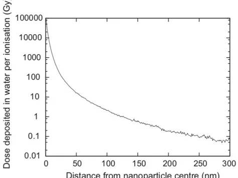
Figure 1.21: Average radial dose deposited following a single ionising event in a 2 nm GNP during a 6 MeV Linac irradiation. ... 29
Figure 1.22: General mechanism of the photocatalysis. ... 33
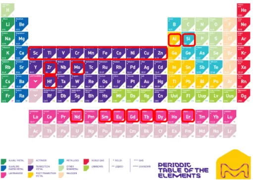
Figure 2.1: Periodic table with the period four metal oxides chosen for this project. ... 42
Figure 2.2: PEI branched structure. ... 42
Figure 2.3: Nanoparticle catalytic ability measurement set-up. ... 48
Figure 2.4: Plate set-up for irradiation at GenesisCare Milton Keynes Elekta LINAC using 6 MeV and 10 Gy. ... 49
Figure 3.1: PEI structures. ... 58
Figure 3.2: Catalytic activity of non-coated (A) and PEI-coated (B) NPs. ... 68
Figure 3.3: Signal of coumarin-3-carboxylic acid for different doses. ... 73
Figure 3.4: HO• production for each type of NP in both conditions. ... 74
Figure 3.5: Significance results for HO• using Prism Graphpad. ... 74
Figure 3.6: HO• production for each type of NP in both conditions after taking the ratio between the irradiated and non-irradiated values. ... 75
Figure 3.7: H2O2 production for each type of NP in normoxia conditions. ... 76
Figure 3.8: Significance results for H2O2 using Prism Graphpad. ... 76
Figure 3.9: H2O2 production for each type of NP in normoxia conditions after taking the ratio between the irradiated and non-irradiated values. ... 77
Figure 3.10: HO• production for each type of NP in both conditions taking into account quenching results. ... 78
Figure 3.11: H2O2 production for each type of NP in normoxia conditions taking into account quenching results. ... 79
Figure 3.12: DHR vs CCA results scatterplots between each variable for all the NPs with the regression line. ... 79
Figure 3.13: ICP-MS uptake results for each type of NP. ... 83
Figure 3.14: ICP-MS metal loading intracellular ratio results for each type of NP. ... 84
Figure 3.15: HSC cell clonogenic assay for each type of NP in both conditions. ... 85
Figure 3.16: HSC cell clonogenic assay for each type of NP in both conditions after taking the ratio between the irradiated and non-irradiated values. ... 85
Figure 3.17: Cell irradiation vs ICP-MS results scatterplots between each variable for all the NPs with
the regression line. ... 86
Figure 3.18: CCA vs cell irradiation results scatterplots between each variable for all the NPs with the regression line. ... 87
Figure 3.19: DHR vs cell irradiation results scatterplots between each variable for all the NPs with the regression line. ... 87
Figure 3.20: ZnPEI and Zn NPs illustration. ... 94
Figure 4.1: Hydroxyl radical production. ... 106
Figure 4.2: Superoxide anion radical production. ... 107
Figure 4.3: Singlet oxygen production. ... 109
Figure 4.4: A) Stacked bar chart summarising the irradiation-induced (‘real’) change in overall radical production for each NP. B) Data from panel A replotted to normalise the proportion of metallic element atoms, based on each NP chemical formula. ... 110
Figure 4.5: A) Superoxide formation in the presence of pH 3.5 10 mM nitric acid or pH 3.5 10 mM formic acid hole scavenger. B) Data from panel (A) replotted as ‘real’ values (10 Gy irradiated value minus 0 Gy control) ±SEM. ... 112
Figure 4.6: DNA damage assays based on the appearance of the relaxed circular form of plasmid DNA. ... 114
Supp Figure 4.1: NP TEM images (low and high magnification) and size histograms. ... 124
Figure 4.7: Scatterplots between each variable for all the NPs with the regression line. ... 128
Figure 4.8: Scatterplots between each variable for the NPs of period 4 of the periodic table with the regression line. ... 130
List of Tables
Table 1.1: Comparison of the oxygenation in organ and respective tumours. ... 14
Table 1.2: Example of radiosensitiser for each radiosensitisation mechanism. ... 17
Table 3.1: Important information of strategies used for designing polymer coated IONPs for optimized stem cell uptake. ... 55
Table 3.2: TEM images at 3000x and 150000x magnification, and the size distribution for all NPs, non-coated and PEI-non-coated ones. ... 61
Table 3.3: Zeta potential for each NP. ... 65
Table 3.4: Mean diameters from TEM analysis and mean zeta potential data for each type of NP. ... 67
Table 3.5: 7OH-3-CCA quenching results for each NP. Each plot compares the coated and non-coated pair of each NP. ... 68
Table 3.6: 7OH-3-CCA quenching significant results.. ... 70
Table 3.7: Rhodamine 123 quenching results for each NP. ... 71
Table 3.8: Rhodamine 123 quenching significant results. ... 73
Table 3.9: Cell toxicity for each type of NP. ... 80
Table 3.10: Loading metal concentration for each type of NP. ... 83
Table 3.11: Summary of the results for each type of NP, where HO• and H2O2 production data include the quenching data. ... 89
Table 3.12: Summary of toxicity results for each type of NP. ... 91
Table 3.13: Summary of the metal oxide NPs and their respective induced damage. ... 97
Table 4.1: NP diameters measured by TEM and DLS. ... 104
Table 4.2: Summary table of total irradiation-induced: ROS, DNA damage and e-/h+ pair formation. ... 117
List of Abbreviations and Symbols
3D-CRT: Three-Dimensional Conformal Radiation Therapy 7OH-CCA: 7-hydroxycoumarin-3-carboxylic acid
A549: Human Alveolar Basal Epithelial Adenocarcinoma cell line AgNP: silver nanoparticles
Balb/c 3T3: Mouse Fibroblast cell line
BEAS-2B: Human bronchial epithelium non-cancerous cell line Caco2: Human Epithelial Colorectal Adenocarcinoma cell line CAT: Catalase
CCA: Coumarin-3-Carboxylic Acid CIT: citrate
DEF: Dose Enhancement Factor DHE: Dihydroethidium
DLS: Dynamic Light Scattering
DMEM: Dulbecco´s Modified Eagle Medium DNA: Deoxyribonucleic Acid
DSBs: Double Strand Breaks
DU145: Human Prostate Adenocarcinoma cell line e-/h+ : electron/hole
EPR: Enhanced Permeability Retention FF: Flattening Filter
H2O2 : Hydrogen peroxide
HaCaT: Human Keratinocyte cell line hMSCs: Human Mesenchymal Stem cells HO. : Hydroxyl radical
HSC-3: Human Tongue Squamous carcinoma cell line IMRT: Intensity-modulated Radiation Therapy
L132: Lung Epithelial carcinoma cell line LINAC: Linear Accelerator
LNCaP: Human Prostate Adenocarcinoma cell line MDA-MB-231: Human Breast carcinoma cell line mMSCs: Mouse Mesenchymal Stem cells NP: Nanoparticle
NPs: Nanoparticles NSs: NanoSheets 1O2 : Singlet oxygen O2-. : Superoxide
PAA: Polyacrylic Acid
PBS: Phosphate-Buffered Saline PEG: Polyethylene Glycol PEI: Polyethylenimine PVA: Polyvinyl Alcohol PVP: Polyvinyl-pyrrolidone
RAW 264.7: Mouse Myeloid cell line rMSCs: Rat Mesenchymal Stem cells ROS: Reactive Oxygen Species RT: Radiotherapy
SABR: Stereotactic Ablative Radiotherapy SEM: Standard Error of the Mean
SNB-19: Human Glioblastoma cell line SOD: Superoxide Dismutase
SOSG: Singlet Oxygen Sensor Green
SPIONs: SuperParamagnetic Iron Oxide Nanoparticles SSBs: Single Strand Breaks
U87MG: Human Glioblastoma cell line XRD: X-ray Diffraction
One of the applications of radiation is to treat cancer, a disease that arises approximately in one among every three individuals. Cancer is a disease that consists of the abnormal growth of cells that do not follow the normal rules of cell division (Hejmadi, 2010).
The cancer treatment that uses radiation to kill cancer cells is called radiotherapy (RT). People have been using ionising radiation to treat cancer for more than 120 years (Nickoloff, 2015). The main goal of RT treatment is the irradiation of a target tumour volume while minimizing the amount of radiation absorbed in the healthy tissue.
In RT there are essentially two modes of treatment: internal radiation therapy and external beam radiation therapy. Internal RT uses radiation sources that are placed inside the body, such as implants (brachytherapy) or as injections, capsules or drinks (radioisotope therapy). External RT uses a machine that produces radiation and directs it into the body, to the tumour.
This project focuses on methods to selectively enhance external RT, which is the most common type of radiation therapy used for cancer treatment.
1.1 Radiotherapy
X-ray beam treatment
In the past 40 years, the combination of high-voltage linear accelerators able to reach deep tissues with computed tomography to allow 3D treatment planning in RT has gained widespread use and minimised exposure to normal tissues (Nickoloff, 2015).
X-rays are produced through the bombarding of a target with energetic electrons. These X-rays consist of bremsstrahlung photons, which result from electron-nucleus Coulomb interactions, and characteristic photons, which result from electron transitions between atomic shells. Different X-ray energies (wavelengths) can be produced. X-ray tubes produce X-rays in the range of kilovoltage energies, called superficial X-rays (50-150 kV) or orthovoltage X-rays (150-300 kV); while linear accelerators (LINACs) produce X-rays with megavoltage energies.
Kilovoltage X-ray beams do not penetrate the body very deeply and deposit most of the X-ray dose close to the surface of the patient and due to the attenuation and scattering of the beam the dose drops-off rapidly with depth. Nowadays, the primary therapeutic clinical use for kilovoltage X-rays is the treatment of skin cancers (Hill et al., 2014). Megavoltage X-rays are more penetrating and have largely replaced kilovoltage treatments. Due to its extensive use and due to its different applications, megavoltage X-rays is the beam energy used in this thesis.
Megavoltage X-rays are produced in a treatment machine called a linear accelerator (LINAC), represented in Figure 1.1. For this high-energy radiation to be produced, an electron beam is needed.
These electrons are produced by an electron gun and then accelerated through a waveguide that increases their energy from keV to MeV range (Podgorsak, 2005). When the electrons hit an X-ray target, X-rays are produced. The LINAC and the treatment couch rotate about a point called isocentre.
Figure 1.1: Design configuration for a medical LINAC.
Megavoltage X-ray physical properties
In a typical treatment, electrons are accelerated to an energy of 6 MV and hit a tungsten target to produce X-ray photons with an energy spectrum from 0 to a maximum value of 6 MV.
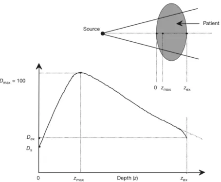
The absorption of X-ray energy per kilogram of matter, the so-called dose measured in units of gray (Gy), varies with depth through the patient. At the skin entry point the dose is Ds, as shown in Figure 1.2. Being at the surface an initial dose build-up occurs due to the contribution of secondary photoelectrons until it reaches a maximum value Dmax at a depth of zmax. This maximum value is followed by an exponential decrease which follows the Beer-Lambert law until it reaches a value of Dex which happens when radiation exits the patient’s body.
Energy fluence decreases exponentially with the Lambert Beer’s law: 𝜓
𝜓&= 𝑒 )*+
where 𝜓 is the energy fluence in MeV/cm2 at a depth 𝑥 in matter, 𝜓
& is the primary beam fluence and 𝜇 is the linear attenuation coefficient in cm-1.
RF power generator Electron gun Accelerating waveguide GANTRY X-ray target Treatment couch STAND Gantry axis Co u c h a x is Electron beam transport Isocentre
Figure 1.2: Typical dose distribution on the central axis of a megavoltage beam. Ds is a superficial dose, at zmax depth dose reach a maximum value Dmax, Dex is the dose delivered to the patient at the beam exit point zex: we can observe a small curve downwards due to missing scatter contribution at the exit point from point beyond the exit dose point (Podgorsak, 2005).
As shown in Figure 1.3, the build-up region increases with increasing photon energy. Also, surface dose, Ds, and dose maximum depth, zmax, depend mainly on the beam energy. For a 6 MV X-ray beam, the first 1.5 mm of tissue (the skin) receives ~15% of the maximum dose while for an 18 MV X-ray skin receives ~10% (Podgorsak, 2005).
6
Figure 1.3: Percentage depth dose (PDD) curves for MV X-ray beams with energies from Co to 25 MV at a surface
to source distance (SSD) of 100 cm for a 10×10 cm2 field. The maximum dose is at 1.5 cm depth for 6 MV and at
5 cm depth for 25 MV (Podgorsak, 2005).
A 6 MeV LINAC beam has contributions from lower photon energies, as it can be observed in Figure 1.4. These lower energies will be very useful for photocatalysis on metal oxide surfaces and DNA damage, as it will be explained later. From Figures 1.4 (a) and (b), one can see that depending on the
(c) (c) (c) (c) (c) (c) (c)
Figure 1.4: Energy spectra due to a 6 MeV LINAC beam at different depths of water. (a): 25.0 cm diameter field (J. L. Robar & Clark, 1999); (b): 2.5 cm diameter field (J. L. Robar & Clark, 1999); (c): simulation of a cylindrical water volume (10 cm long and 10 cm diameter) exposed to a 5 cm diameter beam, directed along the cylinder’s central axis (McMahon et al., 2011).
field size (25 cm vs 2.5 cm) the mean photon energy of 6 MeV X-ray beams varies. For large beams (25 cm) the mean photon energy decreases with depth up to 15 cm due to an increase of phantom scatter (J. L. Robar & Clark, 1999). Figure 1.4 (c), calculated at a depth of 5 cm shows the contribution of both low-energy photons and secondary electrons, significant to dose deposition.
Current methods of improving radiotherapy targeting
Photon (X-ray) RT, known as XRT, can be improved by techniques such as 3D conformation RT (3D-CRT) and intensity-modulated radiation therapy (IMRT). These techniques allow high doses to be shaped to tumours while minimizing doses to surrounding normal tissue (Thariat et al., 2013).
Due to the appearance of three-dimensional radiation treatment planning (3D-RTP) and computer-controlled radiation therapy (CCRT) delivery systems it was possible the implementation of three-dimensional conformal radiation therapy (Purdy, 2001). 3D-CRT technique conforms the radiation into the target volume with the goal of minimizing the amount of radiation that hits the surrounding normal tissue. The increased access of volumetric images of a patient’s internal anatomy has led to the development of 3D-CRT techniques since it allowed the delineation of soft tissue anatomy (King et al., 2017).
A type of 3D-CRT technique is the IMRT was developed to improve RT both through increased tumour control probability and decreased normal tissue complication probability (Podgorsak, 2005). This technique uses multileaf collimators mounted inside the gantry that provide non-uniform radiation beam intensities. These collimators can move during the treatment, dynamically shaping the beams in order to match the shape of the target volume. It relies on inverse treatment planning, in which instead of changing the beams to achieve the dose limits required for each target and organ at risk the user gives the dose limits and the computer calculates the best shape of the beams, and on 3-D multimodality imaging to define the target volumes (Podgorsak, 2005). IMRT is capable of achieving a much higher degree of target conformity and/or normal tissue sparing than the other treatment techniques (Purdy, 2001). This higher degree of target conformity allows the delivery of higher radiation doses than other 3D-CRT techniques, meaning that the conventional 2 Gy radiation dose fractions can be increased. An example of a type of IMRT treatment which uses high radiation doses in a small number of treatment fractions is the Stereotactic ABlative RT (SABR) used to treat metastatic cancer which has spread to a limited number of sites, called oligometastatic disease (King et al., 2017).
Due to IMRT conformal dose distribution with narrower margins, microscopic tumour cells might end up out of the radiation field resulting in tumour reoccurrence. The sharp dose gradient in IMRT requires advanced imaging and accurate delineation of different structures, making it very sensitive to set-up uncertainties and organ motion (Rehman et al., 2018). The most important disadvantage associated with IMRT is the increase in total body irradiation. Although this technique saves organs at risk from receiving high radiation dose, at the same time it spreads out the delivered dose to the whole body
increasing total body exposure (Rehman et al., 2018). To achieve the prescribed dose, IMRT requires a significantly larger number of monitor units than other techniques which results in an increase of radiation scattered and radiation leakage that causes secondary malignancies (Purdy, 2008). Furthermore, this technique is time consuming and requires complex procedures such as its planning, delivery, and quality assurance. Despite all the efforts to improve radiation therapy, healthy tissue surrounding the tumour may still be exposed to levels of radiation that can cause both short and long-term side effects. For this reason, my thesis is focused on the use of radiosensitisers to better target the radiation dose to the tumour and help decrease the side effects of MV X-ray RT treatment.
1.2 Importance of Reactive Oxygen Species (ROS)
One of the main effects of ionising radiation in an aqueous environment, such as a cell, is the formation of highly reactive species that can damage cells. This section describes those reactive species, while the interaction of radiation with cells will be discussed in Section 1.3.
Reactive oxygen species (ROS) play a very important role in aerobic life. The damage that ROS produce to biological molecules, such as DNA, constitutes one of the most efficient pathways used in RT to kill cancer cells (Hosoya & Miyagawa, 2014).
Within the group of reactive chemical species two major types can be distinguished: radicals that have one or more unpaired electron(s) in their outer molecular orbitals; and non-radicals that do not contain unpaired electrons.
Non-radical species, although they do not contain unpaired electrons, are chemically reactive and can be easily converted into radical species.
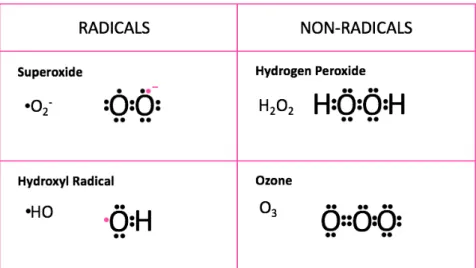
Reactive oxygen species are partially reduced forms of molecular oxygen (O2) and include hydroxyl radical (HO.), hydrogen peroxide (H2O2), superoxide anion (O2-), and ozone (O3), as shown in Figure
Figure 1.5: Radicals and non-radicals from reactive oxygen species.
The radicals serve as the reactive intermediates for the inherent biological action of oxygen and can be found in high amounts in mitochondria, where the respiratory chain from this cell structure liberates electrons that may interact with molecular oxygen producing superoxide. This superoxide can in turn produce other ROS starting a chain reaction (Trachootham et al., 2009).
In aqueous solutions, superoxide exists in equilibrium with its conjugate acid, the perhydroxyl radical (HO2) (Rose, 2012):
HO2 ↔ H+ + O2
-This means that, when talking about superoxide anion in water, both O2- and HO2 are included since
one does not exist without the other.
Under normal physiological conditions, ROS production can promote cell proliferation and differentiation (Boonstra & Post, 2004), but when the levels of these species become too high it starts to damage biological material, such as: nucleic acids, proteins and free amino acids, lipids and carbohydrates (Sies, 1985). For this reason, the maintenance of ROS homeostasis is crucial for normal cell function. Some ways in which cells detoxify ROS include superoxide dismutase (SOD) which converts superoxide to hydrogen peroxide and oxygen; catalase converts hydrogen peroxide into water and oxygen; glutathione peroxidase converts hydrogen peroxide into water or lipid peroxides into alcohols. The loss of these defence mechanisms provokes an abnormal ROS level leading to a condition known as oxidative stress (Toyokuni et al., 1995).
Diseases including cardiovascular disorders, diabetes, chronic inflammation, and cancer can be the product of perturbations in cellular redox homeostasis, since pathological conditions are a result of serious increase in ROS levels which can lead to an irreversible oxidative damage (Alimoradi et al., 2017).
1.3 Effects of radiation in cells
There are two ways of radiation interacting with cells: directly or indirectly, as shown in Figure 1.6. It is estimated that around 40% of X-ray damage is direct and 60% indirect (Baskar et al., 2014).
Through the direct way, X-rays are absorbed in biological material interacting directly with critical targets in the cells (Hall et al., 2016). Biological damage can occur due to the ionized or excited atoms of the target. The most effective constituent is the cell DNA in the nucleus. In this case, the ability of the cell to reproduce and thus survive may be affected. The cell may be destroyed if the chromosomes do not replicate properly or the information carried by the DNA molecule undergoes significant alteration due to the interaction of the radiation with the atoms within the cell.
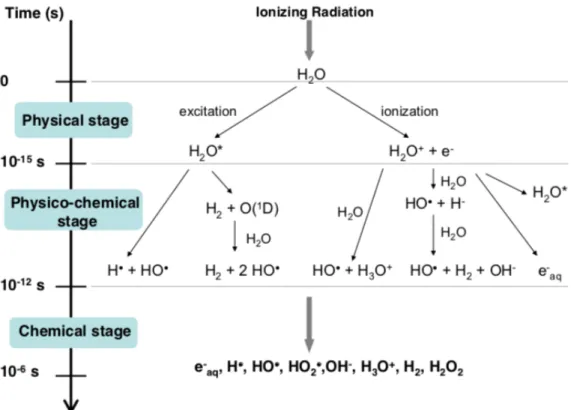
In the indirect way, the radiation damages the cell through the formation of free radical species, which occur due to the excitation and ionization of water molecules inside or in the vicinity of the cell.
Figure 1.7: Main reactions occurring during the three stages of water radiolysis (Le Caër, 2011). Figure 1.6: Direct and indirect actions of
radiation in cell DNA; (a) – single strand break; (b) – double strand break.
+ -
e
-nucleush
n + -e
-nucleush
nHO
. H2OIndirect effect
Direct effect
(a) (b)The interaction of a photon with a water molecule, known as water radiolysis, leads to the formation of radical and non-radical species such as shown in Figure 1.7. The photon can either excite a water molecule or ionize it producing a secondary electron (e-). Both these interactions will lead to the formation of fragments such as hydrogens (H) or hydroxyls (HO). Furthermore, the produced fragments can recombine or interact with other fragments or ions to form either harmless compounds to the cells such as water (H2O) or harmful species, such as radicals, that contribute to cell death. The species more relevant for the process of cell death are hydroxyl radicals (OH.), superoxide anion (O2-) and
hydrogen peroxide (H2O2).
After the initial primary radical formation (i.e., chemical stage in Figure 1.7), both H and e-aq are converted to OH. radicals by reaction with H2O2 (Bielski et al., 1985):
e-aq + H2O2 → OH. + OH -H + -H2O2 → O-H. + H2O
The OH. radicals then react with H2O2 to yield HO2 :
OH. + H2O2 → HO2 + H2O
There is a higher probability of the radiation to interact with the water within the cell than with the DNA inside the nucleus, which takes up only a small proportion of the cell. Most of the radiation damage caused within the cell is due to the indirect pathway since 70% of the cell constitutes of water (Desouky et al., 2015). This means that, since water makes up most of the cell’s volume, the indirect path is the one that should be used and studied to improve RT treatment and is the focus of this thesis.
It is worth mentioning that low energy electrons (e-), showed in Figure 1.7, are species that contribute very efficiently to the damage of the DNA molecule. They can either damage this molecule through the direct pathway or the indirect one through a number of mechanisms such as dissociative electron transfer from water-interface electron traps to DNA bases, quenching of dissociative electron attachment to DNA and quenching of dissociative electronically excited states of H2O in contact with DNA (Alizadeh et al., 2013).
The direct and indirect action of radiation can also cause damage to cellular components other than DNA, such as protein, enzymes, organelles, membranes (Wang et al., 2018). The damage to one of these components can lead to cellular death.
Both direct and indirect radiation mechanisms can cause different types of damages to cell DNA, such as abasic sites, pyrimidine dimers, alkylation adducts, single strand breaks (SSBs) and double strand breaks (DSBs) (Dahlmann et at., 2009). Both SSBs and DSBs are shown in Figure 1.6. DSB is the most effective damage, since it leads to cell death if not repaired (Baskar et al., 2014). Due to mechanical stress or different types of chemical modification DNA structure may be broken in one strand of the double helix, leading to a SSB (Figure 1.6(a)) or broken in two strands of the double helix leading to a DSB (Figure 1.6(b)). It is very difficult to achieve DSB to DNA structure, but the most significant of the external agents that is able to achieve it is ionising radiation (Jeggo & Löbrich, 2007).
Reactive oxygen species primarily induce base damage and SSBs. DSBs arise indirectly from further action of trying to repair some lesions in the strand or can arise from two closely located SSBs (Jeggo & Löbrich, 2007).
DSBs can also occur during the normal life cycle of the cell, more specifically during DNA replication or in meiosis (Mladenov & Iliakis, 2011). Furthermore, within the cell, DSBs arise due to reactive oxygen species generated by normal respiratory metabolism (Featherstone & Jackson, 1999).
When oxygen is present a higher number of reactions will occur (Parveen, 2001) such as: H. + O2 → HO2. → H2O2 + O2 reaction 1
HO2 + e- → HO2- + H+ → H2O reaction 2
X. + O2 → XO2. → X + O2. reaction 3
Reaction 3 refers to the interaction of oxygen with an organic molecule X, such as DNA, RNA, and proteins.
In an oxygenated environment, oxygen molecules can extract an electron from the radical anions turning them into radical cations that have a long life to cause DNA damage. On the other hand, in a hypoxic environment the electron from the radical anion tends to combine with the radical cation, with no damage to the DNA. In conclusion, the presence of oxygen increases the DNA damage produced by a given dose of radiation.
Cells sensitivity increase due to ionising radiation in normal oxygen environment compared to cells sensitivity in hypoxia conditions is called the oxygen effect, and although it has the ability of modifying the dose it is independent of the radiation dose. The ratio between the dose required to achieve a given cell survival in the absence of oxygen compared to the dose required for the same effect under normoxia conditions is called the oxygen enhancement ratio (OER), and its value varies between 2.5-3 for X-ray radiation (Nair et al., 2001).
For radiosensitisation to happen the lowest concentration of oxygen at which normoxia radiosensitisation occurs is as low as 0.25%, since at this concentration dose-response curve is shifted half-way towards the fully aerated condition (Kennedy et al, 1980).
1.4 Tumour environment
It is important to outline the characteristics of the tumour environment to find ways to make these cells weaker and more susceptible to radiation. Increased levels of ROS are a common feature in tumour environment since cancer cells have a higher metabolism and produce more ROS via mitochondrial respiration. Cancer cells adapt to this toxic environment and these adaptation mechanisms also contribute to tumour metastasis (Chen et al., 2007).
Although cancer cells have a higher capability of resisting to high levels of oxidative stress compared to normal cells, their maximum level is still limited. If cancer cells are exposed to agents that overcome that limit, they will die (Fruehauf & Meyskens, 2007).
Figure 1.8: The biology of cancer redox (Trachootham et al., 2009).
Figure 1.8 presents a schematic of what happens in redox cancer cell biology. Redox homeostasis is maintained by normal cells through the balance between generation and elimination of ROS. Exogenous ROS stress can be fought through the reserve antioxidant capacity of normal cells, preventing the ROS level from reaching the cell-death threshold, showed by the horizontal dotted line in the figure. In cancer cells, ROS level is higher than in normal cells but still below the threshold, since these abnormal cells arise from a redox adaptation response that leads to an upregulation of antioxidant capacity and a shift of redox dynamics with high ROS generation and elimination. In this way, cancer cells are more dependent on the antioxidant system and more sensitive to further oxidative stress due to the proximity to the cell-death threshold. This redox characteristic of cancer cells constitutes a possibility for a therapeutic approach to defeat them, since a further increase in ROS production (red bar) will induce cell death (Trachootham et al., 2009).
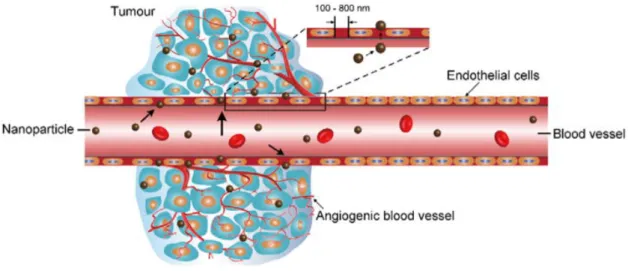
Characteristics such as high metabolic rate, poor blood supply and a ‘leaky’ vascular system (Figure 1.9) lead to low oxygen regions, known as hypoxia regions. Different parts of tumour tissues present heterogeneous areas of oxygenation, where peripheral zones are higher in oxygen levels than the innermost ones (Alimoradi et al., 2017).
Figure 1.9: Schematic representation of tumour vasculature structure (Dai et al., 2017).
Hypoxia is a state of low oxygen levels that in tumours consists of 1-2% O2 compared to the normal tissues (Muz et al., 2015). Tissue normoxia, also known as physoxia, is the oxygenation in healthy tissues that varies between the organs due to blood vessel network diversity and metabolic activity. Table 1.1 summarizes the oxygenation in different organs and their respective tumours. Normoxia conditions performed at the laboratory are far from the oxygen values found in healthy organs and its value in in vitro experiments is 20.9% O2 (Muz et al., 2015).
Table 1.1: Comparison of the oxygenation in organ and respective tumours (Muz et al., 2015).
Tissue/Organ Physoxia (median % O2)
Cancer Hypoxia
(median % O2)
Brain 4.6 Brain tumour 1.7
Breast 8.5 Breast cancer 1.5
Cervix (nullipara) 5.5 Cervical cancer 1.2
Kidney cortex 9.5 Renal cancer 1.3
Liver 4.0-7.3 Liver cancer 0.8
Lung 5.6 Non-small-cell lung cancer 2.2
Pancreas 7.5 Pancreatic tumour 0.3
Rectal mucosa 3.9 Rectal carcinoma 1.8
In these hypoxic regions cells lack molecular oxygen and other nutrients due to the unbalanced growth of the tumour cells and vascular components. Consequently, these cells do not have an adequate blood
supply. At distances more than about 150 µm from the capillary tissue, oxygen tension decreases rapidly with distance falling to a level where cell division cannot happen. Cells deprived of oxygen die, forming necrotic regions, while in the interface between the well-oxygenated and the necrotic regions viable hypoxic cells tend to occur (McKeown, 2014).
The lack of oxygen in the environment of cancer cells makes these cells more resistant to radiation damage than those in a normal oxygen environment. Due to the hypoxic environment of cancer cells, tumours are up to three times less susceptible to radiation damage than normal cells (Paterson, 1981). The reason for this is that oxygen is an important ingredient in the pathway of ROS production. This happened due to oxygen being a potent radiosensitiser in a way that it aids formation of DNA-damaging free radicals increasing the effectiveness of a given dose of radiation.
Cells use glycolysis and oxidative phosphorylation to make ATP for cellular energy. Although glycolysis is less efficient than oxidative phosphorylation, 2 ATP molecules vs 30 ATP molecules respectively, it is a faster process since production of lactate through glucose is 10 to 100 times faster than the complete oxidation of glucose in the mitochondria. Furthermore, using glycolysis to produce energy not only ensures ATP production but this process requires the pentose-phosphate pathway which additionally produces ribose sugars essential for nucleotides required for cell division. Also, this cycle produces a high amount of NADPH essential to produce fatty acids, cholesterol and regenerating gluthione antioxidants. On the whole, the amount of ATP synthesised over any given period of time is comparable between glycolysis and oxidative phosphorylation (Liberti & Locasale, 2016). Otto Warburg, in the 1920s, observed very high amounts of glucose being consumed in tumours compared to the surrounding tissue. This ‘Warburg effect’ describes the metabolism of a cancer cell to produce energy, from the predominantly oxidative phosphorylation to predominantly aerobic glycolysis. It is thought that one of the main reasons why cancer cells use this respiratory mechanism is that upregulated glycolysis constitutes a growth advantage which promotes proliferation and invasion (Gatenby & Gillies, 2004). To overcome this limitation approaches such as hyperbaric oxygen, hypoxic cell radiosensitisers such as misonidazole and nitroimidazole, and hypoxic cytotoxins, such as tirapazamine were used without clinical success (Wardman, 2007). Hyperbaric oxygen is the oxygen released at pressures of 2-4 atmospheres to increase the oxygen in the plasma. This technique was supposed to allow the O2 to diffuse further through tissue than normobaric oxygen breathing, since diffusion distance of oxygen through respiring tissue increases with the initial oxygen tension (Rockwell et al., 2009). Hyperbaric oxygenation demonstrates an overall positive effect on RT treatment but has not been adopted and remains to be validated as a standard treatment (Gérard et al., 2019).
More recently, hypoxia imaging during RT treatment became a technique of study. The level of oxygen present in tumour cells is cyclic, where cells that are hypoxic before RT treatment become oxygenated during or after the treatment. In this way, monitoring the oxygen level during the treatment may help if the RT fraction is given when tumour reoxygenation is expected to be at its maximum so as to optimize the oxygen enhancement ratio (Gérard et al., 2019).
Radio-enhancing NPs and new radiosensitising drugs may be delivered into the tumour to improve oxygenation. In the class of radiosensitising drugs are fluorochemicals able to dissolve considerable amounts of oxygen and deliver it through passive diffusion into the hypoxic regions (Gérard et al., 2019). Although both pre-clinical and clinical studies have shown that hypoxia can be reduced to improve the outcome of radiation therapy, hypoxic modification has still not been established as a standard treatment with RT (Horsman & Overgaard, 2016).
The need to predict and assess the patient’s response to the RT treatment is of extreme importance since RT continues to be the most widely used treatment for cancer, even in conjunction with other types of cancer treatment such as surgery and chemotherapy (Jeggo & Löbrich, 2007). This would lead to an important step for the optimization of this cancer treatment.
RT can be optimized by either the use of radiosensitisers to kill the tumour cells or by radioprotectors to protect the normal tissues from radiation damage.
1.5 Radiosensitisers
The poor response of many tumours to radiation despite the improvement of techniques using conventional sources of radiation have led to the development of other approaches. Three main approaches were considered: hyperbaric oxygen, proton and neutron beams, and radiosensitisers (Paterson, 1981).
The use of hyperbaric oxygen, to increase oxygen tension during irradiation in tumour cells, has not shown an increase in tumour control rates (Henk, 1986). Radiosensitisers have become the most reliable approach since they are inexpensive and widely available and still have a whole potential to be explored.
Radiosensitisers make cancer cells more vulnerable to radiation therapy by enhancing the effect of ionising radiation within the cancer cells while leaving the healthy cells out of the target. The advantage of these materials is that since they passively or actively are conformed to the tumour site the effect of the enhanced radiation leads to an increased dose in the initial treatment within the cancer cells enabling the reduction of the number of treatments for the patient, while still achieving the initially prescribed dose.
The role of these materials can be viewed as the same as the oxygen, in the sense that they can act to restore, at least to some extent, the radiosensitivity of hypoxic tumour cells. In summary, radiosensitisers are oxygen substitutes that are capable of diffusing into hypoxic tumour cells and sensitise them via a similar mechanism to oxygen.
An important feature of radiosensitisers that relates to oxygen is that these compounds are not easily metabolised. In this way, during diffusion to tumour cells they can penetrate further into hypoxic regions (Parveen, 2001).
To define a compound as a radiosensitiser it is crucial to understand the interaction between the compound and the radiation of study. If the combination results in a biological response greater than what would be expected from the radiation by itself, than one can conclude that there is a synergistic effect behind it and the compound is classified as a radiosensitiser.
Radiosensitisers can perform through different mechanisms in order to apply their radiation-enhancing effects. Some of the possible mechanisms of radiosensitisation are (Gill & Vallis, 2019):
1. Inhibition of post-irradiation cellular repair processes: being DNA a critical target for ionising radiation the inhibition of its damage repair pathways will contribute to an increase in radiotoxicity;
2. Cell-cycle dysregulation: compounds that are able to arrest the cell-cycle at a specific phase can be considered radiosensitisers. For example, cells in late G1 or G2/M phases are more sensitive to the effects of radiation while cells in S phase are the most resistant;
3. Radiosensitivity enhancement in hypoxic cells: the radioresistance of hypoxic cells makes it difficult the generation of ROS by ionising radiation. Oxygen-mimetic sensitisers help the increase in ROS in these cells, enhancing the efficacy of the treatment;
4. Production of cytotoxic substances: Radiolysis of the sensitiser produces cytotoxic molecular fragments which in turn contribute to cell-death;
5. Generation of Auger electrons near DNA: Explained in the next section, high atomic number materials after radiation exposure produce Auger electrons due to inner-shell ionization. If the X-rays energy matches the K-shell energy of its electrons, these materials, when internalised by the tumour cells, will enhance the tumour-absorbed dose. This phenomenon may enhance the radiobiological effects of ionising radiation and potentially explains sensitisation by heavy-metal nanomaterials.
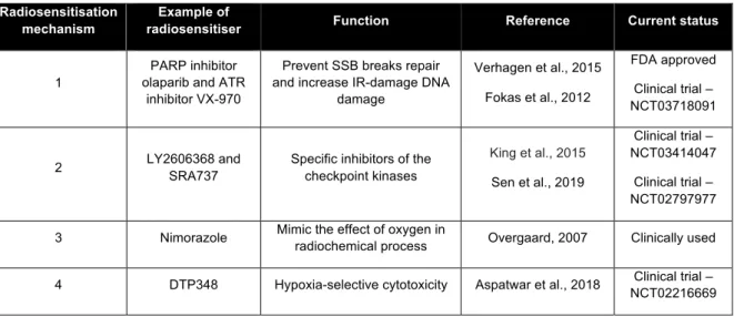
Table 1.2 summarises some examples of radiosensitiser for each of the mechanisms stated.
Table 1.2: Example of radiosensitiser for each radiosensitisation mechanism.
Radiosensitisation mechanism
Example of
radiosensitiser Function Reference Current status
1
PARP inhibitor olaparib and ATR
inhibitor VX-970
Prevent SSB breaks repair and increase IR-damage DNA
damage Verhagen et al., 2015 Fokas et al., 2012 FDA approved Clinical trial – NCT03718091 2 LY2606368 and SRA737
Specific inhibitors of the checkpoint kinases King et al., 2015 Sen et al., 2019 Clinical trial – NCT03414047 Clinical trial – NCT02797977 3 Nimorazole Mimic the effect of oxygen in radiochemical process Overgaard, 2007 Clinically used
4 DTP348 Hypoxia-selective cytotoxicity Aspatwar et al., 2018 Clinical trial –
Physicochemical properties such as, for example, size, shape, coating, and functionalization, control their pharmacokinetics, bioavailability, biodistribution, as well as targeting and intracellular delivery. Tumour vasculature is more leaky than normal vasculature, allowing small particles and larger drugs to enter the tumour environment. Furthermore, the lymphatic drainage of tumour tissue is less efficient than in normal tissues, meaning that particles are not easily removed. Together, these two factors constitute the Enhanced Permeability and Retention (EPR) effect. Thus, specific tumour targeting can be achieved by passive targeting via the EPR effect, active targeting by using high-affinity targeting-molecules, and stimuli-responsive triggered release to endogenous or exogenous stimuli (Liu et al., 2018).
Studies have shown a general relationship between the efficiency of sensitisation and the electron affinity, i.e. oxidizing abilities for chemical radiosensitisers (Adams et al., 1971). Hypoxic cell radiosensitisation happens when one electron is transferred to the radiation-induced lesion, resulting in the fixation of the chemical damage. A lot of studies regarding chemical radiosensitisers have been done (Candelaria et al., 2006; Wardman, 2007) and are still being undertaken (Farhood et al., 2019; Wang et al., 2018).
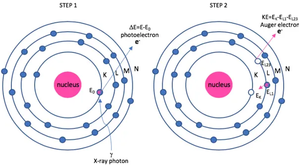
This thesis is focussed on metal-based radiosensitisers with a diameter within the nanometer range (nanoparticles). When an incoming high-energy X-ray photon encounters heavy-metal atom, two important phenomena occur: ejection of photoelectrons and ejection of Auger electrons, as shown in Figure 1.10.
5 AGuIX and NBTXR3 Enhanced absorbed dose
Lux et al., 2018 Bonvalot et al., 2019 Clinical trial – NCT03818386 Clinical trial – NCT03589339
Figure 1.10: Photoelectron and Auger electron schematic in an atom. Step 1: emission of a photoelectron with a kinetic energy (DE) equal to the difference in energy between the incoming photon (E) and the binding energy of
the electron in the K-shell (E0); Step 2: emission of a Auger electron with a kinetic energy (KE) equal to the
difference in energy between the binding energy of the electron in the K-shell (EK) and the binding energies of the
electrons in the L-shell (EL1 and EL23).
In Figure 1.10, if a high energy X-ray photon has enough energy and it is absorbed by an atom in the surface of the metal-based NP, an innermost electron will be ejected. This phenomenon is the photoelectric effect and it results in the ejection of a photoelectron with a kinetic energy equal to the difference in energy between the incoming photon and the binding energy of the electron in the K-shell. An electron from a higher energy state, in this case, the L-shell, will then occupy the empty state in the K-shell. From this transition an X-ray is emitted and if absorbed by another electron in the L-shell, an Auger electron is ejected. In this example, this process is termed a KLL Auger transition. The kinetic energy of an Auger electron is equal to the energy difference of the singly ionized initial state and the doubly ionized final state. The resulting kinetic energy EKLL is given by EKL1L2,3 = EK - EL1 - EL2,3 - F. The term F comes from the electron work function and from the equation it can be concluded that the kinetic energy of the Auger electron is independent of the type and energy of the primary beam.
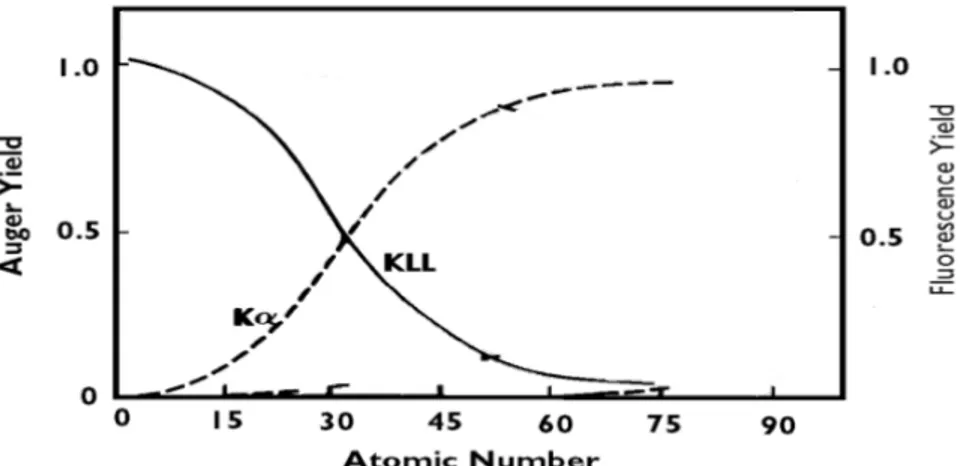
The energy EK - EL1 can be then released as a radiative decay, with emission of an X-ray and in this case an electron vacancy in the K-shell is filled by an electron from the L-shell so the characteristic energy of the emitted photon is called the K-alpha (Ka) spectral line, or as a non-radiative decay, with emission of an Auger electron and the result depends on the relative probabilities of Auger emission and X-ray fluorescence, as shown in Figure 1.11.
As it can be seen from Figure 1.11, the Auger emission is greater for lighter elements, while for heavier elements X-ray yield becomes greater. The energies and relative intensities of Auger transitions show a systematic behaviour with increasing atomic number (Z). In heavier elements, which exhibit more
energy levels, more Auger transitions are possible.
Figure 1.11: Relative probabilities of Auger emission and X-ray fluorescence. The solid lines show the Auger yield; the dashed lines show the fluorescence yield (results obtained from Reinhardt & Kern, 2008 and modified for the purpose of this thesis where irrelevant curves have been removed).
In terms of importance for the RT treatment, the fluorescent photons from X-ray fluorescence are able to travel longer ranges, up to centimetres, while Auger electrons travel much shorter distances, typically 10 nm (Hainfeld et al., 2008). This means that the former, depending on the tumour size, may not provide the desired localized tumour effect, but the latter, due to the fact that the electrons are weakly bound, they can be effective in producing very high local ionization density. For the Auger effect to be useful, the emitting atom should be close to the target molecule to damage the molecule, due to the short distance they are able to travel, although some Auger electrons can travel 50 nm in water. In summary, photoelectrons are the result of the electrons emitted along the primary paths of the ionising radiation which induce inner shell ionization of the metal atoms, whether the Auger electrons result from the relaxation of the excited core of the metal atoms.
Emission of Auger electrons and fluorescent photons can be caused by either photoelectric absorption or by Compton scattering. Basically, in photoelectric absorption the photon is absorbed by the photoelectron, while in Compton scattering the incident photon greatly exceeds the binding energy of the electron to the atom, so the photon does not disappear. Instead it is deflected through a scattering angle q, as shown in Figure 1.12. Part of its energy is transferred to the recoil electron, thus the photon loses energy in the process. The energy transferred
nucleus Incident photon Scattered Photon q, scattering angle Ejected Compton recoil electron
Figure 1.12: Schematic representation of Compton scattering.
does not depend on the density, atomic number, or any other property of the absorbed material (Cherry et al., 2004).
At a photon energy of about 100 keV or more the photon radiation beam attenuation in the target is mostly caused by Compton scattering and not by photoelectric absorption (Kljuev, 2001). The photoelectric effect varies approximately as (Z/E)3, where Z is the atomic number of the target and E is the incident photon energy. For high-Z elements this effect dominates the interaction with matter at energies <0.5 MeV, while for energies >1 MeV pair production dominates (Hainfeld et al., 2008). In pair production electron-positron pairs are created if the incident photon energy exceeds twice the rest mass of the electron (2×0.511 = 1.022 MeV) and it depends on Z2. All these physical interactions of radiation with metals made these materials a possible choice to improve the RT treatment.
Adding to the physical interactions, biocompatibility is a quality that needs to be studied since these metals will be introduced into the human body, specifically that they will need to be injected into the blood flow and reach the tumour site. Luckily, if these metals are produced in the nanometer range, it will allow them to accumulate into the cancer cells due to the leaky nature of the tumour vasculature and its lack of efficient lymphatic drainage as explained before in Figure 1.9. Relying on the EPR effect for NP delivery is a passive strategy while relying on the ligand-receptor binding is an active strategy. The later improves selective accumulation to targeted site and thus discriminates between the cancerous and healthy tissues (Attia et al., 2019).
For a metal-based NP the basic operation of the radiosensitisation process focuses on physical dose enhancement, chemical contribution and biological phase, as shown in Figure 1.13.
Figure 1.13: Schematic illustration of metal-based NPs radiosensitisation process (modified from Liu et al., 2018).
As illustrated in Figure 1.13, the common procedure for the study of metal-based NPs is to load them into the cells for a specific time and irradiate them (Upper-left). Biological mechanisms will happen
within the cells due to the radiation itself, the combined effect of radiation with the NP in study and also due to the NP itself (Upper-right). The basic operation of the radiosensitisation process will then lead to lethal cellular damage where the primary targets, depending on the cellular and subcellular distribution of the NPs, will include cell membrane, cytoplasm, nucleus, mitochondria, endoplasmic reticulum, and other organelles. In this way, the photoelectrons and Auger electrons produced from the combination of radiation with NPs will contribute to a dose enhancement due to the direct interaction of these species with the cells or due to the indirect interaction where these species will increase the amount of ROS damaging even more cells than the amount of ROS produced by radiation alone. Also, HO. production
occurs due to the catalytic-like mechanism/surface-catalyzed reaction present in metal-based NPs (Lower-left). These interactions produce cell damage that leads to cell death and can be probed by the number of cells that survived after a certain period of time. The combination of ionising radiation and metal-based NPs induces physical, chemical and biological mechanisms that contribute for the observed enhanced cell-killing effects (Lower-right) (Liu et al., 2018).
As mentioned before, the increase of ROS in cancer cell can exert the opposite of what it is expected from the RT treatment, such as the initiation and progression of cancer (Wu, 2006). On the other hand, increasing ROS levels can also be toxic to cancer cells which will make them more vulnerable to damage by further ROS attacks induced by external agents such as the ionising radiation used in RT (Trachootham et al., 2009).
This method is not straightforward due to the complexity of redox alterations in cancer cells. A lot of factors are involved in the redox regulation and stress response and they still need to be better understood. In this way, the simple addition of ROS may not lead to cancer cell apoptosis and the persistency of this approach may actually instead contribute to a community of cancer cells that developed an antioxidant capacity making them resistant to exogenous stress (Tiligada, 2006). A lot of investigations have been made using radiosensitisers to cooperate with RT treatment, however, a diversity of challenges such as poor biocompatibility, lack of targeting specificity, and radiosensitisation with clinical energies, mostly MV energies, increase the need for new and optimal radiosensitisers.
1.6 Radioprotectors
Hypoxic cells may limit the response of tumours to conventional RT because of their inherent radioresistance (Kennedy et al, 1980). Although this fact constitutes one of the main limitations for this type of treatment it is not the only one.
Another main challenge of RT is the fact that tumours are located near normal tissues and organs at risk making the doses delivered to the target volumes limited. For this reason, agents that present radioprotective properties have been used to reduce normal tissue complication probability (Liu et al., 2018). These agents are called radioprotectors and have the opposite role of a radiosensitiser.