Supported by
Accepted Article
Title: Impact of a Water-Soluble Gallic Acid-Based Dendrimer on the Color-Stabilizing Mechanisms of Anthocyanins
Authors: Luis Cruz, Nuno Basílio, Johan Mendoza, Nuno Mateus, Victor de Freitas, Maun H. Tawara, Juan Correa, and Eduardo Fernandez-Megia
This manuscript has been accepted after peer review and appears as an Accepted Article online prior to editing, proofing, and formal publication of the final Version of Record (VoR). This work is currently citable by using the Digital Object Identifier (DOI) given below. The VoR will be published online in Early View as soon as possible and may be different to this Accepted Article as a result of editing. Readers should obtain the VoR from the journal website shown below when it is published to ensure accuracy of information. The authors are responsible for the content of this Accepted Article.
To be cited as: Chem. Eur. J. 10.1002/chem.201901912 Link to VoR: http://dx.doi.org/10.1002/chem.201901912
Impact of a Water-Soluble Gallic Acid-Based Dendrimer on the Color-Stabilizing 1
Mechanisms of Anthocyanins 2
3
Luís Cruz,a,* Nuno Basílio,c Johan Mendoza,c Nuno Mateus,a Victor de Freitas,a Maun
4
H. Tawara,b Juan Correa,b Eduardo Fernandez-Megiab
5 6
a
REQUIMTE/LAQV, Departamento de Química e Bioquímica, Faculdade de Ciências,
7
Universidade do Porto, Rua do Campo Alegre, s/n, 4169-007 Porto, Portugal.
8
bCentro Singular de Investigación en Química Biolóxica e Materiais Moleculares
9
(CIQUS) and Departamento de Química Orgánica, Universidade de Santiago de
10
Compostela, Jenaro de la Fuente s/n, 15782 Santiago de Compostela, Spain.
11
c
LAQV, REQUIMTE, Departamento de Química, Faculdade de Ciências e Tecnologia,
12
Universidade Nova de Lisboa, 2829-516 Caparica, Portugal.
13 14
*Corresponding author. Tel.: +351 220402558; fax: +351 220402658. 15
Email address: [email protected] (Luis Cruz)
16 17
ABSTRACT 18
The interaction of two anthocyanins with a water-soluble polyanionic dendrimer was
19
studied through UV-Vis, stopped-flow and NMR spectroscopy. Cy3glc revealed a
20
stronger interaction than mv3glc at pH 1 according to their apparent association
21
constants. A higher color increased was also obtained for cy3glc at pH 3.5 as a result of
22
this stronger interaction. A high-frequency chemical shift of the cy3glc aromatic protons
23
suggest the formation of ionic pairs. The interaction parameters (K~700 M-1, n~295)
24
indicated the binding of approximately two anthocyanin molecules by each sulfate
25
group. The equilibrium and rate constants of cy3glc in the presence of dendrimer
26
showed an increased stability of the flavylium cation and a higher protection of this
27
species from hydration (pK’a and pKh increased almost one pH unit). The tuning and 28
color stabilization of anthocyanins using this dendrimer envisage novel applications as
29
colorimetric sensors for food packaging.
30 31
Keywords: UV-Vis spectroscopy; NMR; gallic acid-based dendrimer; anthocyanins; 32 association constant 33 34
Accepted
Manuscript
Introduction 35
Anthocyanins are glycosylated derivatives of 2-phenyl-benzopyrilium cation being
36
responsible for a pallet of beautiful colors found in many fruits and flowers. This wide
37
range of colors is essentially driven by a pH-dependent multistate involving four different
38
chemical reactions and five species (Scheme 1).[1, 2]
39
40 41
Scheme 1. Multistate of chemical species of cyanidin-3-glucoside (cy3glc). At very acidic pH the system
42
converges to the flavylium cation.
43 44
The reactions shown in Scheme 1 can be described in the following equations (1-4):
45 46 Proton transfer AH+ + H2O A + H3O+ (1) 47 48 Hydration AH+ + H2O B + H3O+ (2) 49 50 Tautomerization B Cc (3) 51 52 Isomerization Cc Ct (4) 53 54
Considering the above chemical equilibria of anthocyanins, a great color fading is
55
expectable at moderated acidic to neutral pH essentially due to the hydration reaction of
56
the flavylium cation to form an uncolored hemiketal species (equation 2). However, in
57
Nature, many plants have found some color-stabilizing mechanisms to maintain their red
58
and blue colors [3-6] at higher pH, such as intermolecular copigmentation,[7] intramolecular
59
copigmentation [8] and self-association [9]. These non-covalent interactions (van der Waals
60
- stacking stabilized by intermolecular hydrogen bonds) partially protects the flavylium
61
cation from the hydration reaction. For example, in wine (pH ~ 3.5) anthocyanins are
62
mainly present in their colorless hemiketal form. However, due to the copigmentation
63
phenomena with other flavonoids[10, 11] and to self-association mechanism[3, 12], the
64
stabilization of the flavylium form of anthocyanins occurs[13]. Other strategies for color
65
stabilization of anthocyanins found by plants is to make acylated derivatives which are
66 Ka Kh Kt Ki 𝐾𝑛 = 𝑘𝑛 𝑘−𝑛 𝑛 = 𝑎, ℎ, 𝑡, 𝑖
Accepted
Manuscript
usually used as food colorants because of superior stability over non-acylated
67
anthocyanins.[14, 15] Supramolecular host-guest interactions have been widely applied in 68
chemical and biochemical fields. Host−guest systems are formed by molecular
69
recognition of a receptor (host) and a ligand (guest) by means of noncovalent interactions,
70
such as electrostatic, hydrogen-bonds, hydrophobic interactions, chemical coordination,
71
van der Waals forces and π−π stacking. Guests gain benefits from the host−guest system
72
by binding to the host to improve their stability, solubility, bioavailability, etc. Several
73
hosts have been described for inclusion of guest molecules such as crown ethers,
74
porphyrins, cyclodextrins, cucurbiturils, nanoparticles and nanotubes, liposomes and
75
dendrimers[16]. The stabilization of natural anthocyanins and flavylium analogues by
76
developing host-guest systems is poorly reported in literature[17-19]. In the case of
77
cyclodextrins, a destabilization of the red flavylium cation generally occurs because the
78
colorless hemiketal species is preferentially encapsulated by the receptor. To the best of
79
our knowledge, dendrimers have never been used as hosts for color tuning of
80
anthocyanins with pH. A recent study demonstrated the use of silica-PAMAM dendrimer
81
nanoparticles to encapsulate anthocyanins and to further evaluated their antiproliferative
82
activity against neuroblastoma (Neuro 2A).[20] Dendrimers are synthetic tree-like
83
macromolecules composed of repetitive layers of branching units that emerge from a
84
central core. They are synthesized in a controlled iterative fashion through generations
85
with nil dispersity, precise molecular weight, and discrete properties. Their high
86
functional surface, globular architecture in the nanometer scale, and inherent
87
multivalency make them ideal candidates for a wide range of applications, from bio- and
88
nanotechnology to catalysis and materials science[21, 22]. Water-soluble dendrimers are
89
recognized as ideal candidates for bioapplications. Furthermore, polyanionic dendrimers
90
have been showed higher biocompatible profiles compared to polycationic ones.[23-25]
91
In this work, a water-soluble GATG (gallic acid-triethylene glycol)
92
dendrimer[26]decorated with 162 terminal anionic sulfate groups was used to study its
93
effect on the color-stabilizing mechanisms of two important anthocyanin monoglucosides
94
found in Nature (malvidin-3-glucoside and cyanidin-3-glucoside) at molecular level, as
95
well as their thermodynamic and kinetic properties (Figure 1). Dendrimers field is a hot
96
topic to explore innovative applications for anthocyanins for example to develop novel
97
pH-sensor systems for food intelligent packaging.[27]
98 99 100
Accepted
101
102 103
Figure 1. Structures of 3[G4]-OSO3Na carrying 162 peripheral sodium sulfate groups and anthocyanins
104 (flavylium salts). 105 106 Experimental Section 107 Materials 108
Theorell and Stenhagen universal buffer [28] was prepared by dissolving 2.25 mL of
109
phosphoric acid (85 % w/w), 3.54 g of boric acid, 7.00 g of monohydrated citric acid and
110
343 mL of NaOH 1M solution in distilled water until 1 L. The other reagents were
111
obtained from Sigma-Aldrich (Madrid, Spain). 3[G4]-OSO3Na, a GATG dendrimer of 112
fourth generation with 162 terminal sodium sulfate groups (MW: 61672 g.mol-1) was
113
obtained from 3[G3]-N3[29, 30] via azide-alkyne cycloaddition. A solution of 3[G3]-N3 (47 114
mg, 1.97 µmol) and ammonium
4,11-dioxo-5,10-dioxa-3,12-diazatetradec-7-yne-1,14-115
diyl bis(sulfate) (145 mg, 0.319 mmol) in tBuOH/H2O 1:1 (160 µL) was stirred at 120 oC 116
for 8 h and then was purified by ultrafiltration (4 x 30 mL 0.1 M NaOH and 2 x 30 mL
117
H2O, Amicon YM3) and lyophilized to afford 3[G4]-OSO3Na as a white foam (108 mg, 118 91%): 1H NMR (500 MHz, D2O) δ: 7.26-7.12 (m, 78H), 6.26 (br s, 3H), 5.43-5.16 (m, 119 324H), 4.71-4.58 (m, 162H), 4.29-4.01 (m, 564H), 4.00-3.51 (m, 1038H), 3.48-3.32 (m, 120 324H). 13C NMR (75 MHz, D2O) δ: 168.8, 157.5, 157.0, 151.8, 141.9, 139.6, 132.6, 121
Accepted
Manuscript
129.3, 106.1, 72.1, 69.9, 69.5, 69.0, 68.8, 68.3, 67.2, 67.1, 57.1, 53.9, 48.6, 40.0. IR
122
(KBr): 3446, 2935, 1718, 1542, 1255 cm-1. Malvidin-3-glucoside (mv3glc) and cyanidin-123
3-glucoside (cy3glc) were obtained by extraction from a young red wine (Vitis vinifera
124
L. cv. Touriga Nacional) and from blackberries (Rubus fruticosus L.), respectively. All
125
the purification procedures were followed as described elsewhere.[31, 32] The purity of the
126
pigments was assessed by HPLC-DAD and 1H NMR.
127 128
pH jumps 129
The equilibrium and rate constants of cy3glc in the presence and absence of
3[G4]-130
OSO3Na dendrimer were studied by UV−visible spectroscopy using the pH jump 131
technique from pH = 1 to higher pH values. To a 1 cm path length cell were added 300
132
μL of 0.1 M NaOH solution, 300 μL of Theorell and Stenhagen universal buffer solution
133
at the desired pH value and 150 μL of a dendrimer stock solution at pH = 1 (0.1 M HCl).
134
At the end, 150 μL of the anthocyanin stock solution at pH 1.0 (0.1 M HCl) was added to
135
the cell giving a final concentration of 19.8 M of cy3glc and 26 M of dendrimer. The
136
same experiment was done without the addition of the dendrimer. In this case, the
137
dendrimer solution was changed by 0.1 M HCl solution. The UV-Vis spectra of the
138
different solutions were taken from 300 to 800 nm in a Thermo Scientific Evolution Array
139
UV−visible spectrophotometer and their kinetics were followed until the equilibrium of
140
the system was reached. The pH values of all solutions were measured in a Radiometer
141
Copenhagen PHM240 pH/ion meter. The fitting of experimental data was carried out
142
using nonlinear least-squares method and the Solver function from Microsoft Excel.
143 144
Stopped-Flow 145
Stopped-flow experiment was conducted in an Applied Photophysics SX20 stopped-flow
146
spectrophotometer provided with a PDA.1/UV photodiode array detector with a
147
minimum scan time of 0.65 ms and a wavelength range of 200 nm to 735 nm. The reverse
148
pH jump was carried out by placing an equilibrated solution of the pigment in the presence
149
of dendrimer at pH 5.58 in one syringe and the respective amount of HCl in the second
150
syringe to obtain the desired final pH ~ 1. A small volume of the sample was recovered
151
after the mixture to confirm the pH of the solution.
152 153 Copigmentation studies 154
Accepted
Manuscript
The solutions were prepared in citrate buffer solution (0.2 M) at pH 3.5, and the ionic
155
strength was set to 0.5 M by addition of sodium chloride. Each pigment:dendrimer
156
solution was obtained by mixing a volume of the pigment stock solution (fixed final
157
concentration of 19.8 M for cy3glc and 10.3 M for mv3glc) with an aliquot of a
158
dendrimer stock solution to give increasing concentrations of dendrimer between 5 and
159
40 M. Each experiment was performed in triplicate and the solutions were left to
160
equilibrate for 30 min before the measurement. UV-visible spectra were recorded from
161
360 to 830 nm (1 nm sampling interval) using a 1 cm path length cell on a BIO-TEK
162
Power Wave XS spectrophotometer at a temperature of 25 ºC.
163 164 Titration experiments 165 UV-Vis spectroscopy 166
The apparent association constants of the anthocyanin−dendrimer complex were
167
estimated by UV−visible spectroscopy in aqueous solutions at pH 1. A solution of
168
anthocyanin (cyglc 19.8 μM; mv3glc 10.3 M) was prepared in 0.1 M HCl (solution A).
169
Similarly, a solution containing a mixture of anthocyanin (cyglc 19.8 μM; mv3glc 10.3
170
M) and dendrimer at the concentration of 24 μM was prepared (solution B). Then, to the
171
solution A was subsequently added a known volume of solution B allowing to achieve
172
increasing concentrations of dendrimer (from 0.59 to 12 μM). UV−visible absorption
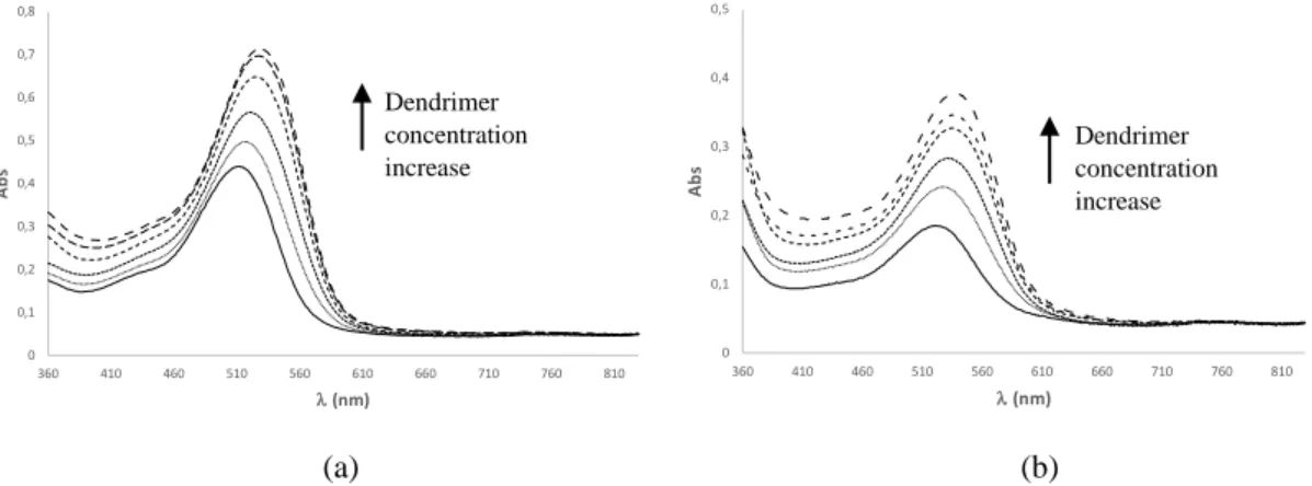
173
spectra were taken in a Thermo Scientific Evolution Array UV−visible spectrophotometer
174
from 360 to 830 nm in a 1 cm path length cell. The absorbance values variations (Abs) as
175
a function of dendrimer concentration [L0] can be expressed by equation (5), previously 176
developed by similar host-guest interactions[33]:
177 178 𝐴𝑏𝑠 = 𝐴𝑏𝑠0+ ∆𝐴𝑏𝑠 𝑛𝐾[𝐿0] 1+𝑛𝐾[𝐿0] (5) 179 180
where nK is the apparent binding constant, n is the number of nonspecific binding sites
181
of the dendrimer where anthocyanin can bind, and [L0] the total concentration of the 182
dendrimer added. The data could be fitted by equation (5) using the nonlinear
least-183
squares method and the Solver function from Microsoft Excel:
184 185 NMR 186
Accepted
Manuscript
For the NMR studies, a 0.124 mM solution of cy3glc was prepared in D2O and the pH 187
was adjusted to 1 (pD 1.4) and transferred into 5 mm NMR tubes. Sodium
trimethylsilyl-188
[2,2,3,3-d4]-propionate (TSP, 5 L, 0.05 mM in D2O) was used as an internal standard 189
for chemical shift measurements. Successive volumes of a dendrimer stock solution in
190
D2O (16.2 M) were added to the NMR tube to obtain different anthocyanin:dendrimer 191
molar ratios during the titration. pH measurements were made in a pH-meter WTW pH
192
320 fitted with a standard glass Crison® 5209 electrode. The calibration was made with
193
standard aqueous buffers at pH 4.0 and pH 1.0 from Crison®. All 1H NMR spectra were
194
recorded at 298.2K on a Bruker Avance III 400 HD spectrometer, operating at 400.14
195
MHz, equipped with 5 mm PADUL and pulse gradient units, capable of producing
196
magnetic field pulsed gradients in the z-direction of 50 G/cm. The measurements were
197
done with standard Bruker pulse sequences at 298.2 K. 1H NMR experiments were
198
performed with water suppression using excitation sculpting with gradients, acquisition
199
time 2.56 s, relaxation delay 1s and 64 transients of a spectral width of 6410.26 Hz were
200
collected into 32 K time domain points,
201 202
NMR data analysis
203
For titration experiments, chemical shift variations (obs) of some cy3glc protons as a
204
function of cy3glc/dendrimer molar ratio can be expressed through equation (6)[16]: 205 206 ∆𝛿𝑜𝑏𝑠 = ∆𝛿𝑚𝑎𝑥 2 {(1 + 1 𝐾[𝐺𝑢𝑒𝑠𝑡]+ 𝑛[𝐷𝑒𝑛𝑑] [𝐺𝑢𝑒𝑠𝑡]) − [(1 + 1 𝐾[𝐺𝑢𝑒𝑠𝑡]+ 𝑛[𝐷𝑒𝑛𝑑] [𝐺𝑢𝑒𝑠𝑡]) 2 −4𝑛[𝐷𝑒𝑛𝑑][𝐺𝑢𝑒𝑠𝑡]] 1/2 } (6) 207 208
Δδmax is the maximum chemical shift variation of the guest molecule in NMR titration 209
experiment K is the binding affinity or association constant The number of binding sites
210
(n) was obtained by fitting the titration data with equation (6) using a nonlinear
least-211
squares method within the software program Microsoft Excel.
212 213
Results and discussion 214
UV-Visible spectroscopy.
215
Preliminary studies to evaluate possible interactions between cy3glc and three types of
216
GATG-based dendrimers, namely cationic amine 3[G4]-NH2·HCl, anionic sulfated 217
3[G4]-OSO3Na and neutral triethylene glycol 3[G4]-OH (each decorate with 162 terminal 218
residues) were performed in aqueous solutions at pH 1 and 3.5. The results showed only
219
Accepted
a significant and interesting stabilization and intensification of the cy3glc color obtained
220
in the presence of the anionic dendrimer at both pH values rather than with the other two
221
dendrimers (supporting information). Bearing this, the interaction of mv3glc and cy3glc
222
with the dendrimer 3[G4]-OSO3Na was studied in detail at pH 1 through UV-Vis 223
spectroscopy by increasing the dendrimer concentration over a solution of anthocyanin at
224
a fixed concentration. Figure 2 illustrates a bathochromic shift with the successive
225
addition of small amounts of dendrimer solution. Upon the binding to the dendrimer, the
226
absorption spectrum of the flavylium cation undergoes a red-shift of ca. 10 nm suggesting
227
the incorporation of the anthocyanin in a microenvironment with lower polarity than
228
water. Similar effects were observed upon binding of flavylium cations to anionic
229
micelles, lignin and cucurbiturils.[33-35] The anionic character of the terminal sulfate
230
groups in 3[G4]-OSO3Na are expected to stabilize the flavylium cation of the anthocyanin 231
by Coulombic interactions. Furthermore, hydrophobic interactions between the 39 gallic
232
acid and 81 triazol residues of the dendrimer and the aromatic framework of the flavylium
233
cation might help stabilizing the interaction. From the absorbance taken at the maximum
234
wavelength of the free pigments as a function of the concentration of the dendrimer and
235
applying the fitting procedures, it was possible to estimate the apparent binding constants
236
of the complexes as nK = 207183 M-1 and nK = 52424 M-1for cy3glc and mv3glc,
237
respectively (Figures 2c and 2d).
238 239 240 241 242 243 244 245 246 (a) (b) 247 248 249 250 251 252 253 254 0,33 0,35 0,37 0 2 4 6 8 10 12 14 A bs (5 1 9 nm ) nK = 52424 M-1 0 0.05 0.1 0.15 0.2 0.25 0.3 0.35 0.4 360 410 460 510 560 610 660 710 760 810 Abs l(nm) 0 0.1 0.2 0.3 0.4 0.5 0.6 0.7 0.8 360 410 460 510 560 610 660 710 760 810 Abs l (nm) Dendrimer concentration increase Dendrimer concentration increase 0.55 0.57 0.59 0.61 0.63 0.65 0.67 0.69 0 2 4 6 8 10 12 14 Abs (5 1 0 n m) nK = 207183 M-1
Accepted
Manuscript
255 256
(c) (d)
257
Figure 2. (a) UV-Visible spectra of cy3glc (19.8 M) and cy3glc (19.8 M) with increasing concentrations
258
of 3[G4]-OSO3Na from 0.59 to 12 M at pH 1 (0.1 M HCl); (b) the same for mv3glc (at 10.3 M); (c)
259
fitting of the absorbance as a function of the concentration of 3[G4]-OSO3Na for cy3glc-dendrimer complex
260
using equation (5) with an estimated error 10 %; (d) the same for mv3glc-dendrimer complex.
261 262
Comparing the two values obtained it can be concluded that cy3glc displays a higher
263
binding affinity to the dendrimer than mv3glc, probably because the presence of the
264
catechol group in cy3glc leads to the establishment of an additional H-bond and/or a
265
stronger bifurcated H-bond. The ability of the dendrimer to interact with anthocyanins
266
was also evaluated at pH 3.5 by means of UV-Vis spectroscopy by adding increasing
267
dendrimer concentrations to a fixed concentration of anthocyanin solution. At this pH,
268
the flavylium and hemiketal forms are the main present species (e.g. pKh oenin = 2.70 269
±0.01)[36, 37], and hence water and the copigment are in competition for the flavylium
270
cation. Usually, in the copigmenation phenomena an increase of the absorbance intensity
271
and a wavelength redshift of the pigment (hyperchromic and bathochromic effects,
272
respectively) occur as a result of the stabilization of the flavylium cation and consequent
273
increase of its mole fraction at the expenses of the neutral species. From Figure 3, it was
274
possible to observe these two effects in the UV-Vis spectra of both anthocyanins with the
275
addition of increasing concentrations of dendrimer.
276
277
(a) (b)
278
Figure 3. (a) UV-Visible spectra of free cy3glc (19.8 M) and cy3glc (19.8 M) with increasing
279
concentrations of 3[G4]-OSO3Na (5, 10, 20, 30 and 40 M) at pH 3.5 (0.2 M citrate buffer); (b) the same
280
for mv3glc (at 10.3 M).
281 282
For the highest concentration of dendrimer (40 M), an increase of the absorption maxima
283
of the anthocyanins was observed compared to that of the free pigments: 29 % for cy3glc
284 0 0,1 0,2 0,3 0,4 0,5 0,6 0,7 0,8 360 410 460 510 560 610 660 710 760 810 A b s l(nm) 0 0,1 0,2 0,3 0,4 0,5 360 410 460 510 560 610 660 710 760 810 A b s l(nm) Dendrimer concentration increase Dendrimer concentration increase
Accepted
Manuscript
and 20 % for mv3glc. This is in good agreement with the higher binding affinity of cy3glc
285
towards the dendrimer, contributing to its great color stabilization.
286 287
NMR spectroscopy.
288
The variations in proton chemical shifts of hosts and guests in 1H NMR can be used to
289
investigate host−guest interactions. The decrease of electron density around a nucleus
290
causes an increase of chemical shift (downfield shift or deshielding), while the increase
291
of electron density leads to a decrease of chemical shift (upfield shift or shielding)[38].
292
When cy3glc was titrated with dendrimer in D2O at pD 1.4, significant downfield shifts 293
of all aromatic protons of cy3glc were observed (Figure 4). This result suggests the
294
formation of ionic pairs between the polyanionic dendrimer and flavylium cation of
295
cy3glc [39-41]. Analysis of the 1H NMR titration data with the proposed equation (2)
296
could be used to estimate the binding parameters of the complex such as the number of
297
binding sites (n), the maximum chemical shift (max) change and the association
298
constant (K) at pH 1.[42-46] To this end, the obs was plotted against the guest/host molar 299
ratio and the data was fitted using equation (16). From the fitting it was possible to
300
achieve a max=0.0365 and to estimate the number of binding sites (n) of the guest to
301
dendrimer around 295, which means that approximately two molecules of flavylium
302
cation of cy3glc could bind to each terminal sulfate group of the dendrimer bearing the
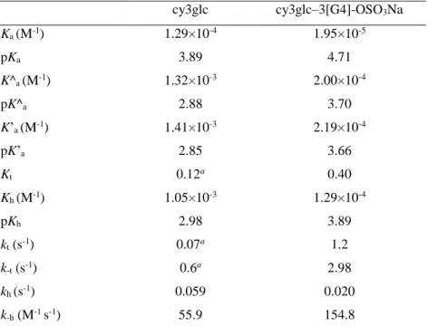
303
fact that dendrimer has 162 termini sulfate groups. The flavylium cation species should
304
be located at the dendrimer periphery, forming reversibly contact ion pairs with the
305
sulfate group in which the anionic charge should be delocalized by the three oxygen
306
atoms of the sulfate group as it has been suggested in literature for similar host-guest
307
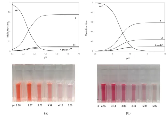
systems (e.g. acetylcholine and benzoate-terminal dendrimers)[41, 42] (Figure 5). From
308
the number of binding sites, the association constant (K) could be estimated to be
309 around 700 M-1. 310 311 312 313 314 315 316 317 0,E+00 5,E-03 1,E-02 2,E-02 2,E-02 3,E-02 3,E-02 4,E-02 4,E-02 0,00 500,00 1000,00 1500,00 2000,00 2500,00 o b s (p p m) [Cy3glc]/[Dendrimer]
Accepted
Manuscript
318
Figure 4. 1H spectra region (9.0–6.5 ppm) of the flavylium cation of cy3glc at initial concentration of 124
319
M with increasing dendrimer concentrations from 0 M (bottom) to 0.47 M (top) recorded in D2O at pD
320
1.4. Representation of the chemical shift variations of H-4C of cy3glc (obs) in function of the guest/host
321
molar ratio (in upper right). Fitting was achieved with equation (6) with an estimated error 10 %.
322 323
324
Figure 5. Schematic representation of the interaction between two cy3glc molecules (flavylium cation)
325
and each sulfate-terminated residue of the 3[G4]-OSO3Na dendrimer.
326 327
Equilibrium and rate constants of cy3glc-3[G4]-OSO3Nacomplex
328
The multistate of chemical reactions showed in Scheme 1, can be conveniently
329
investigated through the pH jump methodology.[47] The direct pH jumps are defined as
330
the addition of base to equilibrated solutions of the flavylium cation while reverse pH
331
H-4C
H-6’B H-2’B H-5’B H-6A H-8A
Accepted
jumps result from addition of acid to equilibrated solutions at moderately acidic to neutral
332
pHs and the relaxation process is follow towards the new equilibrium using spectroscopic
333
techniques. Stopped-flow is a crucial tool to follow the kinetic processes that take place
334
in sub-minutes time scale. After a direct pH jump, the flavylium cation (AH+) transfer a
335
proton to water giving rise to quinoidal base A, which is by far the fastest kinetic step of
336
the multistate, equation (7).
337 338 𝑘1𝑑 = 𝑘𝑎+ 𝑘𝑎[𝐻+] (7) 339 340 341
The second kinetic step is triggered by hydration of the electrophilic flavylium cation
342
followed by the ring opening-closure reaction (tautomerization). The former process is
343
much slower and consequently it is the rate determining step, equation (8).
344 345 𝑘2𝑑= [𝐻+] [𝐻+]+𝐾𝑎𝑘ℎ+ 1 1+𝐾𝑡𝑘−ℎ[𝐻 +] (8) 346 347
At this point the system reaches the so-called pseudo-equilibrium (K^a) because the
348
formation of Ct is much slower, equation (9):
349 350 AH+ + H 2O CB + H3O+ K^a=Ka + Kh+ KhKt (9) 351 with [CB] = [A] + [B] + [Cc] 352 353
After the slowest step of the multistate (isomerization reaction) the system relaxes to the
354
equilibrium according to equation (10)
355 356 𝑘3𝑑= 𝐾ℎ𝐾𝑡 [𝐻+]+𝐾 𝑎^𝑘𝑖 + 𝑘−𝑖 (10) 357 358
The equilibrium of the system is defined by apparent acidic constant (K’a), equation (11):
359 [2, 48-50] 360 361 AH+ + H 2O CB + H3O+ K’a=Ka + Kh+ KhKt + KhKtKi (11) 362 with [CB] = [A] + [B] + [Cc] + [Ct] 363 364 K’a K^a
Accepted
Manuscript
From the equations (1-4, 9 and 11) the mole fraction of each species in function of pH 365 can be deduced: 366 [AH+] = [H+] [H+]+𝐾 a′; (12) 367 [A] = 𝐾a [H+]+𝐾 a′; (13) 368 [B] = 𝐾h [H+]+𝐾 a′; (14) 369 [Cc] = 𝐾h𝐾t [H+]+𝐾 a′; (15) 370 [Ct] = 𝐾h𝐾t𝐾i [H+]+𝐾 a′ (16) 371 372 Direct pH jumps 373
The chemical equilibria network of cy3glc in the presence of the sulfated dendrimer at
374
fixed concentration was studied by UV-Vis spectroscopy through direct pH jumps (from
375
equilibrated acidic solutions to less acidic pH). After a pH jump, the first kinetic process
376
is due to the proton transfer reaction in the time scale of sub-milliseconds (equation 1) in
377
which no other process occurs and, therefore, the flavylium cation (AH+) and the
378
quinoidal base (A) are the only species formed. Then, the spectral variations were
379
monitored with time until the system reached the pseudo-equilibrium (AH+/A, B, and Cc 380
are the species in equilibrium). Fitting the absorbance decay of the pair flavylium
381
cation/quinoidal base as a function of pH allowed the determination of pK^a = 3.70. 382
Finally, the system reaches the equilibrium due to the formation of trans-chalcone Ct 383
which is the slowest step of the multistate. The UV-Vis spectra of the solutions were
384
recorded again and the fitting of experimental data allowed the determination of pK’a = 385
3.66 (Figure 6). The small difference between the pK^a and pK’a accounts for a low mole 386
fraction of Ct in the final equilibrium.
387 388 389 390 391 392 393 394 395 396
Accepted
Manuscript
397
398
Figure 6. Spectral variations of cy3glc (19.8 M) in the presence of 3[G4]-OSO3Na (26 M) after a direct
399
pH jump at the equilibrium, pK’a=3.66. Inset: Fitting of the absorbance values at 530 nm as a function of
400
pH.
401 402
Normally the pKa is determined by stopped-flow technique. Alternatively, the pKa can 403
also be estimated through the pH jump from 1 to 6.06, where the hydration is sufficiently
404
slow, following the absorbance decay of the quinoidal base (Figure 7). From the ratio
405
between Af and Ai and through equation (13) the pKa was determined to be 4.71. 406
The kinetic process of this direct pH jump is showed in inset of Figure 7, which illustrates
407
the second and third kinetic processes described in the introduction (Scheme 1 and eqs.
408
2-4). The initial absorbance is due to the quinoidal base which formation occurs during
409
the mixing time of the base addition in the direct pH jump. The faster decay is coherent
410
with the second kinetic process (hydration and tautomerization reaction, k2) where 411
hydration is the rate-determining step. The third kinetic process is due to the slowest step
412
of the equilibrium (isomerization reaction, k3) and can be observed by the absorbance 413
increase at 350 nm due to the formation of trans-chalcone. Fitting the absorbance values
414
as a function of time allowed determining the observed rate constants: k2 = 4.3×10⁻³ s-1; 415 k3 = 2.5×10⁻⁴ s-1. 416 417 -0,1 0,1 0,3 0,5 0,7 0,9 300 400 500 600 700 800 A l(nm) 0,00 0,20 0,40 0,60 0,80 1,00 2 3 4 5 6 A (5 3 0 n m) pH pH 2.46 pH 6.06
Accepted
Manuscript
418
Figure 7. Spectral variations after a direct pH jump from pH=1 to pH=6.06 of cy3glc (19.8 M) in the
419
presence of 3[G4]-OSO3Na (26 M). Inset: variation of absorbance at 350 nm as a function of time showing
420
the second and third kinetic processes.
421 422
Then, the second observed rate constant obtained for each pH values was fitted by
423
equation (6) that accounts for the pH-dependence of the rate-limiting hydration process
424
of the flavylium cation to reach the pseudo-equilibrium. By fitting the data presented in
425
Figure 8, it was possible to determine the values of kh = 0.020 s-1 and k-h /(1+Kt) = 110.6 426
M-1 s-1.
427 428
429
Figure 8. Representation of the observed rate constant of the second kinetic process as a function of pH.
430 431
Reverse pH jump
432
To determine the dehydration rate constant, the tautomerization equilibrium constant Kt 433
and by consequence, the thermodynamic hydration constant (Kh), it was necessary to 434 -0,1 0 0,1 0,2 0,3 0,4 0,5 0,6 0,7 0,8 300 400 500 600 700 800 A l(nm) 0 0,05 0,1 0,15 0,2 0,25 0,3 0,35 0,4 0,45 2,3 2,8 3,3 3,8 4,3 4,8 5,3 5,8 6,3 k2 (s -1) pH 0,18 0,2 0,22 0,24 0,26 0,28 0 1000 2000 3000 4000 5000 6000 A (3 50 n m) time (s) k2=4.3 10-3s-1 k3=2.5 10-4s-1
Accepted
Manuscript
carry out a reverse pH jump experiment monitored by stopped-flow from an equilibrated
435
solution at pH=5.58 to pH=0.98 (Figure 9a). Immediately after the addition of acid, all
436
quinoidal base initially at pH=5.58 was rapidly converted into the flavylium cation during
437
the mixing time of the stopped-flow experiment. The faster kinetic step with k1=10.4 s-1 438
corresponds to the conversion of the hemiketal (B) initially at pH=5.58 into the flavylium
439
cation (AH+), benefiting from the fact that at sufficiently low pH the hydration is faster
440
than the tautomerization (change of regime).[51] The second kinetic process corresponds
441
to the slower formation of more AH+ from Cc, through B (k2=2.98 s-1). From this 442
experiment it was also possible to determine k-t which is equal to the second kinetic rate, 443
k2. These two kinetic constants were calculated by fitting the absorbance values at 522 444
nm as a function of time considering a biexponential process (Figure 9b). Kt was 445
determined from the ratio of amplitudes of the second process divided by the first
446 process (𝐾𝑡= 𝐶𝑐 𝐵 = 0.4) and kt=1.2 s -1 because 𝐾 𝑡 = 𝑘𝑡
𝑘−𝑡. With Kt value in hand, it was 447
possible do determine k-h=154.8 s-1. The hydration equilibrium constant, Kh=1.29×10⁻⁴ 448
M-1 was then obtained from the ratio of both kinetics constants (𝐾ℎ = 𝑘ℎ
𝑘−ℎ). 449 450 451 452 453 454 455 456 457
Figure 9. (a) Spectral variations after a reverse pH jump from an equilibrated solution of cy3glc (19.8 M)
458
in the presence of 3[G4]-OSO3Na (26 M) from pH=5.58 to 0.98. (b) Fitting of the kinetic processes.
459 460 461
All the kinetic and thermodynamic parameters determined for cy3glc and
cy3glc-462
dendrimer complex are resumed in Table 2. Comparing the values obtained it can be
463
concluded that the presence of the dendrimer has a great effect on the chemical equilibria
464
network of anthocyanins. The pK’a is increased in 0.82 pH units, which indicates a higher 465
stabilization of the colored flavylium species. Moreover, the flavylium cation is more
466
stabilized than the hemiketal form in the presence of the dendrimer due essentially to a
467 0,00 0,10 0,20 0,30 0,40 0,50 300 350 400 450 500 550 600 650 700 A l (nm) 0,00 0,05 0,10 0,15 0,20 0,25 0,30 0,35 0,40 0,0 1,0 2,0 3,0 4,0 5,0 A (5 2 2 n m) time (s) Cc B AH+/A k2=2.98 s-1 k1=10.4 s-1
Accepted
Manuscript
faster dehydration and lower hydration rate constants compared to the absence of
468
dendrimer as observed for other analogous systems.[52] Hence, the hydration constant 469
increases in one pH unit in the presence of the dendrimer. Moreover, for the
cy3glc-470
dendrimer complex, Kt is increased which reveals a preferential interaction of the cis-471
chalcone for the host than the hemiketal species. Finally, the mole fraction distribution of
472
cy3glc 19.8 M in the absence of the dendrimer (Figure 10a) was compared with the one
473
of cy3glc 19.8 M in the presence of dendrimer 26 M (Figure 10b). It was possible to
474
observe that in the presence of the dendrimer the mole fraction of the hemiketal form
475
decreased significantly between pH 4-7.5 (75 % to 58 %), accompanied with an increase
476
of the flavylium cation.
477 478
Table 2. Equilibrium and rate constants obtained by UV–Vis spectroscopy for cy3glc (19.8 M) and for
479
cy3glc (19.8 M) in the presence of 3[G4]-OSO3Na (26 M). Estimated error 10%.
480 cy3glc cy3glc–3[G4]-OSO3Na Ka (M-1) 1.29×10⁻⁴ 1.95×10⁻⁵ pKa 3.89 4.71 K^a (M-1) 1.32×10⁻³ 2.00×10⁻⁴ pK^a 2.88 3.70 K’a (M-1) 1.41×10⁻³ 2.19×10⁻⁴ pK’a 2.85 3.66 Kt 0.12a 0.40 Kh (M-1) 1.05×10⁻³ 1.29×10⁻⁴ pKh 2.98 3.89 kt (s-1) 0.07a 1.2 k-t (s-1) 0.6a 2.98 kh (s-1) 0.059 0.020 k-h (M-1 s-1) 55.9 154.8
Ka, acidity constant; K^a, pseudo-equilibrium constant; K’a, equilibrium constant; Kt, tautomerization
481
constant; Kh, hydration constant; kt, rate of the tautomerization reaction; k-t, rate of the reserve
482
tautomerization reaction; kh, rate of the hydration reaction; k-h, rate of the dehydration reaction. aobtained
483
from Leydet et al., 2012.[53]
484 485 486
Accepted
487 488 489 490 491 492 493 494 (a) (b) 495
Figure 10. (a) Mole fraction distribution of cy3glc 19.8 M in function of pH and (b) the same for cy3glc
496
19.8 M in the presence of dendrimer 26 M.
497 498
Conclusions 499
We have determined the influence of a GATG-based polyanionic dendrimer decorated
500
with 162 sulfate groups on the pH-equilibria network of anthocyanins and studied at a
501
molecular level the non-covalent interactions within the host-guest system by UV-Vis,
502
stopped-flow and NMR techniques. Overall, it can be concluded that the dendrimer exerts
503
a great stabilization effect on the thermodynamic and kinetic parameters of cy3glc,
504
increasing its pKh and pKa in circa one pH unit. By NMR, it was verified that the red 505
flavylium cation is strongly shielded by the host due to the formation of ionic pairs and
506
the number of binding sites and association constant were determined. The set of results
507
obtained for the color stabilization of anthocyanins and respective tuning in function of
508
pH using this dendrimer open novel applications to be explored such as
anthocyanin-509
based sensors for biomedical devices and smart packaging solutions.
510 511
Acknowledgments 512
The authors thank Dr. Mariana Andrade for the NMR analysis. This research was
513
supported by a research project grant (PTDC/OCE-ETA/31250/2017) with financial
514
support from FCT/MCTES through national funds and co-financed by FEDER, under the
515 0 0,2 0,4 0,6 0,8 1 1,5 2,5 3,5 4,5 5,5 6,5 7,5 Mo le Fr a ct io n pH AH+ B Cc A and Ct 0 0,2 0,4 0,6 0,8 1 1,5 3 4,5 6 7,5 Mo le F ra ct io n pH AH+ B Cc A and Ct pH 1.98 2.37 3.06 3.34 4.12 5.69 pH 2.46 3.14 3.88 4.41 5.07 6.06
Accepted
Manuscript
Partnership Agreement PT2020 (UID/QUI/50006/2019 -
516
POCI/01/0145/FEDER/007265). Financial support was also obtained from the Spanish
517
Ministry of Science, Innovation and Universities (CTQ2015-69021-R and
RTI2018-518
102212-B-I00), the Xunta de Galicia (GRC2014/040, ED431C 2018/30, and Centro
519
Singular de Investigación de Galicia Accreditation 2016-2019, ED431G/09) and the
520
European Union (European Regional Development Fund-ERDF). Luís Cruz gratefully
521
acknowledges the research FCT contract.
522
523 524
[1] R. Brouillard, G. A. Iacobucci and J. G. Sweeny, J. Am. Chem. Soc. 1982, 104, 7585-7590. 525
[2] F. Pina, M. J. Melo, C. A. T. Laia, A. J. Parola and J. C. Lima, Chem. Soc. Rev. 2012, 41, 869-908. 526
[3] A. Fernandes, N. F. Bras, N. Mateus and V. de Freitas, New J. Chem. 2015, 39, 2602-2611. 527
[4] F. Di Meo, J. C. S. Garcia, O. Dangles and P. Trouillas, J. Chem. Theory Comput. 2012, 8, 2034-528
2043. 529
[5] M. T. Escribano-Bailon and C. Santos-Buelga, Curr. Org. Chem. 2012, 16, 715-723. 530
[6] F. He, N. N. Liang, L. Mu, Q. H. Pan, J. Wang, M. J. Reeves and C. Q. Duan, Molecules 2012, 531
17, 1571-1601.
532
[7] J. Muller-Maatsch, L. Bechtold, R. M. Schweiggert and R. Carle, Food Chem. 2016, 213, 625-533
634. 534
[8] T. Iwashina, Nat. Prod. Commun. 2015, 10, 529-544. 535
[9] B. J. Qian, J. H. Liu, S. J. Zhao, J. X. Cai and P. Jing, Food Chem. 2017, 228, 526-532. 536
[10] L. Cruz, N. F. Brás, N. Teixeira, N. Mateus, M. J. Ramos, O. Dangles and V. De Freitas, J. Agric. 537
Food. Chem. 2010, 58, 3159-3166.
538
[11] N. Teixeira, L. Cruz, N. F. Brás, N. Mateus, M. J. Ramos and V. de Freitas, J. Agric. Food. Chem. 539
2013, 61, 6942-6948.
540
[12] C. Houbiers, J. C. Lima, A. L. Macanita and H. Santos, J. Phys. Chem. B 1998, 102, 3578-3585. 541
[13] P. Trouillas, J. C. Sancho-García, V. De Freitas, J. Gierschner, M. Otyepka and O. Dangles, 542
Chem. Rev. 2016, 116, 4937-4982.
543
[14] L. Cruz, V. C. Fernandes, P. Araújo, N. Mateus and V. de Freitas, Food Chem. 2015, 174, 480-544
486. 545
[15] L. Cruz, I. Fernandes, M. Guimaraes, V. de Freitas and N. Mateus, Food Funct. 2016, 7, 2754-546
2762. 547
[16] J. Hu, T. Xu and Y. Cheng, Chem. Rev. 2012, 112, 3856-3891. 548
[17] J. Mendoza, N. Basílio, O. Dangles, N. Mora, S. Al Bittar and F. Pina, Dyes Pigm. 2017, 143, 549
479-487. 550
[18] A. Fernandes, G. Ivanova, N. F. Bras, N. Mateus, M. J. Ramos, M. Rangel and V. de Freitas, 551
Carbohydr. Polym. 2014, 102, 269-277.
552
[19] R. Gomes, R. Q. Albuquerque, F. Pina, A. J. Parola and L. De Cola, Photochem. Photobiol. Sci. 553
2010, 9, 991-995.
554
[20] O. Yesil-Celiktas, C. Pala, E. O. Cetin-Uyanikgil and C. Sevimli-Gur, Anal. Biochem. 2017, 519, 555
1-7. 556
[21] D. Astruc, E. Boisselier and C. Ornelas, Chem. Rev. 2010, 110, 1857-1959. 557
Accepted
[22] A.-M. Caminade, C.-O. Turrin, R. Laurent, A. Ouali and B. Delavaux-Nicot, Dendrimers: 558
towards catalytic, material and biomedical uses, John Wiley & Sons, Ltd: Chichester, UK, 2011,
559 p. 560
[23] R. Jevprasesphant, J. Penny, R. Jalal, D. Attwood, N. B. McKeown and A. D’Emanuele, Int. J. 561
Pharm. 2003, 252, 263-266.
562
[24] N. Malik, R. Wiwattanapatapee, R. Klopsch, K. Lorenz, H. Frey, J. W. Weener, E. W. Meijer, 563
W. Paulus and R. Duncan, J. Controlled Release 2000, 65, 133-148. 564
[25] A. Sousa‐Herves, D. Gröger, M. Calderón, E. Fernandez‐Megia and R. Haag in Anionic 565
Dendritic Polymers for Biomedical Applications, The Royal Society of Chemistry, 2013, pp. 56-72.
566
[26] A. Sousa-Herves, R. Novoa-Carballal, R. Riguera and E. Fernandez-Megia, The AAPS Journal 567
2014, 16, 948-961.
568
[27] V. Shukla, G. Kandeepan, M. R. Vishnuraj and A. Soni, Agric. Res. 2016, 5, 205-209. 569
[28] W. F. Küster and A. Thiel, Tabelle per le analisi chimiche e chimico-fisiche. 12 ed.; Hoepli: 570
Milano, 1982, p.
571
[29] S. P. Amaral, M. H. Tawara, M. Fernandez-Villamarin, E. Borrajo, J. Martínez-Costas, A. Vidal, 572
R. Riguera and E. Fernandez-Megia, Angew. Chem. Int. Ed. 2018, 57, 5273-5277. 573
[30] E. Fernandez-Megia, J. Correa, I. Rodríguez-Meizoso and R. Riguera, Macromolecules 2006, 574
39, 2113-2120.
575
[31] J. Pissarra, N. Mateus, J. C. Rivas-Gonzalo, C. Santos-Buelga and V. De Freitas, J. Food Sci. 576
2003, 68, 476-481.
577
[32] M. Guimarães, N. Mateus, V. de Freitas and L. Cruz, J. Agric. Food. Chem. 2018, 66, 10003-578
10010. 579
[33] P. Araújo, N. Basílio, A. Fernandes, N. Mateus, V. de Freitas, F. Pina and J. Oliveira, J. Agric. 580
Food. Chem. 2018, 66, 6382-6387.
581
[34] N. Basílio and F. Pina, Chemphyschem 2014, 15, 2295-2302. 582
[35] J. C. Lima, C. Vautier-Giongo, A. Lopes, E. Melo, F. H. Quina and A. L. Maçanita, J. Phys. Chem. 583
A 2002, 106, 5851-5859.
584
[36] O. Dangles and H. Elhajji, Helv. Chim. Acta 1994, 77, 1595-1610. 585
[37] C. Malien-Aubert, O. Dangles and M. J. Amiot, J. Agric. Food. Chem. 2002, 50, 3299-3305. 586
[38] C. Slichter, P., Principles of Magnetic Resonance; Springer, NewYork, 2010, p. 587
[39] J. Hu, Y. Cheng, Y. Ma, Q. Wu and T. Xu, J. Phys. Chem. B 2009, 113, 64-74. 588
[40] J. Hu, Y. Cheng, Q. Wu, L. Zhao and T. Xu, J. Phys. Chem. B 2009, 113, 10650-10659. 589
[41] C. Ornelas, E. Boisselier, V. Martinez, I. Pianet, J. Ruiz Aranzaes and D. Astruc, Chem. 590
Commun. 2007, 5093-5095.
591
[42] E. Boisselier, C. Ornelas, I. Pianet, J. R. Aranzaes and D. Astruc, Chem. Eur. J. 2008, 14, 5577-592
5587. 593
[43] M. A. C. Broeren, B. F. M. de Waal, M. H. P. van Genderen, H. M. H. F. Sanders, G. Fytas and 594
E. W. Meijer, J. Am. Chem. Soc. 2005, 127, 10334-10343. 595
[44] X.-D. Xu, H.-B. Yang, Y.-R. Zheng, K. Ghosh, M. M. Lyndon, D. C. Muddiman and P. J. Stang, 596
J. Org. Chem. 2010, 75, 7373-7380.
597
[45] A. J. Charlton, N. J. Baxter, M. L. Khan, A. J. G. Moir, E. Haslam, A. P. Davies and M. P. 598
Williamson, J. Agric. Food. Chem. 2002, 50, 1593-1601. 599
[46] C. Simon, K. Barathieu, M. Laguerre, J.-M. Schmitter, E. Fouquet, I. Pianet and E. J. Dufourc, 600
Biochemistry 2003, 42, 10385-10395.
601
[47] F. Pina, J. Agric. Food. Chem. 2014, 62, 6885-6897. 602
[48] R. Brouillard and B. Delaporte, J. Am. Chem. Soc. 1977, 99, 8461-8468. 603
[49] R. Brouillard, B. Delaporte and J. E. Dubois, J. Am. Chem. Soc. 1978, 100, 6202-6205. 604
[50] R. Brouillard and J. Lang, Can. J. Chem. 1990, 68, 755-761. 605
[51] F. Pina, Dyes Pigm. 2014, 102, 308-314. 606
[52] J. Mendoza, N. Basílio, F. Pina, T. Kondo and K. Yoshida, J. Phys. Chem. B 2018, 122, 4982-607
4992. 608
Accepted
[53] Y. Leydet, R. Gavara, V. Petrov, A. M. Diniz, A. Jorge Parola, J. C. Lima and F. Pina, 609 Phytochemistry 2012, 83, 125-135. 610 611
Accepted
Manuscript
Graphical Abstract 612
613
![Figure 1. Structures of 3[G4]-OSO 3 Na carrying 162 peripheral sodium sulfate groups and anthocyanins 104 (flavylium salts)](https://thumb-eu.123doks.com/thumbv2/123dok_br/15148590.1012610/5.892.98.770.143.586/figure-structures-carrying-peripheral-sodium-sulfate-anthocyanins-flavylium.webp)

![Figure 6. Spectral variations of cy3glc (19.8 M) in the presence of 3[G4]-OSO 3 Na (26 M) after a direct 399](https://thumb-eu.123doks.com/thumbv2/123dok_br/15148590.1012610/15.892.145.552.126.376/figure-spectral-variations-glc-presence-oso-na-direct.webp)