Dissertation presented to obtain the Ph.D degree in Cell Biology
Instituto de Tecnologia Química e Biológica António Xavier | Universidade Nova de Lisboa
Oeiras,
UNIVERSIDADENOVA
Sandra Raquel de Oliveira Tavares
The role of actin cytoskeleton downstream of the Src
oncogene in the earlier events of breast tumour
Sandra Raquel de Oliveira Tavares
Dissertation presented to obtain the Ph.D degree in Cell Biology
Instituto de Tecnologia Química e Biológica António Xavier | Universidade Nova de Lisboa
Oeiras, November, 2016
The role of actin cytoskeleton downstream of
the Src oncogene in the earlier events of
breast tumour progression
Declaração/ Declaration
Declaro que esta dissertação é o resultado do meu próprio trabalho desenvolvido entre Abril 2012 e Janeiro 2016 no laboratório da Dra. Florence Janody, Instituto Gulbenkian de Ciência, Oeiras, Portugal. Este doutoramento foi realizado no âmbito do Programa Gulbenkian de Doutoramento (edição 2011-2012). Os capítulos 2, 3 e 4 estão incluídos num manuscrito submetido para publicação com autoria de Sandra Tavares, André Vieira, Anna Verena Taubenberger, Nuno Pimpão Santos Martins; Catarina Brás-Pereira; Margarida Araújo, António Polónia, Joana Vaz, José Pereira Leal, Jochen Guck, Joana Paredes e Florence Janody.
I declare that this dissertation is a result of my own reserach carried out between
April 2012 e January 2016 in the laboratory of Dr. Florence Janody, Instituto Gulbenkian de Ciência, Oeiras, Portugal. Chapters 2, 3 and 4 are part of a manuscript submitted for publication authored by Sandra Tavares, André Vieira, Anna Verena Taubenberger, Nuno Pimpão Santos Martins; Catarina Brás-Pereira; Margarida Araújo, António Polónia, Joana Vaz, José Pereira Leal, Jochen Guck, Joana Paredes and Florence Janody.
Apoio Financeiro/ Financial Support
Esta dissertação teve o apoio financeiro da FCT, no âmbito do Quadro Comunitário de Apoio, bolsa de doutoramento #SFRH/BD/51884/2012, da Fundação Calouste Gulbenkian e Liga Portuguesa contra o Cancro/Pfizer.
This dissertation had the financial support from FCT through the Quadro Comunitário de Apoio, doctoral fellowship #SFRH/BD/51884/2012, Fundação Calouste Gulbenkian and Liga Portuguesa contra o Cancro/Pfizer.
Acknowledgements
What an exciting ride it was!
I guess that is due to the beauty and magic of IGC. There is so much more besides science in these corridors. So many smiles exchanged between people that the only thing they share is the passion for science. Knowledge is our aim and we are happy pursuing it. Well, at least that was how I felt during these 5 years. IGC, the geek wonderland. It didn´t take me long to realize that I had found “my place”.
The main person responsible for this was obviously my supervisor, Florence. You gave me exactly what I was looking for without even knowing it. I said this at the beginning and it is still true. Florence, you gave me the support I needed in tough moments. You were such a cool supervisor that you laughed at most of my jokes, even the ones that were not appropriate between supervisor and student (You really shouldn´t support this kind of behaviour!). Besides giving me the chance to do really cool science, I must say that you taught me something much more inspiring. You showed me the strength and commitment that it takes to stand up for our values. Fighting for our convictions is not just for young teenagers, it’s a feature of courageous adults.
The opportunity to work at the Institute was given to me by Thiago Carvalho and Prof. António Coutinho. I couldn´t thank you more and I hope I have fulfilled your expectations. I must acknowledge the directors of the programme, Élio, and the institute, Jonathan and Jorge. Élio, thank you for keeping our programme up and running; Jonathan, in your own way, you stimulated my most pro-active side and, I thank Jorge for the informal conversations, but most of all, for the consideration and respect that you always had for me, my ideas and my projects.
My dear Communication Team, I had so much fun collaborating with you! Because of you, I met amazing people and had extraordinary experiences
talking about science. If someone would tell me, back in 2011, that I would teach cell biology to Catarina Furtado, I would laugh so hard… For sure, Science sounds better with you!!!
Now, the friends…
Sofia, my ebony goddess, your generosity and devotion to IGC was to me the first evidence that I was in a special place. I wasn´t wrong! You are the glue that keeps us all together and under your embrace we thrive. Looking back and thinking in the way you took care of me gets me so emotional... Fomos primas, somos primas. Adoro-te muito.
There is no way I can thank in the right measure to Bea and Catarina. If I was so happy doing this PhD, is a lot due to you two. You showed me what a group is. It is really hard to thank you. You brought so much to my life that I have no idea how to thank you. For sure I thank you for the laughter and concern. But saying “Thank you for the Friendship” is too vague, “Thank you for the advices” is too short and “Thank you for the patience” is too obvious…
To my dear CCR’s: Carla, Paulo Duarte and Ana Rita Marques. It was fun to be part of your gang. I’ll never forget the advices in times of trouble. Listening to me during this last year was not easy, I know, for this reason, my dear Isa, a big thanks! To my other friends spread across the institute, Marta, Rita, Filipe, Barroso, Catarina Júlio, Inês and Cláudia Bispo; and to the ones that already left IGC: Rui Gardner, Sara, Alex, Ânia, and Cláudia Andrade. You were the walls of “my IGC”, with you I always felt at home.
André and Anna, thank you for being so much more than collaborators. Your help and commitment for this project were crucial. I was really fortunate for having you as partners in this journey. Thank you for teaching me so much and for making me feel like part of your teams! Sweet Margarida, it was a pleasure to work with someone so devoted and enthusiastic as you are.
To my closest family that has always believed in me. You gave me the motivation to do this PhD, even being so far away from you all. I have missed you tremendously, but you made it better with your kind words and support. Obrigada por tornarem a distância mais fácil de suportar.
Finally, to my beloved Ricardo. You have been my rock. Accepting to come with me, to the lab at midnight with a storm alert, it’s something that only a true partner in life would be up for. Can’t wait to be with you again.
If I loved you less, I might be able to talk about it more.
Sumário
A tumorigénese é um processo em que as células progressivamente adquirem alterações genéticas e epigenéticas em genes promotores e supressores tumorais, promovendo a vantagem de crescimento e proliferação das mesmas. Alterações posteriores poderão condenar essas células pré-malignas à transição a malignas com capacidades invasivas e metastáticas. Proto-oncogenes da família das cínases Src foram ligados à tumorigénese, e a sua expressão e sobre-activação foi descrita em cancro da mama. O aumento da actividade de Src foi associado a aumento de proliferação celular, sobrevivência, transição epitélio-mesênquima, migração, invasão e matástases. Apesar de Src ser bem conhecido no despoletar de motilidade de células cancerígenas, migração e invasão, pela regulação de actina filamentosa (F-actina); ainda não se sabe Src também utiliza a F-actina para promover proliferação e sobrevivência de células tumorais em estádios pré-malignos. Em 2013, o nosso laboratório demonstrou que Src promove a acumulação de F-actina apical no epitélio de Drosophila. Por sua vez, a F-actina limita a apoptose ou o crescimento descontrolado de tecido induzidos por Src.
O principal objectivo deste estudo foi investigar se a F-actina desempenha um papel subsequente à activação de Src, na promoção da aquisição de características de cancro da mama pré-maligno. De acordo com a esta hipótese, eu observei que a expressão de genes codificantes de proteínas que se ligam à actina está predominantemente afectada em lesões benignas da mama. Entre estas, Ena/VASP-like (EVL) e cinco outras proteínas também se encontravam
desreguladas numa linha celular indutível por tratamento com tamoxifeno, MCF10A-ER-Src. Adicionalmente, estas também se encontravam envolvidas no crescimento descontrolado de tecido induzido por Src, em Drosophila melanogaster. As minhas observações alegam que muito cedo na progressão do cancro da mama, Src afecta a expressão destas seis proteínas que se ligam à actina e que por sua vez, estas controlam o crescimento tumoral pela regulação de redes de F-actina especializadas (Capitulo 2).
Depois utilizando a linha celular de mama, humana e que através de tratamento com tamoxifeno possibilita a reconstituição in vitro, do percurso natural de cancro da mama induzido por Src (MCF10A-ER-Src), mostrei que a transformação celular pode ser dividida em duas fases. No início da transformação celular, baixos níveis de activação de Src promovem a polimerização transiente de fibras de actina polarizadas, levando a um aumento da rigidez celular e estando associados a capacidade de crescimento independente de factores de crescimento. Posteriormente, a fibras de actina são dissociadas, a células perdem rigidez e adquirem características malignas. Assim, enquanto a perda de rigidez celular permite a invasão celular/metástases, o meu trabalho revela que o enrijecimento das células mediado por fibras de actina poderá levar ao crescimento tumoral durante os estádios pré-malignos (Capitulo 3).
Por fim, eu observei que durante o início da transformação celular da linha celular MCF10A-Er-Src, Src induz o aumento transiente de EVL e a sua mobilização para as extremidades das fibras de actina dependentes de Src. A polarização destas fibras, realizada por EVL, é necessária para a promoção de rigidez celular, proliferação e progressão para o fenótipo maligno. Assim, eu mostro que níveis elevados de EVL são predominantemente encontrados em tumores de mama pré-malignos e estão associados ao sub-tipo molecular Luminal A (Capitulo 4).
Nesta tese, eu demonstro que a F-actina também desempenha um papel consequente à activação de Src, para a promoção da aquisição de características pré-malignas em cancro da mama. Em suma, as minhas observações estão de
acordo com um modelo no qual no inicio da progressão tumoral, baixos níveis de activação de Src promovem a agregação de fibras polarizadas e enrijecimento celular para sustentar a expansão de lesões precursoras de cancro da mama. Enquanto em estádios mais avançados, níveis elevados de Src reduzem a rigidez celular para permitir a invasão de células cancerígenas. Acredito, portanto, que estas observações poderão ter um impacto significativo no desenvolvimento de novas ferramentas de diagnóstico em estádios precoces de cancro da mama.
Summary
Tumorigenesis is a multistep process, by which cells acquire genetic and epigenetic alterations in oncogenes and tumour suppressor genes, which provide growth and/or survival advantage. Subsequent alterations may condemn these pre-malignant cells into malignant ones with invasive and metastatic abilities. Proto-oncogenes of the Src-family kinases have been linked to tumorigenesis and their over-expression and activation described in breast cancer. Increased Src activity has been associated with increased cell proliferation, survival, epithelial-mesenchymal transition (EMT), migration, invasion and metastasis. Although Src is well-known to trigger cancer cell mobility, migration and invasion, through regulation of filamentous actin (F-actin), whether it also uses F-actin to promote proliferation and survival of tumour cells at pre-malignant stages remains unknown. Our lab has demonstrated that Src promotes apical F-actin accumulation in Drosophila epithelia. In turn, F-actin limits Src-induced apoptosis or tissue overgrowth.
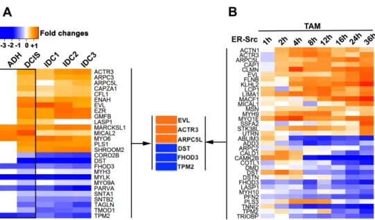
The main goal of this study was to investigate if F-actin plays a role downstream of Src activation to promote the acquisition of pre-malignant breast cancer features. In agreement with this hypothesis, I found that the expression of genes encoding for actin binding proteins (ABPs) is predominantly affected in benign breast lesions. Among those, Ena/VASP-like (EVL), in addition to 5 others ABPs were also misregulated in the Tamoxifen (TAM)-inducible MCF10A-ER-Src cell line, and involved in Src-induced tissue overgrowth in Drosophila
melanogaster. My observations argue that early during breast tumour progression, Src affects the expression of these 6 ABPs that control tumour growth via the regulation of specialized F-actin networks (Chapter 2).
Then, using the Src inducible mammary epithelial cell line MCF10A-ER-Src, which recapitulates the natural history of Src-induced breast cancer, I showed that transformation can be divided into two distinct phases. Early during cellular transformation, low Src induction promotes the transient polymerization of polarized acto-myosin stress fibres that correlate with an increase in cell stiffness and the acquisition of self-sufficiency in growth properties. Following this phase, the acto-myosin stress fibres disassemble, cell soften and acquire malignant features. Thus, while cell softening allows for cell invasion/metastasis, my work reveals that stress fibre-mediated cell stiffening could drive tumour growth during the pre-malignant stage (Chapter 3).
Lastly, I found that early during transformation of the TAM-induced MCF10A-Er-Src cell line, Src induces the transient upregulation of EVL and its mobilization to the tips of the transient Src-dependent acto-myosin stress fibres. Polarization of these fibres by EVL is required to promote cell stiffening, cell growth and the progression toward a malignant phenotype. Accordingly, I show that high EVL levels is predominantly found in pre-malignant breast tumour samples and correlate with the Luminal A breast molecular subtype (Chapter 4).
In this thesis, I demonstrate that F-actin also plays a role downstream of Src activation to drive the acquisition of pre-malignant breast cancer features. All together, my observations are in agreement with a model by which early during breast tumour progression, low Src activity could promote the assembly of polarized stress fibres and cell stiffening to sustain the expansion of cancer precursors, while at later stages of carcinogenesis, higher Src activity reduces stress fibre-mediated cell stiffening to allow for cancer cell invasion. I believe that these findings can have a significant impact for the development of new diagnostic tools in early stages of breast cancer.
List of Abbreviations
Actin Binding Proteins ABPs
Adenosine DiPhosphate ADP
Adenosine Triphosphate ATP
Adherens Junctions AJ
Aldehyde dehydrogenase ALDH
Atomic Force Microscopy AFM
Atypical Ductal Hyperplasia ADH
Bromodeoxyuridine BrdU
Cancer Stem Cell CSC
c-Jun N-terminal kinase c-JNK
Crk-Associated Substrate CAS
C-terminal Src kinase CSK
Cytokeratin 5 CK5
Diaminobenzidine DAB
Diaphanous-Related Formins DRFs
Differentially Expressed DE
Ductal Carcinoma In Situ DCIS
Ena/VASP-Like EVL
Enabled Ena
Epidermal Growth Factor EGF
Epidermal Growth Factor Receptor EGFR
Epithelial-Mesenchymal Transition EMT
Ethanol EtOH
EVL knock-down EVL KD
Extent E
Extra-Cellular Matrix ECM
Extracellular-signal Regulated Kinase/Mitogen-Activated
Protein Kinase ERK/MAPK
F-actin Binding FAB
Filamentous Actin F-actin
Filamin FLN
Focal Adhesion FA
Focal Adhesion kinase FAK
frozen Robust Multi-Array Average fRMA
G-actin Binding GAB
Gene Expression Omnibus GEO
Globular Actin G-actin
Human Epidermal growth factor Receptor 2 HER2
Interleukin 6 IL6
Invasive Ductal Carcinoma IDC
MCF10A-ER-Src ER-Src
MCF10A-PBabe PBabe
Metalloproteinases MMPs
Mitogen-activated protein kinases MAPK
National Center for Biotechnology Information NCBI
N-terminal Ena/VASP Homology 1 EVH1
Nucleation Promoting Factors NPFs
Oestrogen Receptor , ER
Oestrogen Receptor positive ER+
P-cadherin P-cad
Phosphate Buffer Solution PBS
phospho-Myosin Light Chain pMLC
Progesterone Receptor PR
Propidium Iodide PI
Protein Tyrosine Phosphatase 1B PTP1B
Receptor Tyrosine Kinases RTKs
Retinoblastoma RB
Signal Transducer and Activator of Transcription 3 STAT3 Src Family of non-receptor tyrosine Kinases SFK’s
Tamoxifen TAM
Terminal Duct-Lobular Units TDLU
Three-Dimensional 3D
Tris-buffered saline TBS
Tropomyosins TM
Tumour Initiating Cells TICs
α-Cardiac Actin α-CAA
Contents
1 General Introduction ... 1
1.1 TUMOURS OF THE BREAST ... 3
1.1.1 Breast Anatomy and Physiology ... 3
1.1.2 Breast cancer Epidemiology ... 4
1.1.3 Breast cancer Pathology... 6
1.2 HALLMARKS OF CANCER ... 10
1.3 THE SRC PROTO-ONCOGENE ... 13
1.3.1 Historical perspective ... 14
1.3.2 Src and cancer ... 14
1.3.3 Src regulation and activation ... 16
1.3.4 Src functions ... 18
1.3.5 Src phenotypes ... 19
1.4 THE ACTIN CYTOSKELETON ... 20
1.4.1 Regulation of the actin cytoskeleton ... 21
1.4.2 The Diversity of Actin-Filament-Based structures ... 32
1.4.3 F-actin mechanics ... 36
1.5 SRC AND THE ACTIN CYTOSKELETON ... 37
1.6 DROSOPHILA AS A MODEL SYSTEM TO STUDY TUMOURIGENESIS ... 38
1.7 THE MCF10AER-SRC AS A MODEL SYSTEM TO STUDY THE EFFECT OF SRC ON TUMORIGENESIS ... 40
1.8 AIMS AND THESIS SCOPE ... 42
2 Microarray analysis identified Actin cytoskeletal regulators associated with early breast tumours and involved in Src-mediated tissue overgrowth. ... 45
2.1 SUMMARY ... 47
2.2 INTRODUCTION ... 48
2.3 MATERIAL AND METHODS ... 52
2.3.1 Normalization and Statistical Analysis of Microarray Data ... 52
2.3.2 Analysis of Microarray Data ... 53
2.3.4 Candidate ABPs genes... 55 2.3.5 Functional classification of ABPs... 55 2.3.6 Fly strains and genetics ... 55 2.3.7 Quantification of wing disc growth ... 56 2.4 RESULTS ... 56
2.4.1 More than 60% of the total number of ABPs is dysregulated throughout breast cancer progression. ... 56 2.4.2 Misregulation of ABP genes is predominant in non-invasive stages of breast cancer progression ... 59 2.4.3 Non-invasive and invasive breast lesions have different ABPs functional profiles. ... 64 2.4.4 6 common ABPs are differentially expressed in non-invasive tumours and in MCF10A cells with conditional Src induction. ... 67 2.4.5 ABPs deregulated in early breast cancer impact the growth of Drosophila epithelia. ... 69 2.5 DISCUSSION ... 74 2.6 CONCLUSIONS ... 80
2.7 ACKNOWLEDGEMENTS ... 81 2.8 SUPPLEMENTARY DATA ... 82 3 The oncogenic transformation of MCF10A-ER-Src cells involves a pre-malignant and pre-malignant phases associated with striking F-actin-dependent alteration in cell stiffening. ... 83 3.1 SUMMARY ... 85
3.2 INTRODUCTION ... 86 3.3 MATERIAL AND METHODS ... 89 3.3.1 Cell Lines, Culture Conditions and 4OH-TAM treatment ... 89 3.3.2 3D MatrigelTM Cultures ... 89 3.3.3 PCR genotyping ... 89 3.3.4 Immunoblotting Analysis ... 90 3.3.5 CD24/CD44 profiling ... 90 3.3.6 BrdU incorporation profile ... 91 3.3.7 Time lapse imaging ... 91 3.3.8 Wound Healing Motility Assay and quantification ... 91 3.3.9 Soft Agar Colony Assay ... 92 3.3.10 Cell Cycle profile ... 92 3.3.11 Proliferation rate ... 93 3.3.12 Immunofluorescence Analysis ... 93 3.3.13 Quantification of stress fibres anisotropy ... 93 3.3.14 G/F actin assay ... 94 3.3.15 Atomic Force Microscopy (AFM)... 94 3.4 RESULTS ... 95
3.4.1 TAM-treatment induces high levels of Src activation in the MCF10A-ER-Src cell line. ... 95 3.4.2 ER-Src show malignant morphological features 36h after TAM treatment... 97 3.4.3 ER-Src cells upregulate mesenchymal markers 24h after TAM treatment ... 100 3.4.4 ER-Src cells do not show higher ability to migrate in wound healing assay during the 36h of TAM treatment ... 102 3.4.5 TAM-induced ER-Src cells differentiate a pool of cells with Cancer Stem Cell features ... 104 3.4.6 ER-Src cells acquire self-sufficiency in growth properties 12h after Src
activation. ... 108 3.4.7 Parallel Src-dependent stress fibres are transiently assembled in the first 12h of malignant transformation. ... 112 3.4.8 Src promotes the transient polymerization of actin fibres ... 116 3.4.9 Increased F-actin accumulation in the first 12 hours of Src activation is associated with increased cell stiffness ... 117 3.5 DISCUSSION ... 120 3.6 CONCLUSIONS ... 123
3.7 ACKNOWLEDGEMENTS ... 124 4 EVL-mediated orientation of stress fibres promoted malignant
transformation of cells. ... 125 4.1 SUMMARY ... 127 4.2 INTRODUCTION ... 128 4.3 MATERIAL AND METHODS ... 131
4.3.1 Cell Lines, Culture Conditions and 4OH-TAM treatment ... 131 4.3.2 3D MatrigelTM (3D) Cultures ... 131 4.3.3 Immunoblotting Analysis ... 131 4.3.4 shRNA Adenovirus infection and number of cells ... 132 4.3.5 Real-time PCR analysis ... 132 4.3.6 Migration tracking ... 133 4.3.7 Soft Agar Colony Assay ... 133 4.3.8 Cell Cycle profile ... 133 4.3.9 Immunofluorescence Analysis ... 133 4.3.10 Quantification of stress fibres anisotropy ... 134 4.3.11 G/F actin assay ... 134 4.3.12 Atomic Force Microscopy (AFM)... 134 4.3.13 Breast carcinoma series ... 135 4.3.14 Immunohistochemistry (IHC) and quantification ... 135 4.4 RESULTS ... 137
4.4.1 Src activation induces EVL up-regulation and localization at the stress fibres. .... ... 137 4.4.2 Src-induced transformation is dependent on EVL ... 139
4.4.3 EVL is necessary to orientate stress fibres formed transiently in Src-transformed cells ... 141 4.4.4 EVL Inhibits the Src-dependent increase in cell stiffness. ... 144 4.4.5 EVL accumulates in pre-malignant Oestrogen Receptor positive (ER+) DCIS . 145 4.5 DISCUSSION AND CONCLUSIONS ... 149
4.6 CONCLUSIONS ... 153 4.7 ACKNOWLEDGEMENTS ... 155 5 General Discussion... 157 5.1 SRC PATHWAY TRIGGERS FULL TRANSFORMATION OF HUMAN BREAST CELLS ... 159 5.2 SRC HAS DISTINCT EFFECTS ON F-ACTIN-MEDIATED CELL STIFFENING TO PROMOTE PROLIFERATION AND INVASIVENESS ... 161
5.3 EVL ORGANIZES STRESS FIBRES TO CONTROL CELL STIFFNESS AND TUMOUR GROWTH. ... ... 164 5.4 EVL ACTS AS AN ONCOGENE DOWNSTREAM OF SRC ... 165
5.5 EVL IS PART OF A FEED-FORWARD SELF-REINFORCING MECHANO-TRANSDUCER LOOP .... ... 166 Bibliography ... 169
Chapter 1
1
General Introduction
“Para além da curva da estrada
Talvez haja um poço, e talvez um castelo,
E talvez apenas a continuação da estrada.
Não sei nem pergunto.
Enquanto vou na estrada antes da curva
Só olho para a estrada antes da curva,
Porque não posso ver senão a estrada antes da curva.
De nada me serviria estar olhando para outro lado
E para aquilo que não vejo.
Importemo-nos apenas com o lugar onde estamos.
Há beleza bastante em estar aqui e não noutra parte qualquer.
Se há alguém para além da curva da estrada,
Esses que se preocupem com o que há para além da curva da estrada.
Essa é que é a estrada para eles.
Se nós tivermos que chegar lá, quando lá chegarmos saberemos.
Por ora só sabemos que lá não estamos.
Aqui há só a estrada antes da curva, e antes da curva
Há a estrada sem curva nenhuma.”
1.1
T
UMOURS OF THEB
REAST1.1.1
B
REASTA
NATOMY ANDP
HYSIOLOGYThe female breast is a heterogeneous structure that serves as the mammary gland, producing and secreting milk. The mammary gland comprises several branching duct systems built from a series of polarized, bi-layered epithelial ducts. Each duct system drains through an individual lactiferous sinus. Lobules, the functional units of the mammary parenchyma, consist of a cluster of epithelium-lined ductules or acini that arise from proliferation of distal terminal ducts. Each terminal duct and its acini compose the terminal duct-lobular units (TDLU) (Masood & Kameh 2005; Boudreau et al. 2012) (Figure 1.1.A).
The duct systems reside in a complex microenvironment comprised by a laminin-rich basement membrane, that establishes the separation between a collagenous extracellular matrix and a variety of stromal cells types, such as adipocytes, mesenchymal stem cells, etc. (Masood & Kameh 2005; Gudjonsson et al. 2005; Boudreau et al. 2012). The ductal network is composed of two epithelial cell types: an inner layer of cuboidal to low columnar, polarized luminal cells, surrounded by an outer, discontinuous layer of myoepithelial cells, enclosed by the basement membrane. Both cell types are derived from breast epithelial precursor cells positioned within the luminal epithelial compartment (Masood & Kameh 2005; Gudjonsson et al. 2005) (Figure 1.1.B).
The most common form of breast cancer arises in the inner luminal epithelial cells within the TDLU (Masood & Kameh 2005; Gudjonsson et al. 2005).
Figure 1.1: Normal adult female breast anatomy and histology. (A) The human Normal breast architecture is based on a branching ductal network. (B) Each terminal duct and its acini compose the TDLU. TDLU is lined by luminal epithelial cells, surrounded by myoepithelial cells and basement membrane. Adapted from www.thevisualmd.com.
1.1.2
B
REAST CANCERE
PIDEMIOLOGYBreast cancer is the second most common cancer in the world after lung cancer, and by far the most frequent cancer among women. In 2012, a quarter of all cancers diagnosed were from the breast (Ferlay et al. 2014). In Portugal, as in the European Union, the incidence of female breast cancer has been increasing since 1975. On the contrary, the mortality has been decreasing. Although the survival rates were more favourable in Portugal than in the majority of European countries (Figure 1.2.A), breast cancer still stands as the major cause of death from cancer, representing 16.9% from a total of 10.600 cancer-related deaths (Ferlay et al. 2014).
Like most epithelial cancers, invasive breast carcinoma incidence increases rapidly with age. A worldwide age-standardized analysis has shown that there is a significant variation in the distribution of breast cancer, suggesting that there are several causes for breast cancer, besides aging (Figure 1.2.B). The aetiology of breast cancer is multi-factorial and involves diet and diet-related
factors; hormones and reproductive factors; exposure to ionizing radiation; family history of breast cancer; and benign breast disease (Ellis et al. 2003).
Figure 1.2: Global incidence (A) and mortality (B) rates of breast cancer. Age-standardized rates per 100.000 population. From Globocan 2012.
1.1.3
B
REAST CANCERP
ATHOLOGYBreast cancer is a complex and heterogeneous disease at molecular and clinical levels. It encompasses different entities with different risk factors, histological features, clinical behaviour and response to therapy. The commonest type of breast carcinoma is the Invasive Ductal Carcinoma, with a 45-70% incidence (Ellis et al. 2003). Unlike invasive breast cancer, which has been extensively studied, the molecular alterations that lead to the development and progression of breast cancer precursors remain poorly understood (Lopez-Garcia et al. 2010; Polyak 2007).
Breast Cancer progression
A benign lesion is defined by the epithelial growth that is confined to a specific site within a tissue and that has not penetrated through the basement membrane. On the contrary, in malignant lesions (cancer) there is evidence of invasion locally, and there is a possibility of metastasis (Weinberg Robert A. 2007). There are specific morphological and cytological patterns that were consistently associated with distinctive lesions and/or clinical outcomes. These patterns are called “histological types” (Weigelt et al. 2010) and are used to classify tumours.
Benign proliferative lesions of the breast have been reported to be associated with increased risk of breast cancer development. Some of these lesions show neoplastic proliferation with histological, immunohistochemical and molecular features identical to those of matched invasive breast cancers. Lesions that fulfil these criteria are considered breast cancer precursors (Lopez-Garcia et al. 2010).
Different models have been proposed to describe the tumour progression of Invasive Ductal Carcinomas (IDCs) (Ellis et al. 2003; Bombonati & Sgroi 2011). It is believed that these lesions follow a classical mode of progression, in which the normal breast tissue develops atypical ductal hyperplasia (ADH). This type of lesions is characterized by multifocal small clonal populations with monomorphic
cells and generally ovoid to rounded nuclei (Masood & Kameh 2005; Ellis et al. 2003). ADH is associated with risks of 8-10 folds to develop Ductal Carcinoma in Situ (DCIS) and 4-5 folds to evolve to IDC, (Ellis et al. 2003; Hartmann et al. 2005). DCIS is a neoplastic proliferative lesion with complete replacement of normal ducts by atypical cells, confined within spaces bordered by myoepithelium and basement membrane. Considered a precursor lesion (obligate or non-obligate), DCIS has a relative risk of 8-11 folds to acquire malignant features and evolve to IDC. IDC’s are defined by the invasion of the surrounding stroma and a marked tendency to metastasize to distant organs (Lopez-Garcia et al. 2010; Bombonati & Sgroi 2011; Ellis et al. 2003) (Figure 1.3).
Figure 1.3: Classical model of breast cancer progression. Schematic view of Normal, Atypical Ductal Hyperplasia, Ductal Carcinoma in Situ and Invasive Ductal Carcinoma progression based on morphological and molecular features.
Histological grades
Besides the histological type, breast cancers can be classified into biologically and clinically meaningful subgroups according to histological grade. IDCs are routinely graded based on the assessment of their levels of differentiation (tubule formation), their cell morphology (nuclear pleomorphism) and their proliferation rate (mitotic activity). When evaluating tubule formation, only structures exhibiting clear central lumina are counted. Nuclear pleomorphism is assessed by comparing the regularity of nuclear size and shape using normal epithelial cells in adjacent breast tissue as reference. Mitotic activity is evaluated by counting well-defined mitotic figures (Ellis et al. 2003; Lopez-Garcia et al. 2010). Many studies have demonstrated a significant association
between histological grade and patients’ survival. Precursor lesions and a range of invasive lesions may be classified as low (Grade I), intermediate (Grade II) or high grade (Grade III), being the worst prognosis attributed to the highest histological grade (poor differentiation) (Lopez-Garcia et al. 2010; Elston & Ellis 1991; Ellis et al. 2003).
Molecular subtypes
Several transcriptional profiling studies have been applied to the study of breast cancer to unravel the biological and clinical heterogeneity of IDCs but also to our understanding of breast cancer progression. To better understand breast cancer heterogeneity at the molecular level, Perou and colleagues performed cDNA microarray analysis. From this study, they observed the existence of two major classes: oestrogen receptor positive (ER+) and oestrogen receptor negative (ER-) breast cancer. Additionally, they described different molecular subtypes of breast cancer: luminal, HER2 and basal-like (Perou et al. 2000). Posteriorly, the same group showed that ER+ group can be sub-divided into two different groups, luminal A and luminal B, that present different clinical outcomes (Sørlie et al. 2001). Since the existence of these molecular subtypes was confirmed (Sorlie et al. 2003; Hu et al. 2006), the expression status of a set of receptors that includes oestrogen receptor (ER), progesterone receptor (PR) and Human Epidermal growth factor Receptor 2 (HER2) has been used to define the different subtypes (Table 1.1).
Luminal A and luminal B are the most common subtypes, usually representing low- to intermediate-grade tumours characterized by a pattern of expression reminiscent of normal ductal epithelial cells. Lesions up-regulating ER also present increased expression of low molecular weight cytokeratins 8/18 and high levels of expression of genes related to ER and PR (Weigelt et al. 2010). Luminal A lesions present low levels of proliferation genes and lack of expression of HER2. Usually these tumours are of low histological grade and associated with good outcome (Lopez-Garcia et al. 2010; Bombonati & Sgroi 2011; Weigelt et al.
2010). In contrast, Luminal B lesions are frequently associated with higher histological grade, present higher proliferation rates and significant worse prognosis. At the molecular level, these tumours show milder overexpression of ER, over-expression of HER2 and higher expression of proliferation-related genes (Weigelt et al. 2010).
ER+ or ER- tumours are profoundly distinct diseases (Lopez-Garcia et al. 2010). The ER- group is more heterogeneous and comprise the remaining molecular subtypes, HER2 and basal-like (Sørlie et al. 2001; Perou et al. 2000). HER2 tumours are high-grade tumours associated with an aggressive clinical behaviour. Molecularly, these tumours are characterized by the expression of HER2 and HER2 pathway-associated genes, and by the lack of expression of ER and PR. The basal-like subtype expresses cytokeratins and other markers, e.g. P-cadherin, CD44, Epidermal Growth Factor Receptor (EGFR), etc. associated with basal/myoepithelial cells and lack expression of ER, PR and HER2. Basal-like carcinomas are usually of high histological grade, present high proliferation rates and display necrosis and lymphocytic infiltrate (Bombonati & Sgroi 2011; Weigelt et al. 2010; Lopez-Garcia et al. 2010). The association between histological grading and molecular subtypes shows that the tumour grades reflect divergent biological and clinical behaviours (Table 1.1).
Table 1.1: Characteristics of breast cancer molecular subtypes.
Molecular
Subtype ER, PR, HER2
Histology Grade
Proliferation
Rate Incidence*
Luminal A ER+/PR+/HER2- Low Low 73%
Luminal B ER+/PR+/HER2+ Intermediate High 10%
HER2 ER-/PR-/HER2+ High High 5%
Basal-like ER-/PR-/HER2- High High 10–20 %
ER: Oestrogen receptor; PR: progesterone receptor; HER2: Human Epidermal growth factor Receptor 2; -: negative; +: positive. *Anderson et al. 2014
The identification of the molecular subtypes of breast cancers raised the hypothesis of breast cancers can initiate in different cell types, leading to the
emergence of a new area of research, the biology of breast cancer stem cells (CSCs) (Stingl & Caldas 2007).
CSCs were isolated and characterized as tumour initiating cells in several common malignancies. Tumours generated with a subset of cancer cells expressing CD44high/CD24low cells are able to recapitulate the histopathology of the initial tumour demonstrating the ability of these cells to regenerate the full range of tumour heterogeneity. Additionally, CSCs retain their self-renewal ability, generating tumours after serial passages. Notably, higher content of CSCs has been associated with high tumour histological grade and basal-like molecular sub-type. Since, Breast CSCs are also able to resist radiation- and chemotherapy-induced cell death, allowing them to cause tumour recurrence they are also associated with poor clinical outcome (Malhotra et al. 2010; Phillips et al. 2006; Li et al. 2008).
1.2
H
ALLMARKS OF CANCERThe histological progression from breast cancer initiation to metastasis results from the tumourigenic process. Cancer progression of tissues from epithelial origins involves the stepwise acquisition of a number of traits that enable cells to become tumorigenic and ultimately malignant. The set of biological capabilities acquired by tumoural cells includes sustaining proliferative signalling, evading growth suppressors, resisting cell death, enabling replicative immortality, inducing angiogenesis, and activating invasion and metastasis (Figure 1.4) (Hanahan & Weinberg 2011).
Figure 1.4: The hallmarks of cancer. The hallmarks of cancer comprise distinct biological capabilities acquired by tumour cells. The hallmarks enable cells to survive, multiply and invade distant tissues. They include sustaining proliferative signalling, evading growth suppressors, resisting cell death, enabling replicative immortality, inducing angiogenesis, and activating invasion and metastasis (adapted from Hanahan & Weinberg 2011).
The most fundamental feature of cancer cells is the ability to become self-sufficient in growth. Normal tissues exert a tight control on the production and release of growth-promoting signals to ensure tissue homeostasis, while cancer cells use different strategies to deregulate these signals and overcome this control. The acquisition of sustained proliferative signalling can be achieved by self-production of growth factors, resulting in autocrine stimulation. Alternatively, cells can signal neighbour cells. In turn, these cells will synthetize and secrete growth factors. The deregulation of growth signalling pathways can be induce through increasing levels of receptors or by altering their structural conformation, facilitating their direct interaction with ligands. Additionally, constitutive activation of these pathways, through gain of function mutations in
downstream effectors can lead to self-sufficiency in growth (Hanahan & Weinberg 2011).
In addition to sustaining proliferative signalling, cancer cells must also counteract tumour suppressor mechanisms that negatively regulate cell proliferation and tissue growth. Loss of suppressors of proliferation as TP53 and RB (Retinoblastoma) is the most common strategy to overcome these mechanisms. These proteins act as central nodes that control two alternative cell fates, proliferation or activation of senescence and apoptotic programs. Apoptosis, a natural barrier to cancer development, can be triggered by different stresses, including high levels of oncogene signalling and DNA damage associated to over-proliferation. So, tumours that progressed to high-grade malignancy not only have high levels of proliferation rates but also attenuated apoptosis (Hanahan & Weinberg 2011).
Most normal cell lineages undergo a limited number of successive cell divisions. After this number has been reached, cells enter into a non-proliferative viable state, called senescence, or into crisis, characterized by their elimination via cell death. On the contrary, cancer cells acquire an unlimited replicative potential, enabling the development of tumours. In most cases, this trait is attributed to the up-regulation of telomerase, that maintains telomeric DNA length, avoiding senescence or crisis (Hanahan & Weinberg 2011).
To sustain their proliferative status, masses of cancer cells also require access to nutrients and oxygen, as well as get ride of metabolic waste and carbon dioxide. These needs are facilitated by the ability of cancer cells to promote the development of neo-vasculature irrigating the tumour, through an angiogenic process. Another of the key step for malignant transformation is the ability of cancer cells to undergo an epithelial-to-mesenchymal transition (EMT). During EMT, epithelial cells change shape and gene expression programs, acquiring features of mesenchymal cells. This transition is believed to favour cell migration and invasion (Ref). In addition, a large number of evidence suggest that EMT is also involved in the development and maintenance of breast cancer stem cells
(CSCs) or tumour initiating cells (TICs) (Hanahan & Weinberg 2011; Kotiyal & Bhattacharya 2014). Breast CSCs define a small population of cancer cells that share important features with mammary stem cells, namely their ability to self-renew, resist apoptosis, allowing them to survive and cause tumour recurrence. Thus CSCs have been associated with primary tumour initiation and maintenance but also with the seeding and establishment of metastasis at distant sites (May et al. 2011). These cancer cells would also exhibit impaired adhesion to neighbour cells. At this stage, carcinomas had progressed to high pathological grades (Hanahan & Weinberg 2011).
Since 2000, two additional hallmarks of cancer were suggested. The first is the deregulation of cellular energetics to better support neoplastic proliferation. The second is the ability of cancer cells to evade immunological destruction. Additionally, both genomic instability and inflammation facilitate the acquisition of core and emerging hallmarks (Hanahan & Weinberg 2011).
1.3
T
HES
RC PROTO-
ONCOGENEAlteration in the regulation of genes or pathways involved in cancer progression can lead to the acquisition of at least one hallmark of cancer. If the loss or gain-of-function mutations of these genes leads to the acquisition of one or more malignant traits, these genes are called tumour suppressor genes or oncogenes, respectively. The cooperative interaction between these classes of genes are likely to be in the foundation of most carcinomas (Hanahan & Weinberg 2011). Src is one of the oncogenes.
1.3.1
H
ISTORICAL PERSPECTIVEMore than a century has passed, since Peyton Rous has described for the first time an agent that could cause transmissible growth of solid tumours in birds (Rous 1911). 60 years later, the emergence of molecular biology and genetic technics made possible the identification of v-Src in the genome of the Rous sarcoma virus, as the oncogene responsible for cellular transformation. Shortly after, Bishop and Varmus demonstrated that v-Src has a counterpart in human cells, c-Src (Src) (Aleshin & Finn 2010; Yeatman 2004). Src was the first confirmed proto-oncogene ever described and since its discovery, it became the most well studied member of the Src family of non-receptor tyrosine kinases (SFK’s), that includes eight other members: FYN, LYN, LCK, HCK, FGR, BLK, YRK and YES. Besides being the oldest, Src is also the SFK most frequently implicated in cancer (Aleshin & Finn 2010; Yeatman 2004).
1.3.2
S
RC AND CANCERSrc encodes a non-receptor tyrosine kinase, whose increased activity has been associated with the acquisition of several Hallmarks of Cancer (Yeatman 2004; Aleshin & Finn 2010). So far, it has been established that Src is involved in cellular proliferation and EMT, and it also acts as a trigger of cancer cell migration and invasion. In colorectal cancer, low or high levels of Src over-activity are associated with proliferation or invasiveness, respectively. Because overexpressing Src does not provide additional growth potential to the colon cell line KM12C but alters its adhesive properties, it has been proposed that early during tumour progression, low Src activation promotes tumour growth, while, at later stages, higher Src over-activation would promote invasiveness (Yeatman 2004; M S Talamonti et al. 1993; Jones et al. 2002). However, despite strong evidences that Src is implicated in cancer, the mechanism by which Src promotes tumourigenesis is still poorly understood.
Over-expression and specific activation of Src-family kinases have been described in several human solid cancers, including mammary carcinomas (Irby & Yeatman 2000). The clinical significance of Src expression, activation and localization in breast carcinomas has been explored in several translational studies. In the study performed by Elsberger and her colleagues, the assessment of Src mRNA levels showed no differences between normal, non-malignant and malignant tissues. Interestingly, when the analysis was performed considering ER status, a higher expression of Src was correlated with a decreased disease-specific survival, but only when ER was also being over-expressed (Elsberger et al. 2010). Regarding Src activation and localization, different studies have shown a clear correlation between Src kinase activation and poorer survival outcome (Elsberger et al. 2010; Elsberger et al. 2009; Kanomata et al. 2011). In DCIS, moderate to high levels of activated Src were found in 80% of the cases. Additionally, activated Src was associated with HER2 positivity, high tumour grade and elevated proliferation (Wilson et al. 2006). Two distinct studies showed that ER+ breast cancers, presenting a gene expression signature associated with an increase in Src activity, show worse survival rates (Zhang et al. 2009; Bild et al. 2006). Using different cohorts of patients it has also been shown that different cellular localizations of activated Src are correlated with different clinico-pathological features (Elsberger et al. 2009). For instance, cytoplasmic Src was associated with shorter disease-specific survival, increasing grade, tumour size, ER negativity and HER2 positivity (Elsberger et al. 2009; Elsberger et al. 2010). On the contrary, nuclear Src correlates with ER positivity and with the Ki67 proliferation index (Elsberger et al. 2010). Altogether, these studies suggest that Src has distinct functions when localized a specific cellular localization, which affects diverse aspects of tumorigenesis.
1.3.3
S
RC REGULATION AND ACTIVATIONProteins in the Src family have a conserved organization composed of four Src homology (SH) domains and a C-terminal tail containing a negative-regulatory tyrosine residue (Y530 – human and Y427 - chicken) and a unique amino-terminal domain (Figure 1.5.A). Src exists in two conformations open (active) and closed (inactive). The C-terminal tail and the SH2 and SH3 domains are involved in the negative regulation of Src. Phosphorylation at position Y530 promotes the interaction between the C-terminus and the SH2 domains that leads to a closed conformation. Additional interactions between the SH3 and the kinase domains stabilize this close conformation, reduced its ability to interact with its substrates. Transition from a close inactive to open active conformations is achieved by the dephosphorylation at position Y530 and further autophosphorylation within the catalytic domain at position Y419. Unlike, human c-Src, v-Src is constitutively active, because it lacks the C-terminal tail containing the negative-regulatory tyrosine residue. The absence of this region enables v-Src transformation properties, even when its protein levels are low (Yeatman 2004; Aleshin & Finn 2010) (Figure 1.5.B).
The intramolecular activity of Src is regulated by a balance between kinases and phosphatases. Negative regulation of Src, via Y530 phosphorylation is performed by C-terminal Src kinase (CSK). This phosphorylation event can be reverted by the action of specific phosphatases, such as protein tyrosine phosphatase 1B (PTP1B). On the contrary, Src can be activated by direct binding of focal-adhesion kinase (FAK) or its molecular partner Crk-associated substrate (CAS) to the SH2 and SH3 domains. CAS, FAK interaction with Src disrupts the inhibitory intramolecular interactions, leading to its open active conformation. Both FAK and CAS are principal regulators of focal adhesion (FA) complex formation and actin cytoskeleton dynamics (Yeatman 2004; Aleshin & Finn 2010). In addition, Src activation can be triggered by several routes, through integrins, interleukin-6 receptor, amongst others, turning on crucial signalling pathways involved in malignant transformation.
Figure 1.5: Structure and activation of SRC proteins. (A) Structures of human c-SRC, chicken c-SRC and chicken v-Src. All proteins have four SRC homology (SH) domains. The SH1 domain holds the kinase domain and a residue involved in autophosphorylation (Y419 – human and Y416 - chicken). Chicken v-Src lacks the C-terminal containing a negative regulatory tyrosine residue (Y530). (B) Phosphorylation of Y530 leads to a closed and inactive conformational state, stabilized by the interactions between C-terminal and SH2, and the interaction between SH3 and the kinase domain. Dephosphorylation of Y530 displaces thr inhibitory intramolecular interactions, leading to an open conformation of Src. Its full activation occurs with the autophosphorylation of Tyr419. M indicates myristoylation; P indicates phosphorylation.
1.3.4
S
RC FUNCTIONSThe basic function of Src is the transmission of external signals to the cell interior. The control of Src subcellular localization and its co-localization with molecular partners and potential substrates is critical to regulate its activity (Biscardi et al. 2000). Upon activation, Src is translocated to the plasma membrane, where it phosphorylates tyrosine residues on substrates mainly involved in receptor tyrosine kinases (RTKs) signalling and adhesion signalling (Bjorge et al. 2000). RTKs and integrins can act together in several biological processes, such as cell survival, proliferation, cytoskeleton reorganization and invasion (Huveneers & Danen 2009; McLean et al. 2005).
One of the RTKs which its overexpression is associated with Src activity is the epidermal growth factor receptor (EGFR) (Biscardi et al. 2000). The resulting synergistic mitogenicity results from the activation of the Grb2/Sos/Ras/Raf/MEK/MAPK and PI3K/Akt signalling cascades (Belsches et al. 1997; Cantley 2002; Lu et al. 2003; Biscardi et al. 2000). The first pathway, the Ras/MAPK pathway leads to changes in gene expression through the activation of transcription factors responsible for the stimulation of mitosis, survival, and expression of matrix-degrading proteases (Abram & Courtneidge 2000). Src also can act through the activation of PI3K/Akt pathway. The increased activation of Akt leads to the inhibition of pro-apoptotic mediators, protecting cells from death during tumour growth, invasion, and metastasis (Fincham et al. 2000). RTK-induced stimulation of Src results in activation of signal transducers and activators of transcription (STAT) 3. STAT family members are associated with contribute to oncogenesis mainly by promoting cell cycle progression and cell survival. STAT3, in particular, contributes to the Myc mitogenic pathway (Bromann et al. 2004).
The action of Src on substrates involved in migration, invasion and metastasis is a dynamic process, tightly regulated temporally and spatially by cell adhesion and F-actin regulation (Guarino 2010).
1.3.5
S
RC PHENOTYPESTransfection of untransformed cells with v-Src produces striking cellular morphological effects, characterized by the acquisition of elongated, fusiform shape or, in more extreme scenarios, rounding-up and disaggregation. These morphological phenotypes result from the deregulation of cell adhesion complexes that mediate contacts between cells - adherens junctions (AJ) - and with the ECM - focal adhesions (FA). Src plays a pivotal role in the regulation of the assembly and disassembly of these complexes (Figure 1.6). When cells are transformed with Src, they suffer a reduction in FA number (Tarone et al. 1985; Winograd-Katz et al. 2011; Frame et al. 2002), affecting cell shape and mechanics (Yeatman 2004). In addition, Src weakens AJ’s through the phosphorylation of E-cadherin, β-catenin and other AJ proteins, which, in turn, disrupts cell:cell contacts (Frame et al. 2002).
Figure 1.6: Schematic representation of adherens junction and focal adhesion complexes. There are two main types of adhesion complexes in epithelial cells — AJ and FA. Adherens junctions facilitate cell–cell adhesion through binding between E-cadherin molecules on adjacent cells. A cytoplasmic complex consisting of α-catenin, β-catenin and p120 catenin (p120ctn) links E-cadherin homodimers to the actin cytoskeleton. Src associates with this complex and, when activated, is able to promote the disruption of the AJ. At focal adhesions, integrin heterodimers bind to the extracellular matrix. Their cytoplasmic domains bind to protein complexes which connect integrins to the actin cytoskeleton. Several signalling molecules also associate with this complex, including focal-adhesion kinase (FAK) and Src, which can promote the turnover of the focal adhesion when activated, to promote cellular motility. Adapted from (Yeatman 2004)
Src over-expression has been associated with cell cycle progression, particularly in the transition from G2 and M phase. Increased proliferation rates were connected to reduced doubling times and nutrient requirements (Yeatman 2004). Regarding later stages of cancer, high levels of Src activity contribute to the metastatic phenotype mainly through the deregulation of cells adhesion, migration and invasion (Frame et al. 2002; Yeatman 2004). Since these three cellular processes are interconnected and all rely on actin, the Src-mediated acquisition of malignant traits is accompanied by massive changes in organization of the actin cytoskeleton (Winograd-Katz et al. 2011).
1.4
T
HE ACTIN CYTOSKELETONThe cytoskeleton of eukaryotic cells is built of three different types of protein filaments: microfilaments (actin filaments), microtubules and intermediate filaments. The cytoskeleton is responsible for establishing cell shape and their mechanical properties, cell locomotion, chromosome segregation, and intracellular transport of organelles (Uzman et al. 2000). In vertebrates, 6 genes encode for distinct actins, called isoforms (Dugina et al. 2009): two striated muscle actins (α-skeletal, (α-SKA) and α-cardiac, (α-CAA)), two smooth muscle actins (α- and γ-SMA) and two cytoplasmic actins (β- γ-CYA) (Dugina et al. 2009; Baranwal et al. 2012). The 6 actin isoforms share high levels of sequence conservation, with only a few differences in amino-acid at their N-terminus. Particularly, the two cytoplasmic isoforms only differ by four amino acids. Although highly similar, β- and γ-actins have different patterns of cellular distribution and contribute differently in the organization of cell morphology, polarity and motility (Dugina et al. 2009).
1.4.1
R
EGULATION OF THE ACTIN CYTOSKELETONActin filaments are built from the assembly of actin monomers, the glomerular actin (G-actin) that polymerize into filamentous actin (F-actin). Actin filaments are polarized, resembling an arrowhead, with a “barbed end” or fast growing end and a “pointed end” or slow growing end. F-actin polymerization occurs in vitro and can be divided into 3 steps. First, actin monomers slowly associate to form a dimer, followed by the formation of a stable trimmer. This trimmer constitutes the nucleus of polymerization. During the elongation phase, actin monomers bound to adenosine diphosphate (ADP) are charged to form G-actin bound to adenosine triphosphate (ATP). Those can rapidly be added to the growing end of the filament. As the filament ages, ATP decays to ADP+Pi, and then to ADP. Finally, near the pointed end, depolymerisation occurs through dissociation of ADP-bound G-actins from the filament, which can then be reincorporated in a new filament after addition of a phosphate to ADP. The unidirectional growth of filaments due to actin monomers flow from pointed to barbed ends is called “treadmilling” (Figure 1.7). Through these processes, F-actin is in a continuous state of assembly/disassembly (Dos Remedios et al. 2003).
Figure 1.7: Schematic of actin filament treadmilling mechanism. (A) Dynamic equilibrium between the Actin and F-actin pools, which is regulated by ATP hydrolysis and G-actin concentration. At physiological concentrations, ATP-bound G-G-actin-ATP associates with the barbed end of F-actin (Red), while ADP-bound G-actin dissociates from the pointed end of F-actin (yellow). (B) The double-helical actin filament is structurally and kinetically asymmetric, leading to what is known as actin filament threadmilling. Thus, the barbed (or +) end (red) is characterized by net incorporation of actin monomers, while the pointed (or −) end (yellow) is characterized by net dissociation of actin monomers. The oldest subunits of the filaments are in the highest proximity to pointed end (adapted from the webpage of Sichuan University http://cc.scu.edu.cn/G2S).
In vivo, formation of small actin oligomers is an energetically unfavourable event. This is in part due to the presence of proteins, which directly interact with actin to control actin filament polymerization, depolymerisation and organization into distinct ordered networks. These Actin Binding Proteins (ABPs) can be grouped in six functional classes, based on their action on actin. Some 1) promote nucleation and elongation of filaments (e.g., Arp2/3, Ena/Vasp). Others 2) inhibit polymerization or stabilize them (e.g., CapZ, Tropomyosin). In addition, some ABPs 3) depolymerize or sever F-actin (e.g., Destrin, Cofilin). The remaining 3 groups of ABPs 4) promote the cross-linking or bundling of filaments with each other (e.g., Filamin), or 5), or use actin as a trail (e.g., Myosin) or 6), or play a scaffolding role by promoting interactions between actin filaments and cytoplasmic targets (e.g., Cortactin) (Dos Remedios et al. 2003; Michelot & Drubin 2011)
Nucleation and elongation of filaments
The transition from G- to F-actin is tightly regulated in time and space, in response to physical and chemical extracellular stimuli. Actin polymerization can be triggered by one or more mechanisms, including de novo nucleation of actin filaments, resulting from the uncapping of existing barbed ends, or following severing of existing filaments that create new barbed ends. In addition, actin filament nucleation can arise at the side of pre-existing filaments (Wear et al. 2000).
ARP2/3 complex
The ARP2/3 complex is a multimeric complex, which consists of seven subunits: two actin-related proteins Arp2 and Arp3 and five additional subunits ARPC1-5 (Lee & Dominguez 2010). While the Arp2, ARPC2, ARPC3 and ARPC4 subunits are encoded by one gene in vertebrates, Arp3, ARPC1 and ARPC5 are encoded by two genes and appear to have different properties on actin (Abella et al. 2015). ARP2/3 forms stable nucleation centres for new filaments, and assemble a “daughter” filament at an angle of 70º from the side of a pre-existing “mother” filament. Thus, ARP2/3 is localized at the branching point (Y-junction), connecting the pointed end of the newly formed filament with the mother filament, leaving the barbed end of the “daughter” filament available for elongation (Dos Remedios et al. 2003; Lee & Dominguez 2010). By itself, the ARP2/3 complex has very low nucleation activity; in consequence, ARP2/3 complex requires activation by nucleation promoting factors (NPFs). These factors, in addition to bind the first actin subunit of the new filament, promote the conformational change of the ARP2/3 complex and consequently enhance its actin nucleating activity. The most studied NPFs are the Wiskott-Aldrich syndrome protein family (WASP) and WASP and Verprolin homologous protein (WAVE). ARP2/3 complex together with its NFPs plays several functional roles during phagocytosis, cell junction assembly, endocytic structures, membrane
ruffling and lamellipodia dynamics, filopodia formation, and Golgi and tubulovesicular membrane dynamics. The role of WASP/WAVE proteins is critical for cytoplasmic organization during the development in Drosophila and in mammalian cells (Campellone & Welch 2010; Dos Remedios et al. 2003).
It has been shown that the metastatic process correlates with changes in the expression of Arp2/3 complex components. In particular, some studies show that higher levels of some members of the complex in breasts carcinomas, including ARPC2 and ARPC5, are associated with the rise in invasiveness and metastatic potential. Moreover, observations in breast cancer cell lines indicate that ARPC2 and ARPC5 are involved in both cell migration and invasion (Gross 2013).
Formins
Unlike ARP2/3 complex, formins are involved in the elongation of unbranched actin networks, like filopodia and stress fibres. Formins are a large family of proteins, characterized by the presence of the formin homology 1 and 2 (FH1 and FH2) domains. These domains are involved in regulation, dimerization, auto-inhibition, and actin nucleation/elongation. Most formins attach to the growing barbed ends through their FH2 domain and protect them from capping. This way they promote filament elongation (Lee & Dominguez 2010; Ridley 2011; Blanchoin et al. 2014). The most well studied formins are the diaphanous-related formins (DRFs), which include mDia. As most of the cytoskeletal proteins, these are multidomain and multifunctional proteins (Lee & Dominguez 2010).
Ena/VASP proteins
The Ena/VASP (enabled/vasodilator stimulated phosphoprotein) family of proteins comprises three members: Mena, VASP and EVL (Ena/VASP-Like) (Barzik et al. 2005). Ena/VASP proteins have been implicated in several cellular functions such as axon guidance and the migration of cancer cells. Like most
cytoskeletal proteins, Ena/VASP proteins are modular, containing N-terminal Ena/VASP Homology 1 (EVH1) domain, the central proline-rich domain and the C-terminal EVH2 domain. Functional EVH1 domain binds to several important regulators of the actin cytoskeleton, including other ABPs like zyxin, ActA and vinculin (Gentry et al. 2012; Bear & Gertler 2009) (Figure 1.8). The proline-rich domain interacts with SH3-domain–containing proteins, and with profilin (Figure 1.8). This interaction promotes the addition of profilin-actin complexes and is required for filaments barbed end association (Bear & Gertler 2009; Hansen & Mullins 2010; Gentry et al. 2012). The EVH2 domain is the responsible for the interaction with actin. This domain has a G- and an F-actin binding site, GAB and FAB, respectively. Additionally, it mediates the tetramerization of Ena/VASP proteins (Figure 1.8). Interaction between EVH2 and growing ends of actin filaments are required for efficient targeting of Ena/VASP proteins to lamellipodia and filopodia.
Figure 1.8: Domain structure of Ena/VASP proteins. The EVH1 and EVH2 domains and proline-rich region are indicated. EVH1 mediates protein:protein interactions EVH1 domain binds to several other ABPs like zyxin, ActA and vinculin. The proline-rich region harbours binding sites for profilin. The EVH2 domain contains a G-actin-binding site (GAB), an F-actin-binding site (FAB) and a coiled-coil at the very C-terminus that mediates tetramerization.
In vitro experiments have shown that the enrichment of Ena/VASP protein results in long and unbranched filaments, through their anti-capping activity (Winkelman et al. 2014; Barzik et al. 2005) and by delivering actin monomers to the filaments barbed ends (Figure 1.9). In contrast, the depletion of Ena/VASP protein from their normal locations promoted formation of dense actin networks with short, highly branched filaments (Bear et al. 2002; Lebrand et al. 2004; Barzik et al. 2005). The synchronized elongation of filaments by Ena/VASP against the plasma membrane is associated with filopodia tips, lamellipodia ,
cell-cell contacts and focal adhesions (Figure 1.9) (Stevenson et al. 2012). Besides the anti-capping activity, other biochemical activities have been attributed to Ena/VASP proteins. Enabled (Ena) (the sole Drosophila family member) has the ability to promote the clustering of filaments at their polymerizing ends (Figure 1.9) (Winkelman et al. 2014; Lanier et al. 1999). This favours the bundling along actin filament length by fascin (Bear & Gertler 2009). In addition, Ena/VASP proteins appear to display anti-branching activity. Some groups have observed that Ena/VASP proteins reduce the frequency of branches in actin filaments, in an anti-capping independent manner (Bear et al. 2002). This effect has been proposed to result from a competition for actin monomers between Ena/VASP at the barbed ends, and the ARP2/3 complex at the side of the “mother” filaments. Alternatively, Ena/VASP might inhibit directly the docking of the ARP2/3 complex to the side of the “mother” filament. It has also been suggested that Ena/VASP has a catalytic debranching effect of ARP2/3 branches (Bear & Gertler 2009). Although it has not been validated in vivo, Ena/VASP proteins have the ability to nucleate actin filaments in vitro. Altogether, these mechanisms promote the formation of longer, less-branched networks.
Figure 1.9: Schematic model for the role of Ena/VASP mediated actin polymerization on cellular protrusions. Actin filaments polymerize at their barbed ends near the plasma membrane. Filopodia formation result from the elongation of filaments by Ena/VASP proteins, which inhibit the capping of actin barbed ends locally by Capping protein. If the capping activity is high at the barbed-end, capped filaments stop growing and remain short. This favours an actin filament network that is most efficient for lamellipodium expansion (adapted from Schafer 2004).
The Ena/VASP family members have been implicated in cancer progression, in particular the mammalian homologue, Mena, and EVL. Four splice variants of Mena were described and associated with different stages of cancer progression. Mena11a is present in primary tumour cells but lost in invasive ones (Di Modugno et al. 2012). Concomitantly, Mena INV (or Mena++) and Mena+++ become upregulated in these invasive lesions and potentiate carcinoma cell metastasis (Di Modugno et al. 2012). Mena INV was found to be overexpressed in breast and colorectal cancers. Another isoform, Mena∆v6, is restricted to invasive cancer cells (Di Modugno et al. 2012). Mena deficiency decreases invasion, metastasis and tumour progression and impairs normal breast development. EVL has also been shown up-regulated in IDCs (Hu et al. 2008). However, in contrast to Mena, high EVL protein levels are inversely correlated with high invasiveness