July 2011
Escola de Engenharia
Helena Prado Felgueiras
MG63 osteoblast-like cells response to
surface modified commercially pure titanium
U M in ho | 20 11 H el en a P ra do F el gu ei ra s M g 6 3 o st e o b la st -li ke c e ll s re sp o n se t o su rf a ce m o d if ie d c o m m e rc ia ll y p u re t it a n iu m
Master Degree in Biomedical Engineering
Area of Biomaterials, Biomechanics and Rehabilitation
Supervisor:
Professor Luís Augusto Rocha
Co-supervisors:
Professor Véronique Migonney
Professor Didier Lutomski
Professor Mariana Contente Rangel Henriques
July 2011
Escola de Engenharia
Helena Prado Felgueiras
Mg63 osteoblast-like cells response to
ii AUTHOR: Helena Prado Felgueiras
EMAIL: felgueiras.helena@gmail.com
TITLE OF THE THESIS: MG63 osteoblast-like cells response to surface modified commercially pure titanium
SUPERVISOR:
Professor Luís Augusto Rocha
CO-SUPERVISORS:
Professor Véronique Migonney Professor Didier Lutomski
Professor Mariana Contente Rangel Henriques
THESIS CONCLUDED IN: July 2011
Master Degree in Biomedical Engineering
Area of Biomaterials, Biomechanics and Rehabilitation
THE INTEGRAL REPRODUCTION OF THIS THESIS/REPORT IS ONLY AUTHORIZED FOR RESEARCH PURPOSES, PROVIDED PROPER COMMITMENT AND WRITT EN DECLARATION OF TH E INTERESTED PART.
iii
ACKNOWLEDGEMENTS
I sincerely thank my supervisor Professor Luís Rocha from University of Minho for the management of my master project as well as my Erasmus program. I also want to thank him for all his involvement and support during my time abroad.
I would like to thank my co-supervisor Professor Véronique Migonney for all the valuable suggestions and for giving me the opportunity of developing this project in her research group.
I also thank Professor Didier Lutomski for all the support and enthusiasm during my time in Campus de Bobigny, which motivated me to improve and to develop a better work.
A special thank to Professor Mariana Henriques from University of Minho and to Doctor Sylvie Changotade from Université Paris XIII for all their guidance and support. Their dynamics, insight view and pertinent suggestions helped me greatly during this investigation. They were excellent mentors with endless patience to discuss my doubts.
To Professor Ponthiaux from École Centrale de Paris and to Professor Celis from Katholieke Universiteit Leuven for their insightful view and pertinent questions during the discussion meetings.
To Soucunda Lessin, Sophiane Oughlis, Florence Poirier and the rest of the group of the Laboratory of Biomaterials and Special Polymers, from Campus de Bobigny, for their prompt help and great atmosphere at work.
To Alexandra Alves and Fernando Oliveira from the CT2M group for their collaboration on the titanium samples production.
To my family and to all my friends for their unconditional support and constant motivation.
iv
ABSTRACT
The interaction between implanted materials and surrounding tissue, osseointegration, is the critical factor for the successful restoration and reconstruction of damaged body parts. Since the biological response is strongly influenced by the properties of the biomaterials outermost layer, after few minutes of contact a protein film will be formed on the implant surface and its stability will determine the long-term success of the implant. Attending to all the problems related to implantation, it has becoming essential to analyze in detail the response of the human body to different and modified biomaterials surfaces.
In this project, the interaction between the osteoblasts and the commercially pure titanium (CP Ti) was investigated. CP Ti samples were modified by anodic treatment, with calcium (Ca) and phosphorus (P), and all phases involved in the tissue restoration were carefully followed and examined. In parallel, tribocorrosion tests were conducted on that samples, prior to osteoblasts culture, to verify the influence of the chemical and physical properties of the surface on their development and with that extrapolate the possible response of the human body after some time of implantation.
As a result, it was proven that anodic treatment can be effective and can incite osteoblasts MG63 development on titanium surfaces. The adhesion and morphologic tests showed that, even after small periods of time, these cells found their way to interact with the surface and create a bond, which can prevail for longest periods of culture (proliferation). Regarding osteoblasts MG63 differentiation, the results showed a very distinct line of evolution, exposing some important traces of the osteoblasts maturation, with a small but perceptive improvement in the levels of calcium and phosphate, proportioned by the bioactive properties of the anodic film. On the tribocorroded surfaces, it was clear the cells adhesion and progression, although in a slower rate compared to the regular surfaces. Additionally, through this test, it was also verified the MG63 osteoblasts preference for rougher surfaces.
For future investigations, however, the anodic treatment conditions should be changed, starting for instance in the electrolyte composition, in order to achieve a much more significant improvement in the cells behaviour.
v
RESUMO
A interacção entre o implante e o tecido subjacente, osteointegração, é o factor crítico para uma restauração e reconstrução de regiões do corpo danificadas bem sucedida. Uma vez que a resposta biológica é fortemente influenciada pelas propriedades da camada mais externa dos biomateriais, após alguns minutos em contacto um filme proteico é formado à superfície do biomaterial e a sua estabilidade irá determinar o sucesso a longo prazo do implante. Tendo em consideração todos os problemas associados à colocação de um implante, a análise cuidadosa e detalhada da resposta do corpo humano a alterações na superfície dos biomateriais tem-se tornado essencial.
Neste projecto, a interacção entre os osteoblastos e o titânio comercialmente puro (Ti CP) foi investigada. A superfície das amostras de Ti CP foi modificada por tratamento anódico, com cálcio (Ca) e fósforo (P), e todas as fases envolvidas na restauração do tecido ósseo foram meticulosamente seguidas e examinadas. Em paralelo, testes tribocorrosivos foram conduzidos sobre as amostras, num período prévio à cultura celular, de modo a verificar a influência das propriedades físicas e químicas da superfície no desenvolvimento das células ósseas. Com isto pretendeu-se inferir acerca da resposta do corpo humano após algum tempo de implantação.
A partir dos resultados foi provada a eficácia do tratamento anódico e a sua influência positiva sobre o desenvolvimento dos osteoblastos MG63 em superfícies de Ti. Os ensaios de adesão e morfologia comprovaram que, mesmo após curtos períodos de tempo em contacto, as células são capazes de interagir e criar fortes ligações, capazes de prevalecer por longos períodos de cultura (proliferação). Relativamente à diferenciação celular dos osteoblasts MG63, os resultados demonstraram, com grande detalhe, a evolução do desenvolvimento osteoblástico, sendo, ainda, perceptiva uma pequena melhoria nos níveis de cálcio e fosfato proporcionado pelas propriedades bioactivas do filme anódico. Quanto às superfícies tribologicamente modificadas, foi evidente a adesão e progressão celular, contudo de uma forma mais lenta do que nas superfícies consideradas normais. Além disso, foi comprovada a preferência dos osteoblastos MG63 por superfícies mais rugosas.
vi Porém, em investigações futuras, as condições do tratamento anódico deverão ser mudados, começando por exemplo pela composição do electrólito. Deste modo, melhorias mais significativas no comportamento celular poderão ser alcançadas.
vii
CONTENT
Scope and Structure of the Thesis ... ix
List of Figures ... x
List of Tables ... xiii
List of Abbreviations/ Nomenclature ... xiv
Chapter 1 ... 1
General Introduction ... 2
Chapter 2 ... 4
1. History: A Quick Overview ... 5
2. Dental Implants ... 5
2.1 Constitution ... 6
2.2 Implant Systems ... 7
2.3 Success and Failure ... 8
3. The Human Bone ... 9
3.1 Composition ... 9
3.2 Organization ... 10
3.3 Biological Dynamics ... 15
3.4 The Healing Process ... 17
4. Bone-Implant Interaction ... 18
4.1 Biological Mechanisms of Bone Formation in the Interface ... 18
4.2 Bone Adhesion ... 21
5. Biomaterials ... 22
5.1 Biocompatibility ... 23
5.2 Classes of Biomaterials ... 24
5.3 Dental Implants Materials ... 25
6. Titanium as a Biomaterial... 26
6.1 Passive Film ... 28
7. Surface Modification ... 30
7.1 The Anodic Treatment ... 31
7.2 Titanium Surface Modifications: Characterization ... 32
8. Summary ... 39
viii
1. Samples Preparation ... 42
2. Surface Characterization... 42
2.1 Surface Morphology and Chemical Composition ... 43
2.2 Surface Topography ... 44
2.3 Contact Angle and Surface Free Energy ... 45
3. Sterilization ... 47
4. Osteoblasts Culture ... 48
4.1 Culture Expansion ... 48
4.2 Adhesion ... 49
4.3 Spreading and Morphology ... 50
4.4 Proliferation ... 51
4.5 Alkaline Phosphatase Activity ... 52
4.6 Mineralization ... 53
5. Osteoblasts response to tribologically modified surfaces ... 55
6. Statistical Analysis ... 56
Chapter 4 ... 57
1. Surface Characterization... 58
1.1 Surface Morphology and Chemical Composition ... 58
1.2 Surface Topography ... 61
1.3 Contact Angles and Surface Free Energy ... 62
2. Osteoblasts Culture ... 63
2.1 Adhesion ... 64
2.2 Spreading and Morphology ... 67
2.3 Proliferation ... 73
2.4 Alkaline Phophatase (ALP) Activity ... 77
2.5 Mineralization ... 78
3. Osteoblasts response to tribologically modified surfaces ... 80
Chapter 5 ... 86
Conclusions ... 87
Future Perspectives ... 89
ix
SCOPE AND STRUCTURE OF THE THESIS
The interaction between implanted materials and surrounding tissue was always taken as a concerning issue, since it defines the success or falling of an implant. In this investigation the main objective was to study this interaction.
Adhesion, proliferation and differentiation assays were conducted to observe the response of MG63 osteoblasts when in contact with different types of surfaces: anodized and etched treatments. Besides that, the influences of bioactive anodized surfaces on the osteoblasts behaviour were also taken into consideration and followed in detail, with an especial attention to the calcium and phosphate levels (mineralization). This thesis also contributes for a better understanding of the influence of tribocorrosion tests on commercially pure titanium samples and how their changes affect the osteoblasts development.
This thesis is divided in 5 chapters. The Chapter 1 corresponds to the general introduction. Here a small overview over all main subjects involved on this investigation is made and the objectives clearly defined.
Chapter 2 includes a complete literature review about all subjects related to the main theme. The knowledge transmitted in this chapter follows a hierarchical orientation that goes from the simplest aspects about dental implantation to the most complex details.
In the following chapter, Chapter 3, a description of the materials and methods employed in this project is provided. This includes all the steps involved in the development of the bone cells, their proliferation and levels of attachment to the surface as well as differentiation and their response to tribocorrosion modified surfaces.
Chapter 4 deals with the results from all the experiments/assays and each one of them is accompanied by insightful overviews and discussions.
Finally, Chapter 5 is devoted to the main conclusions of this work and to new perspectives for future researches.
x
LIST OF FIGURES
Figure 1. Basic constitution of a dental implant (10). ... 6 Figure 2. Schematic representation of an (A) endosteal and a (B) subperiosteal implant (9). ... 7 Figure 3. Schematic representation of the collagen molecules arrangement to form gap regions for hydroxyapatite deposition, during mineralization (21). ... 10 Figure 4. Representative diagram of the bone tissue organization (22). ... 11 Figure 5. Bone tissue cellular composition: osteoblasts, osteoclasts and osteocytes and lining cells. Totality of cells involved in the bone formation and in the regeneration process (25). ... 12 Figure 6. Stages of bone remodelling (adapted from (31)). ... 16 Figure 7. Osteointegration development on dental implants: (A) Time 0; (B) 1 week; (C) 2 months; (D) 1 year; (E) 10 years (46). ... 20 Figure 8. Mechanisms controlling cell adhesion (adapted from (57)). ... 22 Figure 9. Pourbaix diagram for the titanium-water system (89). ... 29 Figure 10. Simple schematic representation of an electrochemical cell (adapted from (103)). ... 32 Figure 11. Possible classification for contact angles (122). ... 38 Figure 12. Aspect of a CP Ti sample, after anodic treatment application. ... 42 Figure 13. Schematic representation of a contact angle between a liquid and a solid surface. contact angle; sl = solid-liquid interface free energy; lv = liquid free energy; sv = solid free energy (134). ... 45 Figure 14. Osteoblasts culture in a polystyrene culture flask (T75) (CANON A480). ... 48 Figure 15. Six wells plate prepared for culture. A1 and A2 – Anodized samples; E1 and E2 – Etched samples; and O1 and O2 – Control (without sample). To each well, 2 ml of medium with a total of 4 x 10⁴ osteoblasts was added. ... 49 Figure 16. Side view, schematic representation of a plate well with culture medium and sample, showing the three main parts from where the cells are removed and afterwards counted. ... 50 Figure 17. (A) Unidirectional pin-on-disc approach on the samples surface (136). (B) 1. Anodized sample, 0.8 N; 2. Etched sample, 0.8 N. ... 55
xi Figure 18. SEM micrographies of the (A) etched and (B) anodized surface morphology. Magnification of 500X. ... 58 Figure 19. Spectra representative of the (A) etched and (B) anodic surface chemical composition – EDS analysis. ... 59 Figure 20. Evolution of the osteoblasts MG63 adhesion to (A) etched and (B) anodized surfaces from 0.5, 2 and 4 h of culture. (C) Control results obtained by the direct culture on the well (without any sample). ... 64 Figure 21. Evolution of the number of osteoblasts MG63 adhered to the anodized and etched surfaces with time. Trial followed from 0.5 to 4 h of incubation at 37ºC and 5% CO2 in air (p < 0.05 for 0.5 and 4 h). ... 66 Figure 22. Osteoblasts MG63 morphology and geometry after adhesion on etched and anodized samples after 0.5, 2 and 4 h of culture. The images were obtained by fluorescent microscopy using Phaloidin to colour the actin fibres (resolution of 40X and scale = 40 µm). ... 68 Figure 23. SEM micrographies of osteoblasts MG63 on (A) etched and (B) anodized samples with 2000x of magnification. These are representative of the osteoblasts dispersion above the samples’ surface after 4 h of culture. ... 70 Figure 24. (A) Osteobasts conformation and spreading after 4 h of culture above an anodized surface. Image taken using Fluorescent Microscopy and Phaloidin as marker (resolution of 10X and scale = 10 µm). (B) Conversion of the image B to format .tif for shape and size analysis. ... 71 Figure 25. Osteoblasts dimension after (A) 0.5 h, (B) 2 h and (C) 4 h of incubation onto anodized and etched Ti surfaces. ... 71 Figure 26. Evolution of the osteoblasts MG63 dimension on the anodized and etched samples, with time. ... 72 Figure 27. Evolution of the osteoblasts MG63 proliferation on (A) etched and (B) anodized surfaces from 1 to 14 days of culture (5 periods of time). (C) Control results obtained by the direct culture on the plate (without any sample), for the same periods of time. ... 73 Figure 28. Evolution of the number of osteoblasts MG63 on the anodized and etched surfaces, with time. Trial followed from 1 to 14 days of incubation at 37ºC and 5% CO2 in air (no significant differences p > 0.05). ... 75
xii Figure 29. Osteoblasts MG63 confluence on etched and anodized samples after 7 and 14 days of culture. The images were obtained by fluorescent microscopy using
Phaloidin, to colour the actin fibres (resolution of 10X and scale = 10 µm). ... 76
Figure 30. Evolution of the ALP activity with time (from 7 to 28 days of culture; control = culture directly on the plate surface, without sample) – significant results were detected for 14 and 21 days of culture, p < 0.05. ... 77 Figure 31. Evolution of the (A) calcium and (B) phosphate levels with time (from 7 to 28 days of culture). ... 79 Figure 32. SEM micrographies representative of osteoblasts MG63 dispersion on the etched samples after 4 hours of culture (2000x of magnification). (A) and (B) images on the normal surface, and the (C) and (D) on the wear track (centre of the samples). ... 81 Figure 33. SEM micrographies representative of osteoblasts MG63 dispersion on the anodized samples after 4 hours of culture (2000x of magnification). (A) and (B) images on the normal surface, and the (C) and (D) on the wear track (centre of the samples). . 82 Figure 34. SEM micrographies of osteoblasts MG63 cultured on etched and anodized surfaces, for 3 and 7 days (magnification 1000 X). The designation ―TRIBO Surface‖ indicates the visualization on the wear track (→ assembly of osteoblasts)... 85
xiii
LIST OF TABLES
Table 1. Mechanical properties of commercially pure titanium (grade 1 to 4), titanium
alloy Ti6Al4V and cortical bone (78)... 28
Table 2. Composition of the mediums used in the sterilization process. ... 47
Table 3. p-Nitrophenol (p-NP) range of concentrations in the buffer AMP. ... 53
Table 4. BSA range of concentrations in TBS-Triton. ... 53
Table 5. Calcium range of concentrations in TCA (15% (m/v)). ... 54
Table 6. Phosphate range of concentrations in TCA (15% (m/v)). ... 54
Table 7. 2D and 3D topographic (roughness) evaluations of etched and anodized titanium surfaces using Microtopography and Interferometry as techniques... 61
Table 8. Contact angles of water (w), formamide (f) and bromonaphtalene (b) of the etched and the anodized surfaces, measured using a Goniometre, and determination of the total surface free energy (G)... 63
Table 9. Total number of cells presents on each well, considering medium (M), surface of the plate (P) and sample (S), according to the trial duration (0.5, 2 and 4 h). ... 65
Table 10. Analogy between the osteoblasts aspect after 0.5, 2 and 4 h of culture and the cell division introduced by Zhu X. et al (81). ... 70
Table 11. Total number of cells presents on each well, considering medium (M), surface of the plate (P) and sample (S), according to the trial duration (1 to 14 days). ... 74
xiv
LIST OF ABBREVIATIONS/ NOMENCLATURE
CP Ti: commercially pure titanium; SEM: scanning electron microscopy; w/w: weight per weight;
v/v: volume per volume; %: percentage;
mm: millimetres; min: minute; h: hour;
m2: square metre;
BMPs: bone morphogenetic proteins; FGF: fibroblast growth factor;
ALP: alkaline phosphatase; mm2: square millimetre; BMU: basic multicellular unit; H+: hydrogen proton;
ECM: extracellular matrix; nm: nanometre;
MPa: megapascal; g: grams;
g/cm3: grams per cubic centimetre; ºC: degree Celsius;
ASTM: American Society for Testing and Materials;
Ti6Al4V: titanium, aluminium and vanadium alloy;
TiO2: titanium dioxide; GPa: gigapascal;
Ti2O3: titanium oxide/rioxide; Ti3O4: titanium oxide;
OH-: hydroxide ion; O-/O2-: oxygen ions;
Ti2+/Ti4+: titanium ions;
PO43-: orthophosphate or phosphate ion; Ca: calcium;
P: phosphorus;
XRD: X-ray diffraction;
EPMA: electron probe micro analyser; XPS: X-ray photoelectron spectroscopy; FE-SEM: field-emission scanning electron microscopy;
TEM: transmission electron microscopy;
AFM: atomic force microscopy; hMSCs: human bone marrow mesenchymal stem cells;
EIS: electrochemical impedance spectroscopy;
DCA: dynamic contact angle; EDS: energy dispersive x-ray spectroscopy;
HF: hydrofluoric acid; HNO3: nitric acid; H2O: water;
mol/l: mole per litre; V: volts; DC: current continuous; 2D: bi-dimensional; 3D: tri-dimensional; µl: microlitre; µm: micrometre; M: molar mass;
xv PBS: phosphate buffered saline;
UV: ultra-violet;
DMEM: Dulbeco’s Modified Eagle Medium; FBS: Fetal Bovine Serum;
CO2: carbon dioxide; g/l; grams per litre;
DMSO: dimethyl sulfoxide;
EDTA: ethylenediaminetetraacetic acid; BSA: bovine serum albumin;
TBS: tampon tris buffered saline; mM: milimolar mass;
TCA: trichloroacetic acid; CaCl2: calcium chloride;
Na2HPO4: (di) sodium hydrogenophosphate; rpm: rotations per minute;
N: newton;
cm2: centimetre square ϴw: contact angle of water; ϴf: contact angle of formamide; ϴb: contact angle of bromonaftalene;
ΔG: total surface free energy or Gibbs energy; mJ/m2: mili-Joules per metre square.
C
HAPTER
1:
2
GENERAL INTRODUCTION
The technological progresses and advances that characterize our society have led to an outstanding improvement in the dental field. New surgical techniques, instruments and equipments emerged with time and consequently dental implants were developed. Nowadays, they are considered an important strategy (globally) in dental medicine, with approximately one million implantations per year (statistical analysis of the past 20 years) (1).
Premature dental loss or bone defects are some of the most recurrent reasons for dental implantation. In the past, the existing tools only allowed limited esthetical and function recover. Now, with the introduction of the biomaterials, the success rates were enhanced, reaching about 96% (for the year of 2009) (2).
The chemical, physical and mechanical properties of the implantable material have an important role in the implant success, since they influence the behaviour of the bone cells (adhesion, morphology, proliferation and differentiation) and the global response of the human body. Considering that, the material should not produce an abnormal biological response (local or systemic) and suffer degradation when exposed to the surrounding tissues and body fluids.
Currently, there are several options available, namely, metals, polymers, ceramics and composites (most common categories), for tooth replacement. However, the ones that exhibit the best group of prop erties for this specific application are the metals. Their mechanical and physical properties, their corrosion resistance and their biocompatibility are the main points that confer them a grade of excellence for the production of artificial teeth. In the last few years, the titanium has been distinguished, among the metals category, as the best choice for dental implants.
One of the most important aspects about titanium is its ability to react with water and air and to produce a thin oxide layer on the surface, which works as a protective barrier against corrosion and ions release (responsible for inflammatory responses and implant failure). It is also in this film that relies the ability of titanium to interact with the surrounding cells and bone tissue without causing an adverse host response.
Since biocompatibility is determinant for a positive host response, in the last few years different treatments and techniques have been employed to modify the chemistry and tribology of the implantable materials’ surface to enhance this property. Between the alternatives, anodization has been recently reported as the preferred one to form rough, porous
3 and thick oxide films on titanium surfaces, using simple and cheap approaches (3). Due to this technique, it is possible, for instance, to incorporate calcium and phosphate onto the implantable surface or control its roughness or wetability to increase the osteoconductivity and osteointegration, which facilitate the attachment and development of the osteoblasts, responsible for the bone formation at the interface. This way the implant is incorporated without any risks for the human life.
The aim of this project was to investigate the interaction of human osteoblast-like cells (from the cell line MG63) with commercially pure titanium (CP Ti) surfaces, with and without a bioactive anodic treatment. It was intended to observe how the osteoblasts adhesion, morphology, proliferation and maturation was affected by this specific surface treatment and, at the same time, verify the influence of the surface bioactivity on the MG63 differentiation.
C
HAPTER
2:
5
1. HISTORY: A QUICK OVERVIEW
The foundations of the dentistry science (tooth replacement) were only established during the period from 1600 to 1840 (4). In these early days, minerals or animals’ parts shaped to resemble the lost region, teeth extracted from living persons or even from cadavers were the most common choice for teeth replacement (4; 5). Little attention was given to the basic properties of the implant material or their interaction with the tissue surrounding (5).
With the beginning of the twentieth century many refinements and improvements in the quality of various materials and processes used in restorative dentistry were introduced. For the first time a concentrated effort was made to develop and improve products with specific properties and shape (4). This provided the basis for the in-depth considerations of booth design and biomaterials for dental implantation.
The evolution of the dentistry science till nowadays was enormous. In the past 20 years, the number of dental implant procedures has increased steadily worldwide, reaching about one million dental implantations per year (1). The progress and advances introduced in this science have led to an outstanding recognition of the field and to its establishment in our society (5). In a near future, it is hoped that the interactions between different sciences, like biology, tribology, mechanics, physics or chemistry, conduct to new and improved solutions for tooth replacement (4; 6).
2. DENTAL IMPLANTS
Few years ago a long term goal of dentistry was the ability to anchor a foreign material into the jaw to replace an entire tooth. Nowadays the goal is to improve this replacement by testing new and improved materials and designs and increasing the success of the biological interactions. The replication of the natural function and appearance of a lost tooth can be very difficult to accomplish. Although, thanks to the optimization of manufacturing tools and to the conciliation of different science fields, dental implants express now high levels of similarity (5).
Dental implants are small, inert and alloplastic1 (7) structures embedded in the maxilla and/or mandible, and are used for the management of tooth loss and to aid replacement of lost
1 Alloplastic: consisting of inorganic material implanted in living tissue, or involving the implantation of inorganic material into living tissue
6 orofacial structures as a result of trauma, neoplasia and congenital defects. These implants are very durable and can last a lifetime. However, as natural teeth they require maintenance: brushing, flossing and regular dental check-up (4).
2.1 Constitution
A dental implant is composed of three main parts (Figure 1):
- Implant: anchor or foundation for the restoration. It is screwed into the jawbone providing a fixed platform for the abutment. The bone tissue can grow around the implant allowing the regeneration and strengthening of the jaw.
- Abutment: this structure fits over the portion of the implant that protrudes from the gum line (abutment screw) and provides support for the crown.
- Crown: responsible for the replication of the natural teeth appearance and for providing a biting surface. This is the top part of the restoration and the one we see inside the mouth. The crowns are usually cemented or screwed onto the abutment (8; 9; 10).
7
2.2 Implant Systems
There are several kinds of dental implant systems available, which are categorized according to their shape and relation to the bone tissue: endosteal (or endosseous) (Figure 2A) and subperiosteal implants (Figure 2B).
The first type, like the name suggests, is usually placed directly into the jawbone resembling the natural tooth roots. It is the most common type of dental implants used in dentistry (11). Endosseous implant systems include a range of sizes, shapes, coatings and prosthetic components – the choice is dependent on the available bone (2).
The process involved in the endosseous implantation requires surgery, in one or two steps. In the two steps surgery, firstly a hole is drilled into the bone and then the root part of the implant is inserted. Before continuing, it is necessary to make sure that the implant is properly fused with the bone and the tissue is healed (these can take some days or weeks). If the evaluation is positive the second step can be preformed. It consists in a small incision in the gums, which exposes the implant, allowing the attachment of the abutment as well as the crown. Some dentists use a single step surgery; however the only difference is that the implant is left above the gum margin, so the abutment and the crown can be connected without any incision (2; 8; 12; 13).
Figure 2. Schematic representation of an (A) endosteal and a (B) subperiosteal implant (9).
In contrast to the endosteal implants, the subperiosteal are fitted to the bone surface as customize shapes while bone plates are placed onto the bone, under the periosteum, and fixed with endosteal screws. These are mostly used when the bone presents atrophies and/or the jaw structure is limited. In this case, a metal framework, individually designed and very light
8 weight, is fitted over the jawbone providing the equivalent of multiple tooth roots. Nowadays, thanks to sophisticated and accurate technologies, modern CAT scan equipments and advanced computer modelling software, it is possible to produce precise models from a patient jawbone. In this case only one surgery is required, the one necessary to insert the subperiosteal dental implant (8; 9).
2.3 Success and Failure
The success and failure of an implant is defined by its interactions with the tissue surrounding and by the intrinsic response generated. In spite of the recent technological and scientific advances and the remarkable progress in the design and surgical techniques, failures in dental implantation still occur. A review study conducted in the year 2000 showed that approximately 2% of implants failed to achieve osseointegration after placement (14).
One of the most cited reasons for implant removal is fixture failure, also known as ―loosening‖ (11; 15; 16). This problem is usually associated to a deficient integration of the implant by the host tissue, which is unable to establish or maintain the connection, or to an inflammatory response that may lead to the loss of the supporting bone (11; 17).
Besides that, the release and presence of particles/ions, derived from the implant, in the human body is also an important issue that could dictate the implant failure (18). In the oral cavity the materials are subjected to wide changes in the pH and temperature and to the action of acid or alkaline solutions as well as certain chemicals. All of these factors contribute in a high or low level to the materials degradation (particles liberation) and, consequently, to the decrease of their resistance. In this situation two things can happen: inflammatory response and fracture. The most common is the inflammatory response. The free particles are phagocytised by macrophages, which stimulate the release of cytokines (inflammatory mediators) towards bone surface contributing to its resorption by osteoblasts activation. Because of this the osteoblasts function is inhibit, resulting in an eventual osteolysis and in the implant’s loss (11; 18; 19). The other possibility, more extreme and rare, is the fracture of the implant due to the degradation and reduction of the fatigue resistance (20).
Despite these problems the clinical success of oral implantation is real with an estimated rate of 96%, for the year of 2009 (2).
9
3. THE HUMAN BONE
The skeletal system is a collective of many individual bones joined by connective tissues. This structure is responsible for both metabolic supply and biomechanical support for the entire body, including the oral cavity (21).
The bone tissue is formed by inorganic salts embedded in an organic matrix that provides a greatly rigidity and hardness to the bone, when compared to other connective tissues (4; 21; 22). One of the most interesting aspects about bone is that it has the ability of self-repair, which is of extreme importance for a patient recovery after a surgical intervention as well as for the successful integration of an implant into the human body (4).
3.1 Composition
Bone tissue is composed of organic (30% w/w) and inorganic (60% w/w) phases and water (10% w/w).
The organic phase consists predominantly of type I collagen (86%), which gives elastic and viscoelastic qualities to the bone, with a small quantity of types III, V and X collagen. This phase is responsible for the formation and stability of the bone matrix. The collagen molecules (tropocollagens) are organized in fibres, which are further aligned in parallel to each other to produce a lamella sheet. Then, between the ends of these fibres, interfibrillar cross-links are formed providing more stability to the matrix, although numerous gap regions are evident (Figure 3) – during mineralization, the hydroxyapatite crystals (inorganic phase) are firstly deposited into those gaps and then they are extended into other intermolecular spaces resulting in a mineralized fibril. The three-dimensional arrangement of the collagen molecules within a fibril is not yet well understood. However, it is suggested that there are 200 to 800 collagen molecules in the cross section of a fibril with a diameter between 20 and 40 mm. In certain stages of bone matrix formation, the trace amounts of type III, V and X collagen are involved in the regulation of the diameter of the collagen fibrils (21; 22; 23; 24).
10 Figure 3. Schematic representation of the collagen molecules arrangement to form gap regions for hydroxyapatite deposition,
during mineralization (21).
On its turn, the inorganic phase is composed by a ceramic crystalline-type mineral, commonly known as hydroxyapatite. The bone hydroxyapatite contains many impurities such as potassium, magnesium, strontium or sodium (in place of the calcium ions), carbonate (in place of the phosphate ions), and chloride or fluoride (in place of the hydroxyl ions). These impurities can be either incorporated into the crystal lattice or absorbed onto the crystal surface. Since these imperfect crystals are very soluble, the bone is able to re-solubilise and release its calcium, phosphate or magnesium ions into the extracellular fluid as needed – during mineralization, substances that have high bone affinity can also be incorporated into the bone matrix, promoting the equilibrium of the ions (21; 22).
3.2 Organization
The human bone (Figure 4) is divided in two main types: the cortical bone, also known as compact bone, and the trabecular bone, also known as cancellous or spongy bone. The basis for this classification relies on their porosity and microstructure unity and not on their cellular constituents, once they are basically the same (21; 22; 23; 24).
11 Figure 4. Representative diagram of the bone tissue organization (22).
The cortical bone is a dense and hierarchical organization of cylindrical structural unities, the osteons (trabecular bone also possesses osteons), that surrounds the marrow space. The cortical bone is usually find in the shaft of long bones and forms the outer shell that surrounds the spongy bone at the end of joints and the vertebrae. It is composed by a complex vascular system, with blood vessels and nerves, providing this structure with a high sensibility and regenerative capacity. There are approximately 2.1 x 10⁷ cortical osteons in healthy human adults, with a total cortical area of approximately 3.5 m2. The cortical bone porosity ranges about 5 to 10 % while in the trabecular bone it can go from 50 to 90 %. The cortical bone has an outer periosteal surface (periosteum) and an inner endosteal surface (endosteum) (21; 22; 23; 24).
The periosteum is a fibrous connective tissue sheath that surrounds the outer cortical bone, except at joints where bone is lined by articular cartilage. It consists in a dense and irregular connective tissue, which contains blood vessels, nerve fibres and bone cells (osteoblasts and osteoclasts). The periosteum is divided into an outer ―fibrous layer‖ (contains fibroblasts) and in an inner ―osteogenic layer‖ (contains progenitor cells). On the other hand, the endosteum is a soft, thin, membranous structure that lines the inner cavity of long bones. It is in contact with the bone marrow space, blood vessels and trabecular bone (it can also be
12 found covering the inner surface of this bone). It is a highly vascular system and contains osteoblasts and osteoclasts (23; 24).
The trabecular bone, the lightest, possess lots of spaces where is possible to find bone marrow, which is responsible for the production of the majority of the blood cells. It is composed of plates and rods averaging 50 to 400 mm in thickness and its osteons present a semilunar shape. It is estimated that there are 14 x 106 trabecular osteons in healthy human adults, with a total trabecular area of approximately 7 m2. The trabecular bone is usually found in the end of long bones, in vertebrae and in flat bones, like the pelvis. It contributes to about 20% of the total skeletal mass within the body while the cortical bone contributes to the remaining 80% (21; 24).
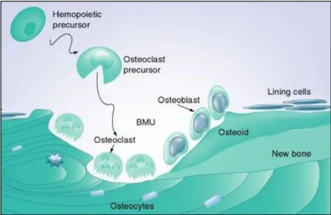
In a histological level, the bone tissue is composed by three major constituents: osteoblasts, osteoclasts and osteocytes (Figure 5). These are responsible for the extracellular matrix formation (organic and inorganic) and each one of those possesses a particular role in this task (22).
Figure 5. Bone tissue cellular composition: osteoblasts, osteoclasts and osteocytes and lining cells. Totality of cells involved
in the bone formation and in the regeneration process (25).
3.2.1 Osteoclasts
Osteoclasts are multinucleated giant cells with a diameter ranging from 20 to over 100 µm. They have acidophilic cytoplasm containing numerous vesicles and vacuoles (lysosomes filled with acid phosphatase) and usually derived from early promonocytes. Under certain
13 circumstances, however, monocytes and macrophages are also capable of osteoclastic differentiation. Some investigations (26; 27) have demonstrated, although without clear details, that osteoblastic cells are involved in the osteoclastogenesis initiation by inducing the activation of the osteoclastic precursors through the ingrowth of blood vessels. The equilibrium between the osteoclasts and osteoblasts activity defines the velocity of bone regeneration.
Osteoclasts are responsible for the resorption of the bone tissue, which consists in the removing of the mineralized matrix followed by the breaking up of the organic bone. After completing their task they migrate into adjacent marrow space, where they undergo apoptosis. They can live for up to seven weeks (21; 22; 23; 24).
3.2.2 Osteoblasts
The osteoblasts are the bone-forming cells, which make them involved in the entire bone formation process. Typically they are 15-30µm cuboidal-shaped cells, with a large nucleus. The cytoplasm is rich in organelles that assure the biological functionality of the cell and maintain the strong cellular activity.
The osteoblasts arise from osteoprogenitor cells (immature progenitor cells2), located in the deeper layer of the periosteum and in the bone marrow, that differentiate under the influence of growth factors such as bone morphogenetic proteins (BMPs), fibroblast growth factor (FGF), and others.
Active osteoblasts exhibit some functional characteristics which includes, for instance, intensive alkaline phosphatase (ALP) activity and the secretion of type I collagen. They can also synthesize osteocalcin and bone sialoprotein that serve as biomarkers for osteoblastic identification and functional evaluation.
The mineralization phase is considered the second stage of osteoblasts evolution and it is defined by the crescent levels of calcium and phosphate. During the bone matrix (osteoid) formation the osteoblasts are responsible for the synthesis and secretion of the collagen fibres. Some can also differentiate into osteocytes and extend out communication processes with neighbouring osteocytes, osteoblasts surface or lining cells. This last type of cells is also derived from osteoblasts (present on the surface), which are already inactivated.
2 Progenitor Cell: biological cell that has a tendency to differentiate into a specific type of cell, a ―target‖ cell (7). In the case of the
osteoblasts, the osteoprogenitor cells derived from self-renewing pluripotent stem-cells stimulated under certain environmental conditions (23).
14 The lining cells (Figure 5) are flat and highly interconnected with each other. They usually form a cellular sheet that covers the entire surface of bone, protecting it and controlling the flux of ions. Besides that, the bone lining cells are very important in the bone remodelling process since they possess hormones and growth factor, both essential for the initiation.
The osteoblasts are also responsible for the secretion of enzymes that lead to the osteoid removal, promoting this way the osteoclasts contact with the mineralized bone surface. Populations of osteoblasts are very heterogeneous, with different osteoblasts having different gene expressions. This may explain the heterogeneity of the trabecular micro-architecture at different skeletal sites, the anatomic site-specific differences in disease states, and the regional variation in the ability of osteoblasts to respond to agents used to treat bone disease (21; 22; 23; 24).
3.2.3 Osteocytes
The osteocytes, star-shaped cells, are the most abundant cell type found in the cortical bone and the only one embedded within the bone matrix. In a mature bone about 95% of the total cells are osteocytes (approximately 20 000 to 30 000 osteocytes per mm2 of bone).
They derive from osteoblasts that became trapped inside the osteoid, in small chambers known as lacunae, during the bone formation. The process of differentiation into osteocytes requires the lost of the osteoblasts organelles and the cytoplasmic extension into long and slight structures. These slight structures are then encased in tiny channels called
canaliculi, which interact and communicate with the surrounding cellular substances,
producing a network for the exchange of ions, nutrients and extracellular fluid.
Since the osteocytes have reduced synthetic activity, and like osteoblasts are not capable of mitotic division, their physiologic function is not completely defined. Although, thanks to their interactive networks, it is believed that they are responsible for detecting microdamages and for initiating the repair process, which indicates, on its turn, that they are involved in the routine turnover of the bone matrix. They are capable of transducing stress signals from bending and stretching of bone into biologic activity. This function is also responsible for the longevity of the osteocytes that can go to 25 years (average half-life), for the slowest turnover rates (21; 22; 23; 24).
15
3.3 Biological Dynamics
The bone dynamics involves three main stages: growth, modelling and remodelling. These are the three major mechanisms that modify the bone mass and the structure of the skeletal system for its adaptation to the mechanical and non-mechanical environments. For a patient recovery after a surgical intervention, such as implantation, the remodelling process is the most important one (21). However, to understand completely the human bone nature and its complete development it is essential to introduce all stages.
3.3.1 Growth
The bone formation is initiated in the first weeks of gestation. However, it is only in the end of the adolescence that the definitive composition of all skeletal bones is completed. Bone grows and models under the influence of metabolic, mechanical and gravitational forces, in a very long and complex process.
This stage is divided in two, the longitudinal and the radial growth. The longitudinal is mainly responsible for increasing bone length, while the radial growth is mainly responsible for enlarging bone cross-sectional area (23).
3.3.2 Modelling
Bone modelling is one of the predominant biological mechanisms that governs the enlargement of each individual bone during growth. It is a process in which bones change their overall shape in response to physiologic influences or mechanical forces, leading to a gradual adjustment of the skeletal. On its turn, bone modelling is divided into bone formation drift (osteoblasts action) and bone resorption drift (osteoclasts action). These two occur separately, although they can work together to guarantee the appropriate shape and size of each individual bone.
In adults, bone modelling is less frequent than remodelling, although in certain circumstances, like hypoparathyroidism, the modelling stage can be increased (21; 23).
16 3.3.3 Remodelling
At a cellular level, the modelling and remodelling processes are not very different. They are both based on the action of the osteoclasts and the osteoblasts.
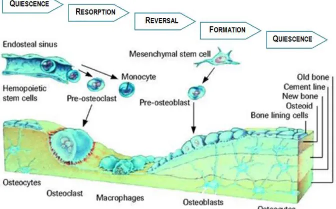
In the remodelling process the osteoblasts and the osteoclasts closely collaborate in a ―Basic Multicellular Unit‖ or BMU (small packets of cells placed in the cortical and trabecular surface).
During childhood, the remodelling process is responsible for the substitution of immature bone for more bio-mechanically and metabolically competent bone, and during growth it is involved in the bone elongation. In the adulthood, this process is responsible for replacing aged bones (damaged or mechanically unfit) – resorbs old bone and forms new bone to prevent accumulation of microdamage bone –, this way maintaining the skeletal mechanical capacity. This process is impelled by specific hormones that control/regulate the calcium concentration in the blood and can only happen thanks to the high level of plasticity of bone.
The remodelling cycle (Figure 6) is composed of four sequential phases: quiescence phase; resorption phase; reversal phase; and formation phase (21; 23; 28; 29; 30).
17 Quiescence or Activation Phase: recruitment and activation of osteoclasts (haematopoietic
origin) by hormonal stimuli.
Resorption Phase: the osteoclasts adhere to the bone surface and start to erode the mineral structure, which is followed by digestion of the organic matrix. In this phase, small cavities in the surface of the trabecular bone are created by the solubilisation of the mineral structure. This acidifies (pH=4) the surrounding microenvironment inducing the liberation of H+ ions against the bone surface. Since osteoclasts have limited life span (≈ 12.5 days) the progression of bone remodelling requires the continual addition of osteoclasts. After completed their task, the osteoclasts die by apoptosis.
Reversal Phase: intermediary between the resorption and the bone formation phases, and the responsible for the transmission of the bone inducing signal. During this phase the osteoclasts disappear and macrophage-like cells are seen on the bone surface. These latter cells can release factors that inhibit the osteoclasts action and stimulate the osteoblasts.
Formation Phase: the bone formation results from a complex cascade of events. It, basically, involves proliferation of primitive mesenchymal cells, differentiation into osteoblasts precursor cells, maturation of osteoblasts that adhere to the previously resorbed surface, formation of the bone matrix, and finally mineralization (28; 29; 32).
3.4 The Healing Process
During dental implantation, the bone tissue that surrounds the area is subjected to tensions and forces. These aggressions can damage the tissue, inducing a healing response.
Usually, the healing process depends majorly on the vascularised system that surrounds the area, which works as an oxygen and nutrients supplier, and on the local stability, which requires the absence of biomechanical actions/forces for a faster recover (33; 34)
Immediately after the surgical intervention, the first healing phase (reactive phase) starts. It involves the formation of a blood clot or hematoma, resultant from the vessels constriction. This hematoma helps stopping the bleeding and at the same time serves as a
18 building block for the rest of the environment. During this phase, the surrounding region experiences inflammation and the patient suffers pain.
The second phase is the reparative one. This starts few days after implantation, with the fibroblasts and osteoblasts action. The fibroblasts secret collagen fibres that are arranged into layers, giving origin to a fibrocartilaginous callus. Then, the capillaries in the extremities start connecting the tissue and, at the same time, the immune system (macrophages) demolishes the hematoma. On its turn, the osteoblats produce trabecular bone and soft callus, restoring most of the original local strength.
The third phase, remodelling, consists in the transformation of the callus into bone callus. In other words, it is the process responsible for the substitution of the spongy bone for compact bone, like was defined in the previous section. During that time, the callus is remodelled, re-establishing the original properties and characteristics of the local tissue (35; 36).
4. BONE-IMPLANT INTERACTION
4.1 Biological Mechanisms of Bone Formation in the Interface
The bonding between the implant and the bone tissue is established based on physical and chemical processes induced by three biological mechanisms: osteoinduction, osteoconduction and osteointegration. Each one of those depends on the others and that dependence defines the success or failure of the interaction, at the interface.
4.1.1 Osteoinduction
The osteoinduction is the act or process that induces the osteogenesis. This phenomenon consists in the phenotype conversion of the soft tissue cells into bone tissue precursors, through appropriate stimulation (37). In the 70’s, Marshall Urist defined for the first time this stimulus as dependent on the cells presence. Nowadays, after more investigations, it is considered to be determined by the presence of some high molecular weight glicoproteins, like the BMP – among all the available donor areas in the jaw, the cortical bone presents the higher concentration of BMP (38).
19 This process allows the immature or undifferentiated mesenchymal cells to become a cellular line capable of producing bone, like the pre-osteoblasts. Thanks to this property, some materials (bone grafts, most common) are able to create the conditions necessary to induce the bone tissue formation in places without it, like muscles or ligaments (37; 39; 40).
4.1.2 Osteoconduction
The osteoconduction is a three-dimensional process, observed immediately after the implant contact with the bone tissue. It is characterized as the ability of growing bone in apposition to the existing one or above it.
Contrary to the osteoinduction, where the material possesses elements that stimulate the bone tissue formation, in the osteoconduction the material works as a passive support ―waiting‖ for the tissue response. In this case, the material is defined as a physical structure that favours bone progression by allowing cellular and vascular local invasion. It is expected that the cellular organisms adhere, grow and invade all the material structure. However, for this to happen, the presence of differentiated mesenchymal cells or bone tissue it is obligatory, in other words it depends on the osteoinduction phenomenon.
In implantology, the most common osteoconductive materials are the natural hydroxyapatites and the bioceramics. Since these can be both resorbable (preferred for implants) or non resorbable, depending on the objective of the medical intervention, they are able to adapt (37; 40; 41).
4.1.3 Osteointegration or Osseointegration
There has been much discussion about the meaning of osteointegration, since it was introduced in the 70’s. Branemark (1977) was the first one defining this phenomenon. He described it as a ―direct structural and functional connection between the living bone and the
surface of a load-carrying implant‖ (42). On its turn, the Williams Dictionary of Biomaterials
(43) offered a similar description, although a little bit more formal: osteointegration is ―the
concept of a clinically asymptomatic attachment of a biomaterial to bone, under conditions of functional loading‖. Neither of these two definitions elucidates the fact that, in some cases,
osteointegration can occur when just a physical contact is observed and there is not a real and direct connection between the implant and the bone. However, since the concept has been
20 generalized, these definitions are still accepted and used by the majority of the specialists in clinical implantology, nowadays (44; 45).
Figure 7. Osteointegration development on dental implants: (A) Time 0; (B) 1 week; (C) 2 months; (D) 1 year; (E) 10 years (46).
Osteointegration has been intensively studied since Branemark. Currently, it is well accepted that an implant is considered osteointegrated when there is no progressive relative movement between the implant and the bone and the anchorage between them is such that can persist under all normal conditions of loading (47). In dentistry, osteointegration can be defined as a biological state where the bone of the mandible or maxilla grows into physiological contact with the implant itself (Figure 7) (48).
The creation and maintenance of osteointegration is dependent on the understanding of the tissue’s healing, repair and remodelling capacities, since they are all involved in a later stage of osteointegration – consolidation of the bone at the implant site and maintenance of the normal bone conditions – and since their basic principle is similar. For instance, during the healing process a bond is formed between bone tissues, without intermediate fibrous tissue or fibrocartilage formation. In the case of osteointegration the same thing happen, although instead of attaching two biological structures, the bone tissue is connected to an implant (47; 49).
To achieve a good osteointegration some parameters must be follow: (i) the bone must be viable (should not cause necrosis or inflammation); (ii) the space between the bone and the implant must be small and contain no fibrous tissue; (iii) the material should be properly
A B C D E A B C D E
21 choose (biocompatibility is required as well as mechanical stability and resistance similar to the natural bone); and (iv) the bone-implant interface must be able to survive loading by a dental prosthesis. To guarantee a successful implantation, the implant must be allowed to heal for a time without load, after the surgical intervention (4; 42; 48; 50). Vascularisation is, also, essential during osteointegration, as it influences tissue differentiation and ossification.
This phenomenon is an absolute requirement for the successful implant-supported dental prosthesis (51). In humans, the osteointegration of an implant is a slow process that can take up several months.
4.2 Bone Adhesion
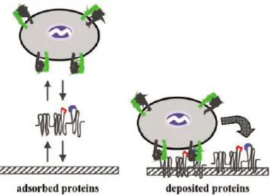
To guarantee osteointegration the adhesion of osteoblasts to the biomaterials surface should occur in a very short period of time, during which multiple steps must be complete: focal adhesion (discrete regions in the cell membrane, intimately associated with the substrate); combination between the proteins and the substrate; and cell spreading (52).
Due to the presence of a support matrix or extracellular matrix (ECM) secreted by the cells, the attachment to the material surface is possible. This matrix is composed by a vast group of proteins, proteoglycanes and glicoproteins, which will determine the cell shape and ultimately the proliferation, as well as, the proper function and tissue integrity (53; 54). Between those, the fibronectin (high-molecular weight protein and the first one activated by the ECM during bone-biomaterial contact (55)) and the vitronectin are considered the major proteins responsible for cell-substrate adhesion interaction. They have, also, an important paper in the promotion of the osteoblasts proliferation and differentiation (53; 54).
According to Yang et al (2002) (56) the presence of fibronectin on Ti surfaces plays an important role in governing osteoblasts attachment.
In living systems, the blood is the first component to contact with biomaterials and, immediately after, rapid adsorption of plasma proteins occur. When a substrate contacts with a biofluid, a considerable number of events take place, in order to modify the materials state to promote the interaction with the cells. The first step is the material hydration. The water molecules bond to the biomaterial surface originating an ionic layer, which will allow the adsorption of proteins (like fibronectin). Thus, the cells that reach the surface establish contact with the protein-coated substrate and attach to the extracellular matrix of those proteins. For instance, fibronectin proteins can bind to the integrins (membrane-spanning receptor of
22 proteins) on the osteoblasts and activate signalling pathways that induce cell-cycle progression, gene expression, matrix mineralization and regulates osteoblasts survival. This contact put in evidence that, in reality, this is not a direct attachment between bone and material but a protein intermediate interaction (Figure 8) (52; 53; 55).
Figure 8. Mechanisms controlling cell adhesion (adapted from (57)).
In the end an effective cell adhesion is completed, allowing a cascade of cellular events to take place, like proliferation and cellular spreading over the surface, in a dynamic environment.
During the previous events (especially protein adsorption) the implant’s surface is significantly changed, which occurs both in vivo and in vitro. Even if the selected material for implantation possesses already a stable oxide film, under these circumstances, it still suffers electrochemical changes. For instance, considering that commercially pure titanium implants possess an oxide from 2 to 6 nm before implantation, after retrieving them from the human body the thickness seems to be two or three times higher (53). That is why the implantable material should respect an entire list of demands before being used in contact with the human body.
5. BIOMATERIALS
Over the years the medical field have suffered astronomic changes. The surgical and medical techniques were improved and the devices used were constantly challenged to achieve higher levels of quality. Those changes led to the emergence of a new range of
23 materials, the biomaterials. According to the Williams Dictionary of Biomaterials (43), “a
biomaterial is a nonviable material used in a medicine device, intended to interact with biological systems (...) to evaluate, treat, augment or replace any tissue, organ or function of the body”. Ideally it should be able to sustain a positive interaction with the surround tissues
without causing an abnormal response (58; 59).
The idea of preserving the human body integrity and comfort for the longest time possible and restoring lost functions and damaged tissues/organs were the main reasons that motivated the biomaterials development (48; 58).
In medical applications, they are rarely used as isolated materials but are more commonly integrated into devices or implants. Although they are primarily employed in this field, they can also be used in biological investigations, for instance, to grow cells in culture, to assay for blood proteins in the clinical laboratory, in equipments for processing biomolecules for biotechnological applications and others. In both cases, the biomaterials must always be considered in the context of their final fabricated, sterilized form (59; 60).
5.1 Biocompatibility
Although the selection of the best biomaterial for dental applications relies on a substantial range of requirements, there is one that is considered the most important – the biocompatibility (60).
The understanding and measurement of biocompatibility is unique to biomaterials science. It was firstly defined by William (1987) as ―the ability of a material to perform with
an appropriate host response in a specific application‖ (43; 59). In 2008, considering all the
changes and improvements in the biomedical field, William proposes a new definition, ―biocompatibility refers to the ability of a biomaterial to perform its desired functions with
respect to a medical therapy, without eliciting any undesirable local or systemic effect in the recipient or beneficiary of the therapy, but generating the most appropriate beneficial cellular or tissue response in that specific situation, and optimizing the clinically relevance of the therapy‖ (61). According to these definitions a biocompatible material must not: irritate the
surrounding structures, provoke an abnormal inflammatory response, incite allergic or immunologic reaction and cause cancer (58; 59; 60).
Inherent to this, however, was the idea that a single material may not be biologically acceptable in all applications. For example, a material that is satisfactory as a full cast crown
24 may not be adequate as a dental implant. Also implicit is an expectation for the biological performance of the material. In a bone implant, the expectation is that the material will allow the bone to integrate with the implant. Thus an appropriate biological response for the implant is osteointegration. In a full cast crown, the expectation is that the material will not cause inflammation of pulpal or periodontal tissues, but osseointegration is not. Whether a material is biocompatible or not is therefore dependent on what physical function we ask of and what biological response we require from it (4; 60).
5.2 Classes of Biomaterials
The search for more sophisticated devices to replace and treat damaged body parts have led to a wide range of high quality biomaterials. Although they are usually distinguished in three basic categories – metals, ceramics and polymers –, in this study another one will be taken into consideration, the composites.
5.2.1 Metals
Metals and metallic alloys play a prominent role in dentistry and are used in almost all aspects of dental practice (implants, dental restoration and manipulation instruments). Thanks to their optical, physical, chemical, thermal and electrical properties, these materials can be favourably exploited in dentistry (4; 48; 22). In this category the most distinguished types of metals for medical applications are: titanium, cobalt, stainless steel, nickel, chromium and the noble metals, like gold, tantalum, platinum, palladium, silver, iridium and niobium (59; 62; 63).
5.2.2 Ceramics
The use of ceramics in dentistry was initially based upon the relative biological inertness of ceramic materials. Nowadays, the bioinert and bioactive ceramics, materials that induce normal tissue formation and assure an intimate bond with it, are the preferred choice.
There are lots of ceramics applied in the biomedical field. The most common are the carbon, alumina and zirconia – bioinert ceramics – and the bioactive glasses and glass
25 ceramics, calcium phosphate ceramics and the combination of the two previous – bioactive ceramics (59; 63; 64).
5.2.3 Polymers
The first polymer used in dentistry was vulcanized rubber for denture bases. Nowadays, other polymers were already introduced in this field: vinyl acrylics, polystyrene, epoxies, polycarbonates, silicones, polyethers, polysulfids and polyacrylic acids. These are used in the construction of prosthetic appliances, artificial teeth, tooth restoratives, implants, temporary crowns, cements, and others (22; 65).
5.2.4 Composites
Although, there is a rich history associated with the development of dental composites and their prominent position in dentistry, it is still evident the discussion around the composite materials definition.
Composite materials have a bulk continuous phase, called matrix, and one or more non-continuous phases, called the reinforcement, which usually have superior mechanical or thermal properties than the matrix. Although, inorganic materials (titanium, steel, carbon...), thermoplastics (polyesters, polycarbonate...), thermosets (epoxy, silicone...) and resorbable polymers (chitosan, collagen) can be used as matrix, some of them can also be used as reinforcement. The choice is dependent on the material application (59; 63; 66).
5.3 Dental Implants Materials
Dental implants have been manufactured in a wide variety of shapes and materials. According to their design (implant systems), cost and aesthetic purpose, the materials applied to their production can change; however all of them must possess some requirements, like resistance to high intensity mechanical and physical efforts as well as chemical attacks and simultaneously be recognized by the human body as ―friendly‖ substrates (67; 68; 69).
The first implants used were made by precious materials like gold, platinum, palladium or iridium (22). However, the high costs and the resistance deficiencies of those conducted to new categories, such as the ones described in 5.2 Classes of Biomaterials.