Computed Tomographic evaluation of a young
adult treated with the Herbst appliance
Savana Maia*, Dirceu Barnabé Raveli**, Ary dos Santos-Pinto**, Taísa Boamorte Raveli***, Sandra Palno Gomez***
Introduction: The key feature of the Herbst appliance lies in keeping the mandible continu-ously advanced. Objective: To monitor and study the treatment of a patient wearing a Herbst appliance by means of Cone-Beam Computed Tomography (CBCT) images for 8 months after pubertal growth spurt. The subject was aged 16 years and 3 months and presented with a Class II, Division 1 malocclusion associated with mandibular retrognathia. Results: The CBCT images of the temporomandibular joints suggest that the treatment resulted in the remodeling of the condyle and glenoid fossa and widening of the airway. Conclusions: The Herbst appliance constitutes a good option for treating Class II malocclusion in young adults as it provides patients with malocclusion correction and improves their aesthetic profile.
Abstract
Keywords: Temporomandibular joint. Computed Tomography. Orthopedic appliances.
* MSc in Orthodontics, PhD Student in Orthodontics, Araraquara School of Dentistry (UNESP). ** Associate Professor, Department of Orthodontics, Araçatuba School of Dentistry (UNESP). *** MSc Student, Araraquara School of Dentistry (UNESP).
introduction
Despite the availability of a wide range of Class II malocclusion treatment options, the actual ac-tion mechanism behind these orthopedic devices remains controversial. The effectiveness of the Herbst appliance in treating Class II malocclusions has been studied for decades. However, despite the obvious effectiveness of this therapy, the possibil-ity of manipulating mandibular growth potential beyond what is genetically determined still fuels the debate between proponents and opponents of dentofacial orthopedics.l Some researchers,
grounded in Functional Matrix theory, believe that local environmental factors ultimately determine the final size of the craniofacial skeleton, which
could therefore be subjected to some regulation by changing its functional pattern.1 Opponents of this
view advocate that control is predominantly ge-netic, alterations are restricted to the dentoalveolar component and do not affect basal bone growth. It is suggested that the use of functional appliances for stimulating mandibular growth would have only a temporary impact on the dentofacial pattern and that over the long term the morphogenectic pattern would prevail.1,2
of the condyle, glenoid cavity and posterior mandibular ramus in adult rhesus monkeys. The results showed adaptation of the condyle and glenoid fossa during treatment with the Herbst appliance.
Advances in Imaging Technology in Den-tistry and the advent of Computed Tomography (CT) scans ensure accurate diagnoses with great reliability, enabling the three-dimensional anal-ysis of structures. As well as specific CT soft-ware, which allows measurements to be carried out in tomographic slices, a new methodology has emerged which makes for the assessment of inclinations and angulations of individual teeth, and bone remodeling, accurately reproducing the various structures.
Computed Tomography is the exam of choice for analyzing bone components and dental struc-tures.12 The development of this new technology
has provided dentistry with the reproduction of three-dimensional images of mineralized maxil-lofacial tissues with minimal distortion and sig-nificantly reduced radiation doses.13
Its diagnostic reliability is due to the ac-curacy of the measurements used in different methods, which is of great importance to ortho-dontists since orthodontic treatment diagnosis, prognosis and planning, among other factors, depend on such measurements.
cLinicAL cASE rEPort
The case described in this article is part of a
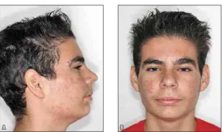
re-A Brazilian patient, male, 16.3 years old, sought orthodontic treatment at the Araraquara School of Dentistry (UNESP) complaining that his chin was positioned backwards. Front view facial analysis showed a mesofacial pattern and absence of lip seal. Lateral view analysis dis-closed a convex profile associated with man-dibular retrognathia (observed clinically), and a short chin-neck line (Fig 1).
Intraoral examination showed that the pa-tient presented permanent dentition, a Class II malocclusion and 7.3 mm overjet (Fig 3). At di-agnosis, functional changes were noted in swal-lowing. Morphological analysis of the cepha-lometric radiograph confirmed a convex facial pattern (Fig 2).
Skeletal age was verified by means of carpal X-ray using skeletal maturation indicators according to the Greulich and Pyle atlas.16 The patient was
nearing the end of the descending growth curve (FPut – Complete epiphyseal union in the proxi-mal phalanx of the 3rd finger; FMut – Complete
epiphyseal union in the middle phalanx of 3rd
fin-ger; and/or Rut - Complete epiphyseal union of the radius bone), i.e., at the end of pubertal growth.
using the Herbst appliance
A
A
B
B C
habitual intercuspation (MHI): At T1, beginning of treatment, and T2, eight months after treat-ment. The Cone-Beam CT scans were performed at the beginning of treatment and after removal of the Herbst appliance, and analyzed using spe-cific software (Dolphin 10.5, Dolphin Imaging & Management Solutions, USA).
The anchorage system used in the upper and lower arches was a banded Herbst (Figs 4, 5 and 6). To cement the anchorage structures we used light cure glass ionomer cement (3M Unitek).
A telescopic mechanism was used (Flip-Lock - TP Orthodontics), composed of the following accessories: a) Tube, determines the amount of mandibular advancement, b) Piston, adapted to the length of the tube, c) Connectors, with a spherical shape.
FIGURE 1 - Initial extraoral photographs profile (A) and front (B) views.
FIGURE 3 - Initial intraoral photographs right (A), front (B) and left (C) views.
FIGURE 2 - Initial lateral cephalometric radio-graph.
ct examination and measurements
The CT scans were obtained with an i-CAT scanner with the patient’s mouth shut and in maximal intercuspation (MHI). Scanners pro-vide standardized images in a single 360-degree rotation, 20-second scan. It reconstructs the data in real time, automatically and immediately, yielding 460 individual 0.5 mm slices in each or-thogonal plane. Data were exported in DICOM format and evaluated using Dolphin software.®
plane comprises the line where the Porion crosses the Frankfort horizontal plane.
ct scan image assessment
The CT scans revealed a 0.8 mm increase in condyle diameter on the right side and 0.7 mm on the left side (Figs 7, 8 and 9). The subjec-tive analysis of the region suggests that the area of the glenoid fossa and condyle experienced remodeling. However, analysis of a single case does not allow meaningful assessment.
Studies14 report this change and show, by
means of magnetic resonance imaging in patients treated with Herbst appliance, an adaptation of the temporomandibular joint, concluding that such remodeling of the glenoid fossa and condyle does take place. Another investigation,7 this time
using MRI in 20 adolescent patients treated with Herbst, pointed out changes in TMJ disc posi-tion and concluded that during treatment with Herbst there is an alteration in the position of the articular disc, but within normal limits. Treat-ment with Herbst in young adults provides bone remodeling and formation of new condylar bone. Furthermore, this newly formed bone has been shown to be stable.3,6
CT examinations allow anatomical areas to be reconstructed and viewed in three dimen-sions, disclosing information about size, shape
and texture of the area under analysis. CT scanners capture body images in slices using radiation and export them to a dedicated soft-ware. Given its accuracy, CBCT contributes to scientific investigations of remodeling in the TMJ region through the use of orthopedic ap-pliances. Studies conducted in adult monkeys treated with the Herbst appliance showed by means of histological sections that the treat-ment produces significant bone formation in the glenoid fossa or remodeling in the area of the fossa and condyle.
In assessing an individual’s airway, the initial volumetric value of 4324.5 mm3 can be found,
whereas after treatment with Herbst, such value rises to 5108.5 mm3 (Fig 9), indicating an
in-crease in the nasopharyngeal region after treat-ment. A study15 conducted with 26 individuals
A B FIGURE 7 - TMJ examination method using Dolphin software.
A B
A B
of mandibular advancement devices, as in the treatment presented in this study.
After eight months of treatment with Herbst the results show (Fig 10) correction of Class II and Class I malocclusion as well as improved fa-cial aesthetics (Figs 11 and 12) with no changes
in muscles and joints.
CT studies on the influence of Herbst in the TMJ region and airways are scarce. The findings show assessments made using resonance and disc positioning since CT examinations are a more re-cent phenomenon.7
FIGURE 9 - Airway tomogram analysis: A) initial and B) final.
FIGURE 10 - Final intraoral photographs: Right side (A) and left side (B) views.
concLuSionS
CT scans provide better diagnosis and orth-odontic treatment planning, making it possible to view the problem in three dimensions in space. Furthermore, CBCT allows structures such as the condyle and glenoid fossa to be analyzed while en-abling the evaluation of remodeling in this region
after treatment with orthopedic appliances. Treat-ment with the Herbst appliance produces satisfac-tory results, providing patients with malocclusion correction and improving their aesthetic profile. After treatment with the Herbst appliance CT evaluation is suggestive of remodeling in the TMJ region and condyle, and a widened airway.
1. Ursi W, McNamara JA, Martins DR. Alteração clínica da face em crescimento: uma comparação cefalométrica entre os aparelhos extrabucal cervical, Fränkel e Herbst, no tratamento das Classes II. Rev Dental Press Ortod Ortop Facial. 1999 set-out;4(5):77-108.
2. Pancherz H, Fackel U. The skeletofacial growth pattern pre and post-dentofacial orthopaedics. A long-term study of Class II malocclusions treated with the Herbst appliance. Eur J Orthod. 1990 May;12(2):209-18.
3. Konik M, Pancherz H, Hansen K. The mechanism of Class II correction in the late Herbst treatment. Am J Orthod Dentofacial Orthop. 1997 Jul;112(1):87-91.
4. Ruf S, Pancherz H. Orthognathic surgery and dentofacial orthopedics in adult Class II division 1 treatment: mandibular sagittal split osteotomy versus Herbst appliance. Am J Orthod Dentofacial Orthop. 2004 Aug;126(2):140-52.
5. Paulsen HU, Karle A, Bakke M, Hersink A. CT-scanning and radiographic analysis of temporomandibular joints and cephalometric analysis in a case of Herbst treatment in later puberty. Eur J Orthod. 1995;17(3):165-75.
6. Paulsen HU, Karle A. Computer tomographic and radiographic changes in the temporomandibular joints of two young adults with occlusal asymmetry, treated with the Herbst appliance. Eur J Orthod. 2000 Dec;22(6):649-56.
7. Aidar LA, Abrahão M, Yamashita HK, Dominguez GC. Herbst appliance therapy and temporomandibular joint disc position: a prospective longitudinal magnetic resonance imaging study. Am J Orthod Dentofacial Orthop. 2006 Apr;129(4):486-96. 8. Paulsen HU, Rabøl A, Sørensen SS. Bone scintigraphy of
human temporomandibular joints during Herbst treatment: a case report. Eur J Orthod. 1998 Aug;20(4):369-74.
9. McNamara JA Jr, Peterson JE, Pancherz H. Histologic changes associated with the Herbst appliance in adult Rhesus Monkeys (macaca mulatta). Semin Orthod. 2003;9:26-40.
10. Voudouris JC, Woodside DG, Altuna G, Kuftinec MM, Angelopoulos G, Bourque PJ. Condyle-fossa modiications and muscle interactions during Herbst treatment, Part 1. New technological methods. Am J Orthod Dentofacial Orthop. 2003 Jun;123(6):604-13.
rEfErEncES
11. Voudouris JC, Woodside DG, Altuna G, Angelopoulos G, Bourque PJ, Lacouture CY, et al. Condyle-fossa modiications and muscle interactions during Herbst treatment, Part 2. Results and conclusions. Am J Orthod Dentofacial Orthop. 2003 Jul;124(1):13-29.
12. Firooznia H, Golimbu CN, Raii M, Rausching W, Weinreb JC. MRI and CT of the musculoskeletal system. St. Louis: Mosby Year Book; 1992. 443-64.
13. Scarfe WC, Farman AG, Sukovic P. Clinical applications of cone-beam computed tomography in dental practice. J Can Dent Assoc. 2006 Feb;72(1):75-80.
14. Ruf S, Pancherz H. Temporomandibular joint remodeling in adolescents and young adults during Herbst treatment: a prospective longitudinal magnetic resonance imaging and cephalometric radiographic investigation. Am J Orthod Dentofacial Orthop. 1999 Jun;115(6):607-18.
15. Haskell JA, McCrillis J, Haskell BS, Scheetz JP, Scarfe WC, Farman AG. Effects of Mandibular Advancement Device (MAD) on airway dimensions assessed with cone-beam computed tomography. Semin Orthod. 2009 Jun;15(2):132-58. 16. Greulich WW, Pyle SI. A radiographic atlas of skeletal
development of the hand and wrist. 2nd ed. Stanford: Stanford University; 1959.
Submitted: June 2010 Revised and accepted: August 2010
contact address
Savana Maia