w w w . r b o . o r g . b r
Original
Article
Use
of
superficial
peroneal
nerve
graft
for
treating
peripheral
nerve
injuries
夽
Samuel
Ribak
a,b,∗,
Paulo
Roberto
Ferreira
da
Silva
Filho
a,b,
Alexandre
Tietzmann
a,
Helton
Hiroshi
Hirata
a,b,
Carlos
Augusto
de
Mattos
a,
Sérgio
Augusto
Machado
da
Gama
aaPontifíciaUniversidadeCatólicadeCampinas,Campinas,SP,Brazil
bHospitalNossaSenhoradoPari,SãoPaulo,SP,Brazil
a
r
t
i
c
l
e
i
n
f
o
Articlehistory:
Received1March2015 Accepted6April2015
Availableonline13January2016
Keywords:
Peripheralnerve Nerve/transplantation Peronealneuropathies
a
b
s
t
r
a
c
t
Objective:Toevaluatetheclinicalresultsfromtreatingchronicperipheralnerveinjuries usingthesuperficialperonealnerveasagraftdonorsource.
Methods:Thiswasastudyonelevenpatientswithperipheralnerveinjuriesintheupper limbsthatweretreatedwithgraftsfromthesensitivebranchofthesuperficialperoneal nerve.Themeantimeintervalbetweenthedatesoftheinjuryandsurgerywas93days.The ulnarnervewasinjuredineightcasesandthemediannerveinsix.Therewerethreecases ofinjurytobothnerves.Inthesurgery,alongitudinalincisionwasmadeontheanterolateral faceoftheankle,thusviewingthesuperficialperonealnerve,whichwaslocatedanteriorly totheextensordigitorumlongusmuscle.Proximally,thedeepfasciabetweentheextensor digitorumlongusandtheperoneallongusmuscleswasdissected.Next,themotorbranch oftheshortperonealmuscle(oneofthebranchesofthesuperficialperonealnerve)was identified.Theproximallimitofthesensitivebranchwasfoundatthispoint.
Results:Theaveragespacebetweenthenervestumpswas3.8cm.Theaveragelengthofthe graftswas16.44cm.Thenumberofsegmentsusedwastwotofourcables.Inevaluatingthe recoveryofsensitivity,27.2%evolvedtoS2+,54.5%toS3and18.1%toS3+.Regardingmotor recovery,72.7%presentedgrade4and27.2%grade3.Therewasnomotordeficitinthedonor area.Asensitivedeficitinthelateraldorsalregionoftheankleandthedorsalregionofthe footwasobserved.Noneofthepatientspresentedcomplaintsinrelationtowalking.
Conclusions: Useofthesuperficialperonealnerveasagraftsourcefortreatingperipheral nerveinjuriesissafeandprovidesgoodclinicalresultssimilartothosefromothernerve graftsources.
©2016SociedadeBrasileiradeOrtopediaeTraumatologia.PublishedbyElsevierEditora Ltda.Allrightsreserved.
夽
WorkdevelopedintheHandSurgeryandMicrosurgeryGroup,PontifíciaUniversidadeCatólicadeCampinas,Campinas,SP,Brazil, andHospitalNossaSenhoradoPari,SãoPaulo,SP,Brazil.
∗ Correspondingauthor.
E-mail:[email protected](S.Ribak).
http://dx.doi.org/10.1016/j.rboe.2015.04.010
Emprego
do
enxerto
do
nervo
fibular
superficial
para
tratamento
de
lesões
de
nervos
periféricos
Palavras-chave:
Nervosperiféricos Nervo/transplante Neuropatiasfibulares
r
e
s
u
m
o
Objetivo:Avaliarresultadosclínicosdotratamentodaslesõescrônicasdenervosperiféricos comonervofibularsuperficialcomofontedoadoradeenxerto.
Métodos: Estudode11pacientescomlesõesdenervosperiféricosnosmembrossuperiores tratadoscomenxertodoramosensitivodonervofibularsuperficial,comintervalomédiode 93diasentreadataderegistrodalesãoeacirurgia.Foramobservadaslesõesdonervoulnar emoitopacientesedonervomedianoemseis.Emtrêsambososnervosforamlesados. Nacirurgiafaz-seincisãolongitudinalnafaceanterolateralnotornozelo,visualiza-seo nervofibularsuperficial,situadoanteriormenteaomúsculoextensorlongodosartelhos. Proximalmentedisseca-seafásciaprofundaentreosmúsculosextensorlongodosartelhos eofibularlongo.Aseguir,identifica-seoramomotordomúsculofibularcurto,umdos ramosdonervofibularsuperficial.Olimiteproximaldoramosensitivoencontra-senesse ponto.
Resultados: Amédiadoespac¸oentreoscotosnervososfoide3,8cm,comprimentomédio dosenxertosde16,44cm,númerodesegmentosusadosdedoisaquatrocabos.Naavaliac¸ão darecuperac¸ãodasensibilidade,27,2%evoluíramparaS2+,54,5%paraS3e18,1%paraS3+. Quantoàrecuperac¸ãomotora,72,7%apresentavamgrau4e27,2%,grau3.Nãohouvedéficit motordaáreadoadora,observou-sedéficitsensitivonaregiãodorsolateraldotornozeloe dorsaldopé.Nenhumpacienteapresentouqueixasàdeambulac¸ão.
Conclusões:Ousodonervofibularsuperficialnotratamentodaslesõesdenervosperiféricos comofontedeenxertoéseguro eproporcionaresultadosclínicossemelhantesaoutras fontesdeenxertodenervos.
©2016SociedadeBrasileiradeOrtopediaeTraumatologia.PublicadoporElsevier EditoraLtda.Todososdireitosreservados.
Introduction
Intreatingperipheralnerveinjuries,theobjectiveistoachieve primaryrepairwithouttensiononthesuture.Situationsin whichthereisnopossibilityofsuturing,orincasesoflossof nervesegments,suchaslateinjuries,orincomplexcases,the treatmentconsistsofreconstructionofthenerve.1
Overrecentdecades,avarietyofexperimentalstudieshave beendevelopedtodeterminethebestmethodsforfillingthe gapbetweenthestumpsofinjurednerves.2
Althoughresearchusingautogenoustubes(frommuscles orvessels)3,4andsynthetic(non-autogenous)tubes5hasbeen developed,graftsfromautogenousnervesarestillthematerial mostindicatedandused.1,2
Inchoosingthenervegraft,themattersthatneedtobe takenintoconsiderationincludewhetheritissufficientlylong toensuretension-freeanastomosis;whetherthenumberof fasciculiiscoincidentwiththoseofthereceptornerve;and whetherthesequelaeinthedonorareaareminimal.6
Giventhesecharacteristics,thedonornervesaregenerally limitedtothecutaneousnervesoftheextremities.
In the upper limbs, the nerves that are used most are themedialcutaneousnerve oftheforearm andthe lateral cutaneousnerveoftheforearm.1,2,6Theadvantageofthese nervesistheirlocation(inthesamelimbthatistobe oper-ated),whiletheirdisadvantageistheirsmall diameterand limitedlength,whichisofteninsufficienttoadequatelyfillthe gap.6
Thesuralnerve,inthelowerlimbs,isconsideredtobethe standardfornervegrafts.7,8Itistheonemostusedbecauseof itsmoresuitablediameterandlength(upto30cminlength). However,despitetheabovecharacteristics,eventhismaynot alwaysbesufficientwhenlargergapsneedtobefilledorin casesofmultipleinjuries.Italsohastheinconvenienceof sen-sorylossonthelateralfaceofthefootorothercomplications inherenttothesurgicalprocedure.
In seekingalternatives,the superficial fibular nervehas emergedasaninterestingoption.Thisisalateralbranchof thecommonfibularnervethatinnervatesthelongandshort fibularmuscles.Itsuppliessensitivitytothelateraland infe-riorfacesoftheskinofthelowerleganddorsumofthefoot.9 In the lower third of the lower leg, it perforatesthe deep fascia andpenetrates into thesubcutaneouscellulartissue atthejunctionofthemiddleandlowerthirds.Atthislevel (i.e.themalleolusoftheankle),itdividesintotwobranches (medialandintermediatedorsalcutaneousbranches),which arebothresponsibleforthesensitivityofthedorsalsurface ofthefoot.10Thisisthecommonestbranchingpatternthat hasbeendescribed.Inalesscommontype,thesebranches passindependentlythroughthedeepfascia,whichindicates thestartingpointforbranchingthatismoreproximal,2butit presentsthesameareaofsensitivityonthefoot.
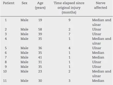
Table1–Dataonthe11patients:patientnumber,sex, age,timebetweentheoriginalinjuryandthesurgical treatment,andnerveaffected.
Patient Sex Age
(years)
Timeelapsedsince originalinjury
(months)
Nerve affected
1 Male 19 9 Medianand
ulnar
2 Male 58 2 Ulnar
3 Male 39 7 Ulnar
4 Male 35 1 Medianand
ulnar
5 Male 36 4 Ulnar
6 Male 35 1 Median
7 Male 41 1 Median
8 Male 31 1 Ulnar
9 Male 35 1 Ulnar
10 Male 23 2 Medianand
ulnar
11 Male 30 3 Median
Theobjective ofthe present study was to evaluatethe resultsfromclinicaluseofthesuperficialfibularnerveasa graftsourcefortreatingperipheralnerveinjuries.
Materials
and
methods
InthisretrospectivestudyconductedbetweenJune2011and January2013,11patientswithdiagnosesofperipheralnerve injuriesunderwentoperations.Directrepairstotheseinjuries duringtheoperationwerenotpossible.Inallofthesecases, thesensorybranchofthesuperficialfibularnervewasusedas agraftdonorsource.
Allthepatientsweregivenexplanationsandsigneda state-mentoflegalresponsibility,forthestudytobeconducted.The studyreceivedpriorapprovalfromtheethicscommitteefor researchonhumanbeings.
Allthepatientsweremale,withameanof4.7years(range: 19–58), and the time that had elapsed between the initial injuryand thesurgical treatmentrangedfrom onetonine months(meanof2.9).Theupperlimbswereaffectedinall cases,withwoundsinthevolarregionoftheforearm:seven ontheleftsideandfourontherightside,andthedominant sidewasaffectedinfivecases.Theulnarnervewasinjuredin eightcasesandthemediannerveinsixcases.Inthreecases (Table1),therewasconcomitantinjurytobothofthesenerves.
Surgicaltechnique
Generalanesthesiawasusedinallthecases.Thepatientwas positionedinsupinedecubitus,ahandtablewasusedand exsanguinationwasperformedusingapneumaticcuff.The elbowandforearmwerekeptextendedinordertomarkout theincision.
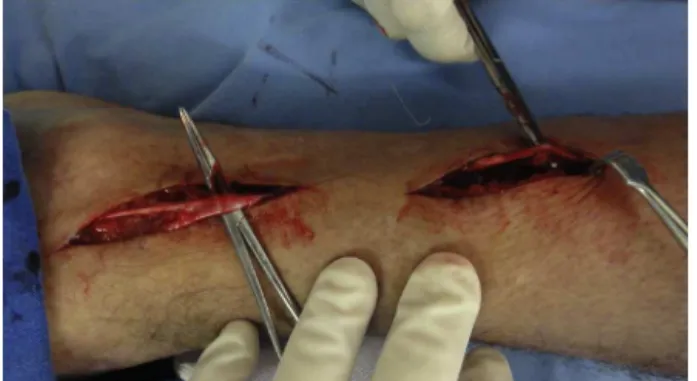
Afterthenerveinjuryhadbeenidentified,thetechnique consistedofresectingthedamagednervetissueuntilhealthy tissue was encountered. The fascicles of this tissue were identified.Atthispoint,thegapbetweenthestumpswas mea-sured,alongwiththesizeofthegraftthatwastobeharvested (Fig.1).
Fig.1–Measurementincentimetersofthespacebetween thestumpsinacaseofulnarnerveinjury.
With the limb positioned, the anterior subcutaneous courseofthesuperficialfibularnervetothelateral malleo-luscouldbeviewed(Fig.2).Thesubcutaneouscourseofthis nervecouldbeviewedinninepatientsbeforetheoperation.
Withthepatientinthesamedecubituspositionandwith atourniquetonthelowerlimbthatwastobeoperated,a lon-gitudinalincisionwasmadeinthelateralfaceoftheankle, 4cmanteriorlytothemidlineofthemalleolus.
Afterthesubcutaneoustissuehadbeenopened,the super-ficialfibularnervecouldbeviewed.Thiswaslocatedanteriorly tothelongextensormusclesofthetoes.Atthislocation, prox-imal dissectionwasperformedbymeans oflongitudinalor continuousincisions,whichfollowedthesubcutaneouspath ofthenerveasfarasthelowerthirdofthelowerleg,whereit perforatedthedeepfascia(Fig.3).
Depending onthe size ofgraft required, dissectionwas thenperformedintheproximaldirectionatadeeperlevel, inwhichthedeepfasciawassectionedalongthelongaxisof theincisionandthelayerbetweenthelongextensormuscle ofthetoesand thelong fibularmuscleswasseparatedout laterally(Fig.4).
Followingthis,thebranchtotheshortfibularmusclewas identified.Theproximallimitofthesensitivebranchwasset atthispoint.Thedistaldissection,atthelevelofthelateral malleolus,followedthemedialandintermediatedorsal cuta-neousbranches(Fig.5).
Toharvestthenerve,sectioningoftheproximalportionof thenervewaspreferred.Followingthis,thenervewasraised
Fig.3–Viewingofthefibularnerveanditsanatomical references:4cmanteriorlytothelateralmalleolusand dissectionmoreproximallyalongitssubcutaneouscourse.
Fig.4–Identificationofthesuperficialfibularnerveina moreproximaldissection,throughopeningthefascia.
proximallyandalongitsentirelength,includingthetwodistal branches(Fig.6).
Independentofthebranchingpatternobserved,themain trunk of the superficial fibular nerve penetrated into the deepfascia,orthemedialandintermediatedorsalcutaneous branchespenetrateditseparately.Thedissectionwassimilar towhatwasdescribedabove,sinceinidentifyingthebranches, theywerefollowedtothestartofthemostproximalbranching, alongthedeepfascia(Fig.7).
Inthereceptorarea,thelengthanddiameterofthe inter-fascicular grafts that would be necessary in order both to
Fig.5–Identificationofthemedialandintermediatedorsal cutaneousbranchesafterdistaldissection.
Fig.6–Viewoftheproximalharvestingofthenerveand itselevationalongitsentirecourse.
Fig.7–Identificationofthemostproximalbranchingofthe sensoryfibularnerve.
fill the gap between the stumps and to cover the entire cross-sectional area ofthe injured nervewere ascertained. Inpreparingthe finalgraft,cablesofappropriatesizewere arrangedinparallelandwerejoinedusingfibringlue(Fig.8).
Followingthis,thegraftwassuturedbothproximallyand distally (Fig. 9)usingfibrin gluetogether withthesuturing (Ethilon 8 or 9-0 nylon thread). In cases in which associ-atedtendoninjurieswerepresent,thetendonsweresutured first.
Fig.9–Nervegraftpositionedbetweenthenervestumpsin anappropriatepositionforsuturingwithouttension.
Criteriaforassessingtheresults
- Measurementofthegapbetweenthenervestumpsafter excisionoftheneuroma,usingarulerwithascalein mil-limeters,withthejointsadjacenttotheinjurymaintained intheneutralposition.
- Identificationoftheanatomicalpatternofthebranchingof thefibularnerve,definedastype1,whenthemaintrunkof thesuperficialfibularnervepenetratedintothedeepfascia; ortype2,withseparatepenetrationofthemedialand inter-mediatedorsalcutaneousbranchesintothedeepfascia.11 - Lengthofthesuperficialfibularnerveharvested.
- Numberofcablesneededtoachieveadequatethicknessfor thecross-sectionalareaoftheinjurednerve.
- Evaluation oftherecoveryofsensitivity(measuredusing thescaleoftheBritishMedicalCouncilSystemof Assess-ment), inwhichS0represented lackofsensory recovery; S1, recoveryregardingdeep cutaneouspain; S2, recovery regardingsuperficialcutaneouspain;S2+,exacerbationof theresponse;S3,recoveryregardingpainandtouchwithout exacerbationanddiscriminationoftwopoints>15mm,S3+, goodlocalizationofstimulianddiscriminationoftwopoints at7–12mm;andS4,completerecoveryanddiscrimination oftwopointsat2–6mm.
- Evaluationofmotorrecovery,usingthescaleoftheBritish MedicalCouncilSystemofAssessment,inwhichgrade5 representednormalstrengthagainsttotalresistance;grade 4,musclestrengthisreduced,butthereismuscle contrac-tion against resistance; grade3, joint movement is only achievedagainstgravityand withoutresistancefromthe examiner;grade2,thereismusclestrengthandjoint move-mentonlywithoutresistancefromgravity;grade1,muscle contractionwithoutmovementisseenorfelt,or fascicula-tionisobservedinthemuscle;andgrade0,nomovement isobserved.
- Sensoryandmotordeficitsinthedonorarea. - Complaintsaboutabnormalitiesofwalking. - Complaintsaboutthehealinginthedonorarea.
Results
Themeandurationofthepostoperativefollow-upwas11.18 months(range:sixto18).
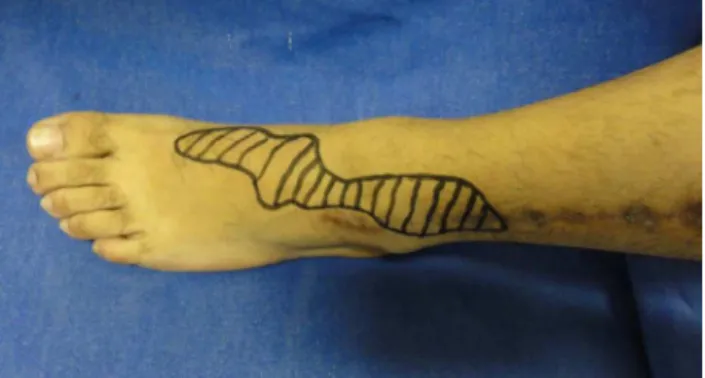
Fig.10–Areaofresidualanesthesia,sixmonthsafterthe operation.
Themeandistancebetweenthenervestumpsafter exci-sion oftheneuroma was 3.8cm (range: 3–5.5cm). Incases ofinjuryonlytotheulnarnerve,itwas3.57cm;andtothe mediannerve,4.08cm.Whenbothnerveswereinjured,the meansizeofthegraftneededwas4.13cm.
Regardingtheanatomicalpatternoffibularnerve branch-ing,90.9%(tencases)presentedthetype1patternandonly onecaseshowedtype2(9.09%).Themaximumlengthofthe graftharvestedwas26cmandtheminimumwas9cm(mean of16.9cm).
Themeannumberofcablesusedinordertoachieve ade-quatethicknessofthecross-sectionalareawasthreetofour cablesforthemediannerveandtwotothreefortheulnar nerve.
Inevaluatingtherecoveryofsensitivity,27.2%presented S2+ (three cases),54.5% S3 (six cases) and 18.1% S3+ (two cases). Regarding motor recovery, 72.7% (eight cases) pre-sentedgrade4and27.2%(threecases),grade3.
Innocasewasmotorlossinthedonorlimbobserved. Sensorydeficitinthedonorareawasobservedinthe dor-solateral region of the ankle and the dorsal region of the foot (Fig. 10). There was no sensory deficit in the plantar region.Noneofthepatientspresentedcomplaintsinrelation towalking.Regardingthedonorarea,therewerenocaseson complaintsabouthealing,eveninthecasesinwhichalarge quantityofgraftwasnecessary,withagreaternumberof inci-sionstoharvestit.
Onlyonecasepresentedsuperficial infectious complica-tionsoftheskininthedonorarea,whichwasseenoneweek afterthesurgery.Itwastreatedwithoralantibiotic,withgood evolution.
Discussion
Despitedecadesofadvancesinnerveresearch,12 treatment ofperipheralnerveinjuriescontinuestobeasignificant chal-lenge.
For filling the space between the nerve stumps, grafts fromautogenousnervesremainthegoldstandardfornerve reconstruction,sincetheyprovidesupportarchitecture, neu-ralgrowthguides,neurotrophicfactorsandSchwanncells.13
ofthelocationsofthemedialandlateralcutaneousnervesof theforearm,thesenervesareoflimitedthicknessandlength.6 Thesuralnerveistheonemostused,anditisconsidered tobethestandardasagraftdonorsource.7,8 However, har-vestingthisnervepresentssomeinconveniencesintermsof itspositioning,theneedtochangedecubitusandtheareaof lossofsensitivityinthelateralregion.Thissourceislimited whenalargequantityofgraftisneeded.
Theidealwouldbeto haveanoptionalgraft sourcefor whenthisisnecessary,orevenasthefirstchoicetobeused. Thesuperficialfibular nerve hasbeen shownto beagood optionasadonorsource,sinceitsuppliesalonggraftofgood caliberthatisanatomicallypredictable.Itcanbeharvested withthepatientindorsaldecubitusandiseasilyaccessible, withouttheneedforchangesofdecubitus.
In an anatomical study, Buntic et al.6 reported that in theirsample,themeanlengthharvestedwas14.7cm(range: 3–25cm),andthat40cmwouldbepossible.Inourstudy,we achievedasimilarmean,of16.44cm(range:9–26cm),which wascomparabletothesizesusedwhenthesuralnervewas chosen.7,13
Lossofsensitivityintheregionsuppliedbythedonornerve isa formofmorbidity common toany graft source.What maydifferentiatethe sourcesisthe extentofthe areaand itslocation,whichmightbeclosetoaninconvenientregion. Inthisregard,useofthelateralcutaneousnervecanbecited, whichgivesrisetolossofsensitivityalongthelateralfaceof theforearmthatmayextendoverthethenarregion,whichis undesirableincasesofinjuriestothemediannerveorfinger nerves.
Inthecaseoftheradialsensorynerve,compromisingthe dorsolateralregionofthehandisalsoundesirable.
Inthelowerlimbs,preservationoflateralandplantar sen-sitivityisextremelyimportantforpreventingulcersandother wounds.Inthisregard,useofagraftfromthesuperficial fibu-larnervehasanadvantagebecause onlyanareaofdorsal anesthesiaoccurs.
Inrelationtopossiblecomplicationsinthedonorarea, for-mationofapainfulneuromawouldbeoneofthese.Buntic etal.6 observedthe presenceofacaseofpainfulneuroma ofthesuperficialfibular nervethathad tobeoperated. No presenceof neuromas was observed in our series. Forma-tionofneuromasuponharvestingthesuralnervehasbeen reportedintheliteratureatratesrangingfrom22%to42%of thecases.14,15
Inourseries,noinjuriestomotorbranchestothefibular musclesweredetectedinanyofourcases.
Knowledgeoftheanatomicalvariationsofbranchingofthe fibularnerveisofprimeimportanceforsurgeonswhowish tousethisnerveasagraftsource,sothatinjurywhile rais-ingitsdistalbranchescanbeavoided.Inthepresentstudy, thetype1anatomicalpatternofbranchingwasmore preva-lent.Themaintrunkofthesuperficialfibularnervepenetrated thedeepfascia,asalsoseeninotherstudiesintheliterature, whichconfirmstheeaseofharvestingofthisnerve.However, itshouldbenotedthatoccurrencesoftype2arepossible.
Thepossibilityofsubcutaneousviewingofthefibularnerve inmostpatientsmakesiteasiertomaketheinitial identifi-cationandtoperformthedissection.Intheliterature,some methodsfor viewingthis nerve havebeen described, such
asplantarflexionoftheanklecombinedwithinversion.Its courseinthedistalsegmentofthelowerlegcanbemarked outbeforetheoperation,evenifitslocationmaychangewith differentpositionsofthefootandankle.16Thisisanadvantage indissectingit.
Accurate and reproducible assessment of the evolution iftreatmentsfornerveinjuriesisdifficult,giventhatmany variables are involved, in relation to both the patient’s comorbiditiesandthesurgicaltechnique,typeoflesionand postoperativerehabilitationprotocols.
Theresultsfromthisstudyonclinicaluseofgraftsfrom the superficialfibularnervewerecomparablewiththosein theliterature.Useofthisnerveremainslow,buttheoverall resultsweresimilartothosefromseriesthatusedothernerves asgraftdonorsources.6,7
Thesuperficialfibularnervethereforeemerges asasafe and valuabledonornerve source,particularlyincasesthat require long grafts.Not onlydoesit constitute anoptional source,butalsoitcould formthe firstchoiceforuseasan autologousnervegraft,becauseofitsadvantages.
Conclusions
Useofthesuperficialfibularnerveasanervegraftsourcefor treatingperipheralnerveinjuriesissafeand providesgood clinicalresults.
Conflicts
of
interest
Theauthorsdeclarenoconflictsofinterest.
r
e
f
e
r
e
n
c
e
s
1.BirchR.Nerverepair.In:WolfeSW,HotchkissRN,Pederson WC,KozinSH,editors.Green’soperativehandsurgery.6thed. Philadelphia:Elsevier/ChurchillLinvingstone;2011.p. 1035–74.
2.MafiP,HindochaS,DhitalM.Advancesofperipheralnerve repairtechniquestoimprovehandfunction:asystematic reviewofliterature.OpenOrthopJ.2012;6Suppl1:M7:60–8.
3.ChiuDT,StrauchB.Aprospectiveclinicalevaluationof autogenousveingraftsusedasanerveconduitfordistal sensorynervedefectsof3cmorless.PlastReconstrSurg. 1990;86(5):928–34.
4.NorrisRW,GlasbyMA,GattusoJM,BowdenRE.Peripheral nerverepairinhumansusingmuscleautografts.Anew technique.JBoneJtSurgBr.1988;70(4):530–3.
5.MackinnonSE,DellonAL,HudsonAR,HunterDA.Nerve regenerationthroughapseudosynovialsheathinaprimate model.PlastReconstrSurg.1985;75(6):833–41.
6.BunticRF,BunckeHJ,KindGM,ChinBT,RuebeckD,Buncke GM.Theharvestandclinicalapplicationofthesuperficial peronealsensorynerveforgraftingmotorandsensorynerve defects.PlastReconstrSurg.2002;109(1):145–51.
7.LeeYH,ChungMS,GongHS,ChungJY,ParkJH,BaekGH. Suralnerveautograftsforhighradialnerveinjurywithnine centimeterorgreaterdefects.JHandSurgAm.
2008;33(1):83–6.
9. NarendiranK,RaoMohandasKG,SomayajiSN,KoshyS, RodriguesV.Clinicallyimportantanatomicalvariationof cutaneousbranchesofsuperficialperonealnerveinthefoot. OpenAnatJ.2010;2:1–4.
10.PachaD,CarreraA,LlusaM.Clinicalanatomyofthe superficialperonealnerveinthedistalleg.EurJAnat. 2003;7(1):15–20.
11.AgthongS,HuanmanopT,SasivongsbhakdiT,RuenkhwanK, PiyawacharapunA,ChentanezV.Anatomyofthesuperficial peronealnerverelatedtotheharvestingfornervegraft.Surg RadiolAnat.2008;30(2):145–8.
12.FornazariAA,deRezendeMR,MattarJúniorR,TairaRI,Dos SantosGB,PaulosRG.Effectofneuro-trophicfactorMDPon rats’nerveregeneration.BrazJMedBiolRes.
2011;44(4):327–31.
13.PayneSHJr.Nerverepairandgraftingintheupperextremity. JSouthOrthopAssoc.2001;10(3):173–89.
14.StaniforthP,FisherTR.Theeffectsofsuralnerveexcisionin autogenousnervegrafting.Hand.1978;10(2):187–90.
15.OberleJ,RichterHP.Painfulparesthesiaafterremovalofthe suralnerveforautologousnervetransplantation.Zentralbl Neurochir.1998;59(1):1–3.