Shear bond strength of metallic and
ceramic brackets using color change adhesives
Aisha de Souza Gomes Stumpf1, Carlos Bergmann2, José Renato Prietsch3, Juliane Vicenzi4
Objective: To determine the shear bond strength of orthodontic brackets using color change adhesives that are sup-posed to aid in removing excess of bonding material and compare them to a traditional adhesive. Methods: Ninety metallic and ninety ceramic brackets were bonded to bovine incisors using two color change adhesives and a regular one. A tensile stress was applied by a universal testing machine. The teeth were observed in a microscope ater debond-ing in order to determine the Adhesive Remnant Index (ARI). Results: The statistical analysis (ANOVA, Tukey, and Kruskall-Wallis tests) demonstrated that the mean bond strength presented no diference when metallic and ceramic brackets were compared but the bond resistance values were signiicantly diferent for the three adhesives
used. The most common ARI outcome was the entire adhesive remaining on the enamel. Conclusions: The bond
strength was similar for metallic and ceramic brackets when the same adhesive system was used. ARI scores demon-strated that bonding with these adhesives is safe even when ceramic brackets were used. On the other hand, bond strength was too low for orthodontic purposes when Ortho Lite Cure was used.
Keywords: Orthodontic brackets. Dental materials. Adhesives.
How to cite this article: Stumpf ASG, Bergmann C, Prietsch JR, Vicenzi J. Shear bond strength of metallic and ceramic brackets using color change adhesives. Dental Press J Orthod. 2013 Mar-Apr;18(2):76-80.
Submitted: November 07, 2008 - Revised and accepted: June 19, 2009
Contact address: Aisha de Souza Gomes Stumpf
Rua Oswaldo Aranha, 99 – sala 705c – Centro – Porto Alegre/RS, Brazil CEP: 90.035-190 – Email: [email protected]
1 Assistant Professor, UFRGS.
2 Professor of Materials Science, School of Engineering, UFRGS. 3 Professor of Orthodontics, Dental School, UFRGS.
4 Assistant Professor, School of Engineering, UFRGS.
» The authors report no commercial, proprietary or financial interest in the products or companies described in this article.
Objetivo: determinar a resistência adesiva à tração de braquetes ortodônticos usando resinas coloridas que se propõem a ajudar na remoção do excesso de material adesivo e compará-las com um adesivo tradicional. Métodos: Noventa braquetes metálicos e 90 cerâmicos foram colados com dois adesivos coloridos e com um adesivo tradicional em incisivos inferiores bovinos, sendo aplicada tração com uma máquina de ensaios universal. Após a descolagem, os dentes foram observados em microscópio para a determinação do índice de adesivo remanescente (ARI). Resultados: a análise esta-tística (testes ANOVA, de Tukey e de Kruskall-Wallis) demonstrou que a força de união média foi signiicativa entre os adesivos usados. Os ARIs mais comuns foram aqueles onde o adesivo permaneceu no esmalte. Conclusão: a resis-tência adesiva foi similar entre braquetes metálicos e cerâmicos quando o mesmo adesivo foi usado. Os resultados do ARI demonstraram que esses adesivos são seguros, mesmo com o uso de braquetes cerâmicos. A resistência adesiva foi muito baixa para Ortodontia no grupo colado com Ortho Lite Cure.
INTRODUCTION
Brackets bonding on tooth enamel is one of the most important procedures in orthodontic practice. The adhesive force should be enough to keep the bracket in position throughout the orthodontic treat-ment, but not strong enough to cause damage on its
debonding. Lopez,14 as well as Reynolds,21 suggests
that shear strength should be 6-8 MPa. Equivalent
traction would be about 5 MPa.1 This adhesive force
would be clinically effective and would minimize the risk of enamel fracture. Unions stronger than 14 MPa
can be disastrous to human enamel.20
Bonding strength depends on the design of the bracket base, the adhesive used, the bonding tech-nique, the adhesive thickness, the bracket geometry
and experience of the professional.2,6,8,23 Experimental
procedures used in the test brackets may affect the
re-sults.12 Bonding force may vary according to the
ma-terial used in manufacturing the bracket. Currently, the use of ceramic brackets is becoming increasingly
common as more patients seek esthetic appearance.5
Unfortunately, the bond strength between ceramic brackets and enamel can be very high, causing
dam-age to the enamel during debonding.4
Besides the adhesive strength, while fixing orth-odontic appliances, it is important to consider that they may increase the accumulation of dental plaque, enabling the development of gingival inflammation
and carious lesions.10,16 In a short period of 3 months,
orthodontic appliances can alter the subgingival
mi-crobiota.16 Thus, removal of any excess of adhesive
material around the bracket is highly recommended. It is sometimes difficult to remove all material since the color of resins is very similar to enamel. Recently, colored resins that aid in the removal of excess ma-terial were introduced in the orthodontic market. These adhesives lose their color after polymerization.
The objective of this study is to evaluate the ten-sile strength of metal and ceramic brackets when two colored resins and a traditional one are used.
MATERIAL AND METHODS
Ninety metallic brackets (Morelli, Sorocaba, SP, Brazil) and 90 ceramic brackets (Virage, American Orthodontics, Sheboygan, WI, USA) were bonded in the center of the buccal surface of the lower inci-sors of healthy bovines, according to the
manufac-turer’s instructions. Next, the teeth were placed in plastic tubes which were filled with acrylic resin.
The teeth were randomly divided into 6 groups: Group 1 (G1) = metallic brackets bonded with Ortho Lite Cure (Orthosource, N. Hollywood, CA, USA); Group 2 (G2) = ceramic brackets bonded with Ortho Lite Cure; Group 3 (G3) = metallic brackets bonded with Transbond Color Change (3M, Monrovia, CA, USA); Group 4 (G4) = ceramic brackets bonded us-ing Transbond Color Change; Group 5 (G5) = metal-lic brackets bonded with Transbond XT (3M, Mon-rovia, CA, USA); and Group 6 (G6) = ceramic brack-ets bonded with Transbond XT. All teeth received prophylactic pumice stone and rubber cup for 10 seconds, washed and dried, etched with 37 % phos-phoric acid according to the manufacturer’s recom-mendations, then the primer was applied. The resin was applied at the base of the bracket and the same were placed in the center of the buccal surface of the teeth with slight hand pressure. The excess material was removed with an explorer probe. In G1, the ad-hesive was polymerized for 20 seconds on incisal and lingual aspects. In G2, the cure time was 20 seconds on the buccal aspect. In G3 and G5, the polymeriza-tion time was 10 seconds on mesial and distal on each bracket. In G4 and G6, the cure time was 10 seconds applied to the buccal surface. After the bonding pro-cedure, the samples were stored in a closed container
with 100% relative humidity at 23 oC for one hour
and then immersed in distilled water for 23 hours. The samples were tensioned with two segments of 0.010-in orthodontic wire tied on proximal wing and bent in the central region of the buccal surface to apply force perpendicular to the base of the bracket. The opposite end of the wire was connected to a
uni-versal testing machine (Applied Test Systems®,
ATS-1105c, Butler, PA, USA). Displacement rate used was 0.5 mm/min. Ater bonding, all samples were analyzed by optical microscope (Olympus S7CTV, Center Val-ley, PA, USA) with a magniication of 10X to evaluate the adhesive remnant index (ARI) according to Artun
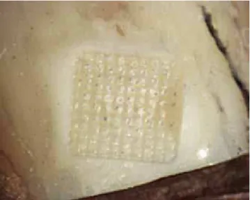
and Bergland3 (Table 1). To illustrate the debonding
pattern, photographs taken with the optical micro-scope with a 20X magniication were made (Fig 1).
Statistical analysis was performed using SPSS®
compare the results of the bond strength, analysis of variance (ANOVA) and the Tukey test were used. To compare the results of the ARI between different groups it was used the Kruskal-Wallis test.
RESULTS
The results of this study are shown in Tables 2 and 3. In G1 and G4, five specimens were excluded because of discrepancies in the obtained values.
The results show no statistical difference be-tween the groups where the same adhesive was used for bonding ceramic and metallic brackets. How-ever, there was significant difference between the three brands of adhesive used. The groups bonded with Transbond XT (G5 and G6) showed the high-est adhesion, followed by the groups bonded with Transbond Color Change (G3 and G4). The worst results were obtained with the Ortho Lite Cure resin (G1 and G2). The results for the ARI showed no significant difference between the groups and the most frequent result was the resin completely bonded to enamel surface after debonding (Fig 1).
DISCUSSION
The bond strength between brackets and enamel has been extensively researched over the past two de-cades. However, some issues still persist for the choice of adhesive by the orthodontist, especially due to the large amount of materials available in the market. The main issues fall back on the safety of the procedure,
Table 1 - Adhesive remnant index (ARI).
Table 3 - ARI comparison between groups. Figure 1 - Illustration of the ARI score 3 (20X magniication).
Score Quantity of adhesive bonded to enamel
0 No adhesive left on the tooth
1 Less than half of the adhesive left on the tooth 2 More than half of the adhesive left on the tooth
3 All adhesive left on the tooth with a distinct impression of the bracket mesh
* Diferent letters represent statistical diference (p <0.01).
Table 2 - Comparison of bond strength (MPa) between the groups (p < 0.01).
Group n Mean* SD
G1 25 2.47a 1.66
G2 30 2.75a 1.64
G3 30 4.26b 1.20
G4 25 4.22b 1.12
G5 30 5.62c 0.75
G6 30 5.95c 0.91
Group Index 0 Index 2 Index 3 Total
n % n % n % n %
G1 5 20.0 2 8.0 18 72.0 25 100.0 G2 8 26.6 4 13.3 18 60 30 100.0 G3 1 3.3 6 20 23 76.6 30 100.0
G4 - - 4 13.3 21 70 25 100.0
G5 2 6.6 5 16.6 23 76.6 30 100.0 G6 1 3.3 3 10 26 86.6 30 100.0 Total 17 10 24 14.11 129 75.88 170 100.0
cost-benefit and efficacy of the product. Plaque ac-cumulation around the brackets is an important issue, since the orthodontic appliances may be responsible
for caries and gingival disease.2,17 Thus, the colored
adhesives can be an important tool in reducing the excess of material and dental plaque.
In the present study, when the results of the ARI are evaluated, we note that for all groups the most common result was the score 3, where all the adhesive
remains in the enamel ater debonding. Olsen et al18
reported that the ARI index 3 is the safest, where the chance of dental damage is less likely. Therefore, we can conclude that the tensile strength obtained with both resins, traditional and colored, is safe.
A clinical observation noticed by the operator was that the Ortho Lite Cure orange pigment does not difer-entiate from tooth enamel color and it is not very useful in removing the excess of material. The pigments present in the Color Change Transbond are quite distinct from enamel and assist in the removal of resin excess, but their sandy consistency is not very pleasant to work with.
Further studies with these materials must be con-ducted to obtain more information about the shear strength. The impact of these adhesives on plaque ac-cumulation must also be thoroughly investigated in order to determine whether or not they have a posi-tive effect on plaque reduction during orthodontic treatment. Since these adhesives are more expensive than traditional ones, the benefits should be signifi-cant. It is recommended that more studies be con-ducted to investigate the ideal adhesion strength of orthodontic accessories in human enamel.
CONCLUSIONS
The results of this study demonstrated:
a) Tensile strengths of metallic and ceramic brack-ets were similar when the same adhesive was used.
b) Transbond XT adhesive had the highest bond strength, followed by Transbond Color Change ad-hesive, which presented clinically acceptable resis-tance values.
c) The worst results in terms of tensile strength were obtained with the Ortho Lite Cure adhesives, which were considered low for orthodontic use.
d) Further studies must be conducted on the clini-cal use of colored adhesives.
was used, which gives the orthodontist the possibil-ity to safely bond ceramic brackets. This fact brings tranquility, due to the increasing demand for esthetic orthodontic brackets in clinics. The bond strength obtained in G1 and G2 was too low to orthodontic
use.9,14,19 The adhesive strength for G3 and G4 was a
bit low for bonding orthodontic accessories, but it was
acceptable.22 The tensile strength shown in G5 and
G6 was adequate according to Aasrum et al.1
How-ever, while Reynolds21 recommends shear strength of
7 MPa, he also relates the diiculty of establishing the optimal adhesive strength between bracket and tooth enamel. Other studies have also demonstrated the dif-iculty of establishing a numerical value for bonding strength when it depends on many factors such as
ex-perimental procedures,11 adhesive thickness,8
bond-ing procedures,6 type of adhesive used,2,18 and the
di-rection and type of applied force,13 and other factors.
Therefore, the comparison of results becomes dii-cult. This raises some questions as: Are we basing our clinical choices properly, or should further studies be conducted to clarify these issues? Murray and
Hob-son15 as well as Eliades et al,7 report the diiculty of
comparing the results obtained in in vitro tests to the
in vivo reality. According to these authors, methods for
1. Aasrum E, Ng’ang’a PM, Dahm S, Ogaard B. Tensile bond strength of orthodontic brackets bonded with a luoride-releasing light-curing adhesive. An in vitro comparative study. Am J Orthod Dentofacial Orthop. 1993 Jul;104(1):48-50.
2. Abu Alhaija ESJ, Al-Wahadni AMS. Evaluation of shear bond strength with diferent enamel pre-treatments. Eur J Orthod. 2004;26(2):179-84. 3. Artun J, Bergland S. Clinical trials with crystal growth conditioning as an alternative to acid-etch enamel pretreatment. Am J Orthod. 1984;85(4):333-40.
4. Bishara SE, Fonseca JM, Fehr DE, Boyer DB. Debonding forces applied to ceramics brackets simulating clinical conditions. Angle Orthod. 1994:64(4):277-82.
5. Britton J, McInnes P, Weinberg R, Ledoux W, Retief D. Shear bond strength of ceramic orthodontic brackets to enamel. Am J Orthod Dentofacial Orthop. 1990;98(4):348-53.
6. Chung CH, Cuozzo PT, Mante FK. Shear bond strength of a resin-reinforced glass ionomer cement an in vitro comparative study. Am J Orthod Dentofacial Orthop. 1999;115(1):52-4.
7. Eliades G, Eliades T, Brantley WA, Watts DC, editors.
Dental materials in vivo: aging and related phenomena. Chicago: Quintessence; 2003. p. 3-23.
8. Evans LB, Powers JM. Factors afecting in vitro bond strength of no-mix orthodontic cements. Am J Orthod. 1985;87(6):508-12.
9. Habibi M, Nik TH, Hooshmand T. Comparison of debonding
characteristics of metal and ceramic orthodontics brackets to enamel: an in vitro study. Am J Orthod Dentofacial Orthop. 2007;132(5):675-9. 10. Huser MC, Baehni PC, Lang R. Efects of orthodontic bands on
microbiologic and clinical parameters. Am J Orthod Dentofacial Orthop. 1990;97(3):213-8.
11. Kanemura N, Sano H, Tagami J. Tensile bond strength to and SEM evaluation of ground and intact enamel surfaces. J Dent. 1999;27(7):523-30.
REFERENCES
12. Katona TR, Chen J. Engineering and experimental analyses of the tensile loads applied during strength testing of direct bonded orthodontic brackets. Am J Orthod Dentofacial Orthop. 1994;106(2):167-74. 13. Klocke A, Kahl-Nieke B. Inluence of force location in orthodontic shear
bond strength testing. Dent Mater. 2005;21(5):391-6.
14. Lopez JI. Retentive shear bond strengths of various bonding attachment bases. Am J Orthod. 1980;77(6):669-78.
15. Murray SD, Hobson RS. Comparison of in vivo and in vitro shear bond strength. Am J Orthod Dentofacial Orthop. 2003;123(1):2-9.
16. Naranjo AA, Triviño ML, Jaramillo A, Betancourth M, Botero JE. Changes in the subgingival microbiota and periodontal parameters before and 3 months after bracket placement. Am J Orthod Dentofacial Orthop. 2006;130(3):275.e17-22.
17. Oesterle LJ, Shellhart WC, Belanger GK. The use of bovine enamel in bonding studies. Am J Orthod Dentofacial Orthop. 1998;114(5):514-9. 18. Olsen ME, Bishara SE, Damon P, Jakobsen JR. Evaluation of
Scotchbond multipurpose and maleic acid as alternative methods of bonding orthodontic brackets. Am J Orthod Dentofacial Orthop. 1997;111(5):498-501.
19. Pickett KL, Sadowsky PL, Jacobson A, Laceield W. Orthodontic in vivo bond strength: comparison with in vitro results. Angle Orthod. 2001;71(2):141-8.
20. Retief DH, Harris BE, Bradley EL, Denys FR. Pyruvic acid as an etching agent in clinical dentistry. J Biomed Mater Res. 1985;19(3):335-48. 21. Reynolds IR. A review of direct orthodontic bonding. Brit J Orthod.
1975;2(3):171-8.
22. Sharma-Sayal SK, Rossouw PE, Kulkarni GV, Titley KC. The inluence of orthodontic bracket base design on shear bond strength. Am J Orthod Dentofacial Orthop. 2003;124(1):74-82.